|
Topographical Disorientation
Topographical disorientation is the inability to orient oneself in one's surroundings, sometimes as a result of focal brain damage. This disability may result from the inability to make use of selective spatial information (e.g., environmental landmarks) or to orient by means of specific cognitive strategies such as the ability to form a mental representation of the environment, also known as a cognitive map. It may be part of a syndrome known as visuospatial dysgnosia. Classification Topographical disorientation is the inability to find one's way through an environment due to cognitive impairment. Topographical disorientation has been studied for decades using case studies of patients who have selectively lost their ability to find their way within large-scale, locomotor environments. Several dozen case reports of topographical disorientation have been presented over the last century. Studying these people will aid in the understanding of the complex, multi-component behavior ... [...More Info...] [...Related Items...] OR: [Wikipedia] [Google] [Baidu] |
Focal And Diffuse Brain Injury
Focal and diffuse brain injury are ways to classify brain injury: focal injury occurs in a specific location, while diffuse injury occurs over a more widespread area. It is common for both focal and diffuse damage to occur as a result of the same event; many traumatic brain injuries have aspects of both focal and diffuse injury. Focal injuries are commonly associated with an injury in which the head strikes or is struck by an object; diffuse injuries are more often found in acceleration/deceleration injuries, in which the head does not necessarily contact anything, but brain tissue is damaged because tissue types with varying densities accelerate at different rates. In addition to physical trauma, other types of brain injury, such as stroke, can also produce focal and diffuse injuries. There may be primary and secondary brain injury processes. Focal A focal traumatic injury results from direct mechanical forces (such as occur when the head strikes a windshield in a vehicle a ... [...More Info...] [...Related Items...] OR: [Wikipedia] [Google] [Baidu] |
Posterior Parietal
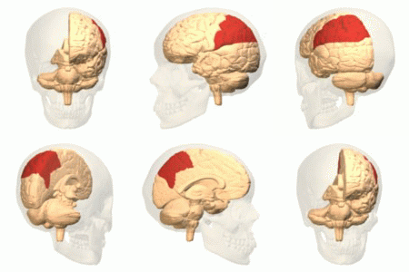
The parietal lobe is one of the four major lobes of the cerebral cortex in the brain of mammals. The parietal lobe is positioned above the temporal lobe and behind the frontal lobe and central sulcus. The parietal lobe integrates sensory information among various modalities, including spatial sense and navigation (proprioception), the main sensory receptive area for the sense of touch in the somatosensory cortex which is just posterior to the central sulcus in the postcentral gyrus, and the dorsal stream of the visual system. The major sensory inputs from the skin (touch, temperature, and pain receptors), relay through the thalamus to the parietal lobe. Several areas of the parietal lobe are important in language processing. The somatosensory cortex can be illustrated as a distorted figure – the cortical homunculus (Latin: "little man") in which the body parts are rendered according to how much of the somatosensory cortex is devoted to them. The superior parietal lobule and in ... [...More Info...] [...Related Items...] OR: [Wikipedia] [Google] [Baidu] |
Declarative Memory
Explicit memory (or declarative memory) is one of the two main types of long-term human memory, the other of which is implicit memory. Explicit memory is the conscious, intentional recollection of factual information, previous experiences, and concepts. This type of memory is dependent upon three processes: acquisition, consolidation, and retrieval. Explicit memory can be divided into two categories: episodic memory, which stores specific personal experiences, and semantic memory, which stores factual information.Tulving E. 1972. Episodic and semantic memory. In Organization of Memory, ed. E Tulving, W Donaldson, pp. 381–403. New York: Academic Explicit memory requires gradual learning, with multiple presentations of a stimulus and response. The counterpart to explicit memory is known as implicit memory, refers to memories acquired and used unconsciously such as skills (e.g. knowing how to get dressed) or perception. Unlike explicit memory, implicit memory learns rapidly, even ... [...More Info...] [...Related Items...] OR: [Wikipedia] [Google] [Baidu] |
Temporal Lobe
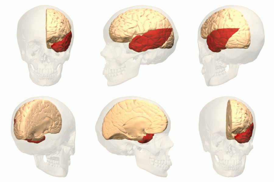
The temporal lobe is one of the four Lobes of the brain, major lobes of the cerebral cortex in the brain of mammals. The temporal lobe is located beneath the lateral fissure on both cerebral hemispheres of the mammalian brain. The temporal lobe is involved in processing sensory input into derived meanings for the appropriate retention of visual memory, language comprehension, and emotion association. ''Temporal'' refers to the head's Temple (anatomy), temples. Structure The Temple (anatomy)#Etymology, temporal Lobe (anatomy), lobe consists of structures that are vital for declarative or long-term memory. Declarative memory, Declarative (denotative) or Explicit memory, explicit memory is conscious memory divided into semantic memory (facts) and episodic memory (events). Medial temporal lobe structures that are critical for long-term memory include the hippocampus, along with the surrounding Hippocampal formation, hippocampal region consisting of the Perirhinal cortex, perirhinal, ... [...More Info...] [...Related Items...] OR: [Wikipedia] [Google] [Baidu] |
Hippocampus
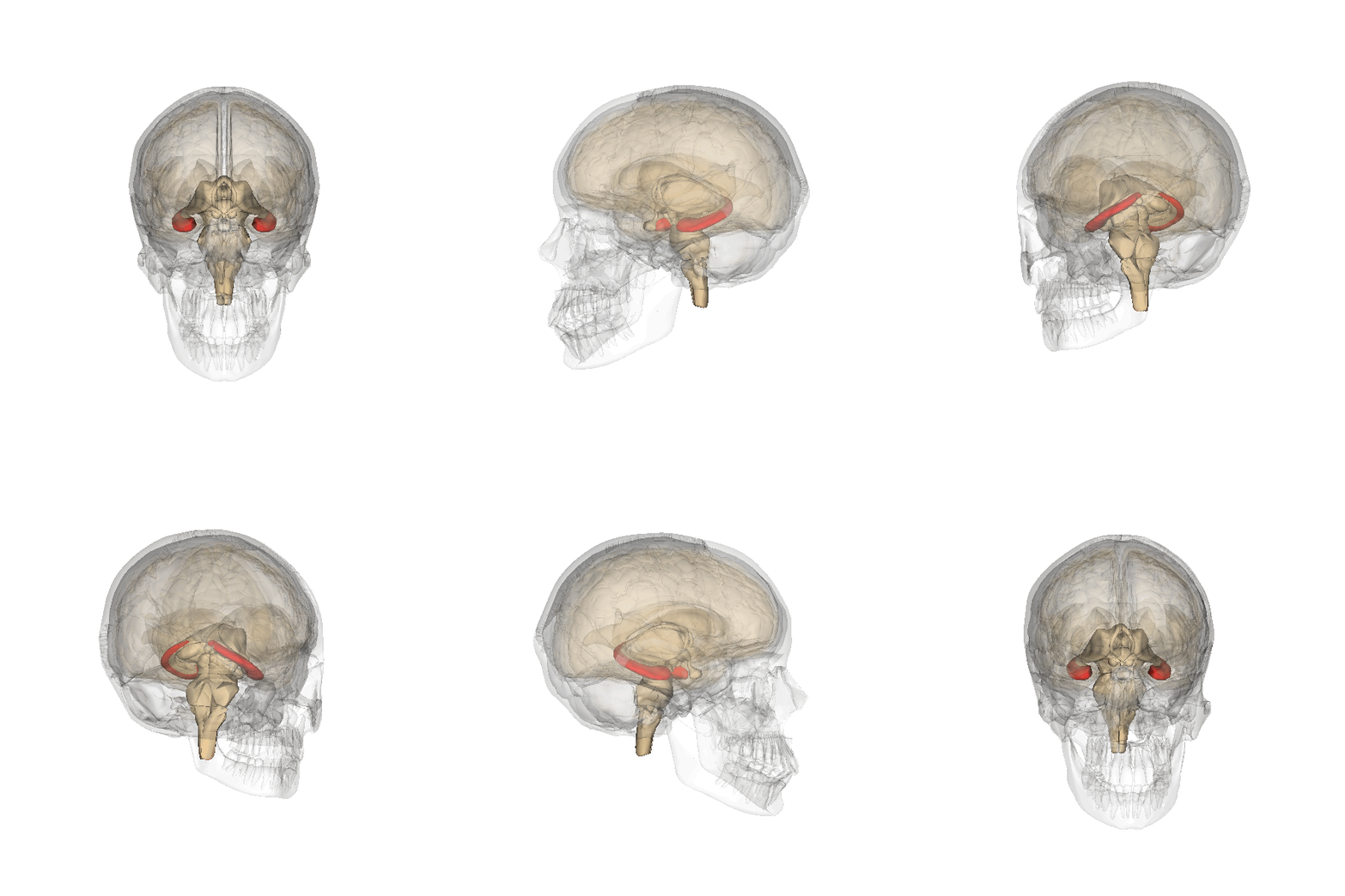
The hippocampus (via Latin from Greek , 'seahorse') is a major component of the brain of humans and other vertebrates. Humans and other mammals have two hippocampi, one in each side of the brain. The hippocampus is part of the limbic system, and plays important roles in the consolidation of information from short-term memory to long-term memory, and in spatial memory that enables navigation. The hippocampus is located in the allocortex, with neural projections into the neocortex in humans, as well as primates. The hippocampus, as the medial pallium, is a structure found in all vertebrates. In humans, it contains two main interlocking parts: the hippocampus proper (also called ''Ammon's horn''), and the dentate gyrus. In Alzheimer's disease (and other forms of dementia), the hippocampus is one of the first regions of the brain to suffer damage; short-term memory loss and disorientation are included among the early symptoms. Damage to the hippocampus can also result from ... [...More Info...] [...Related Items...] OR: [Wikipedia] [Google] [Baidu] |
Parahippocampus
The parahippocampal gyrus (or hippocampal gyrus') is a grey matter cortical region of the brain that surrounds the hippocampus and is part of the limbic system. The region plays an important role in memory encoding and retrieval. It has been involved in some cases of hippocampal sclerosis. Asymmetry has been observed in schizophrenia. Structure The anterior part of the gyrus includes the perirhinal and entorhinal cortices. The term parahippocampal cortex is used to refer to an area that encompasses both the posterior parahippocampal gyrus and the medial portion of the fusiform gyrus. Function Scene recognition The parahippocampal place area (PPA) is a sub-region of the parahippocampal cortex that lies medially in the inferior temporo-occipital cortex. PPA plays an important role in the encoding and recognition of environmental scenes (rather than faces). fMRI studies indicate that this region of the brain becomes highly active when human subjects view topographical scene sti ... [...More Info...] [...Related Items...] OR: [Wikipedia] [Google] [Baidu] |
Anterograde Amnesia
In neurology, anterograde amnesia is the inability to create new memories after the event that caused amnesia, leading to a partial or complete inability to recall the recent past, while long-term memories from before the event remain intact. This is in contrast to retrograde amnesia, where memories created prior to the event are lost while new memories can still be created. Both can occur together in the same patient. To a large degree, anterograde amnesia remains a mysterious ailment because the precise mechanism of storing memories is not yet well understood, although it is known that the regions of the brain involved are certain sites in the temporal cortex, especially in the hippocampus and nearby subcortical regions. Signs and symptoms People with anterograde amnesic syndromes may present with widely varying degrees of forgetfulness. Some with severe cases have a combined form of anterograde and retrograde amnesia, sometimes called global amnesia. In the case of drug-i ... [...More Info...] [...Related Items...] OR: [Wikipedia] [Google] [Baidu] |
Visual Perception
Visual perception is the ability to interpret the surrounding environment through photopic vision (daytime vision), color vision, scotopic vision (night vision), and mesopic vision (twilight vision), using light in the visible spectrum reflected by objects in the environment. This is different from visual acuity, which refers to how clearly a person sees (for example "20/20 vision"). A person can have problems with visual perceptual processing even if they have 20/20 vision. The resulting perception is also known as vision, sight, or eyesight (adjectives ''visual'', ''optical'', and ''ocular'', respectively). The various physiological components involved in vision are referred to collectively as the visual system, and are the focus of much research in linguistics, psychology, cognitive science, neuroscience, and molecular biology, collectively referred to as vision science. Visual system In humans and a number of other mammals, light enters the eye through the cornea and is ... [...More Info...] [...Related Items...] OR: [Wikipedia] [Google] [Baidu] |
Cerebral Hemorrhage
Intracerebral hemorrhage (ICH), also known as cerebral bleed, intraparenchymal bleed, and hemorrhagic stroke, or haemorrhagic stroke, is a sudden bleeding into the tissues of the brain, into its ventricles, or into both. It is one kind of bleeding within the skull and one kind of stroke. Symptoms can include headache, one-sided weakness, vomiting, seizures, decreased level of consciousness, and neck stiffness. Often, symptoms get worse over time. Fever is also common. Causes include brain trauma, aneurysms, arteriovenous malformations, and brain tumors. The biggest risk factors for spontaneous bleeding are high blood pressure and amyloidosis. Other risk factors include alcoholism, low cholesterol, blood thinners, and cocaine use. Diagnosis is typically by CT scan. Other conditions that may present similarly include ischemic stroke. Treatment should typically be carried out in an intensive care unit. Guidelines recommend decreasing the blood pressure to a systolic of 1 ... [...More Info...] [...Related Items...] OR: [Wikipedia] [Google] [Baidu] |
Retrosplenial Region
The retrosplenial cortex (RSC) is a cortical area in the brain comprising Brodmann areas 29 and 30. It is secondary association cortex, making connections with numerous other brain regions. The region's name refers to its anatomical location immediately behind the splenium of the corpus callosum in primates, although in rodents it is located more towards the brain surface and is relatively larger. Its function is currently not well understood, but its location close to visual areas and also to the hippocampal spatial/memory system suggest it may have a role in mediating between perceptual and memory functions, particularly in the spatial domain. However, its exact contribution to either space or memory processing has been hard to pin down. Anatomy There is large variation in the region's size across different species. In humans it comprises roughly 0.3% of the entire cortical surface whereas in rabbits it is at least 10% and in rats it extends for more than half the cerebrum dor ... [...More Info...] [...Related Items...] OR: [Wikipedia] [Google] [Baidu] |
Agnosia
Agnosia is the inability to process sensory information. Often there is a loss of ability to recognize objects, persons, sounds, shapes, or smells while the specific sense is not defective nor is there any significant memory loss. It is usually associated with brain injury or neurological illness, particularly after damage to the occipitotemporal border, which is part of the ventral stream. Agnosia only affects a single modality, such as vision or hearing. More recently, a top-down interruption is considered to cause the disturbance of handling perceptual information. Types Visual agnosia Visual agnosia is a broad category that refers to a deficiency in the ability to recognize visual objects. Visual agnosia can be further subdivided into two different subtypes: apperceptive visual agnosia and associative visual agnosia. Individuals with apperceptive visual agnosia display the ability to see contours and outlines when shown an object, but they experience difficulty if ask ... [...More Info...] [...Related Items...] OR: [Wikipedia] [Google] [Baidu] |
Posterior Cingulate
The posterior cingulate cortex (PCC) is the caudal part of the cingulate cortex, located posterior to the anterior cingulate cortex. This is the upper part of the "limbic lobe". The cingulate cortex is made up of an area around the midline of the brain. Surrounding areas include the retrosplenial cortex and the precuneus. Cytoarchitectonically the posterior cingulate cortex is associated with Brodmann areas 23 and 31. The PCC forms a central node in the default mode network of the brain. It has been shown to communicate with various brain networks simultaneously and is involved in diverse functions. Along with the precuneus, the PCC has been implicated as a neural substrate for human awareness in numerous studies of both the anesthesized and vegetative (coma) states. Imaging studies indicate a prominent role for the PCC in pain and episodic memory retrieval. Increased size of the ventral PCC is related to a decline in working memory performance. The PCC has also been strongly ... [...More Info...] [...Related Items...] OR: [Wikipedia] [Google] [Baidu] |


.jpg)
