|
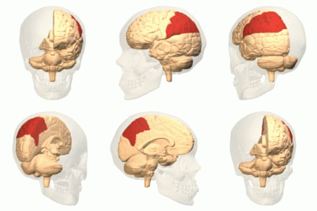
Posterior Cingulate
The posterior cingulate cortex (PCC) is the caudal part of the cingulate cortex, located posterior to the anterior cingulate cortex. This is the upper part of the "limbic lobe". The cingulate cortex is made up of an area around the midline of the brain. Surrounding areas include the retrosplenial cortex and the precuneus. Cytoarchitectonically the posterior cingulate cortex is associated with Brodmann areas 23 and 31. The PCC forms a central node in the default mode network of the brain. It has been shown to communicate with various brain networks simultaneously and is involved in diverse functions. Along with the precuneus, the PCC has been implicated as a neural substrate for human awareness in numerous studies of both the anesthesized and vegetative (coma) states. Imaging studies indicate a prominent role for the PCC in pain and episodic memory retrieval. Increased size of the ventral PCC is related to a decline in working memory performance. The PCC has also been strongly ... [...More Info...] [...Related Items...] OR: [Wikipedia] [Google] [Baidu] |
Cingulate Gyrus
The cingulate cortex is a part of the brain situated in the medial aspect of the cerebral cortex. The cingulate cortex includes the entire cingulate gyrus, which lies immediately above the corpus callosum, and the continuation of this in the cingulate sulcus. The cingulate cortex is usually considered part of the limbic lobe. It receives inputs from the thalamus and the neocortex, and projects to the entorhinal cortex via the cingulum. It is an integral part of the limbic system, which is involved with emotion formation and processing, learning, and memory. The combination of these three functions makes the cingulate gyrus highly influential in linking motivational outcomes to behavior (e.g. a certain action induced a positive emotional response, which results in learning). This role makes the cingulate cortex highly important in disorders such as depression and schizophrenia. It also plays a role in executive function and respiratory control. Etymology The term ''cingul ... [...More Info...] [...Related Items...] OR: [Wikipedia] [Google] [Baidu] |
Parieto-occipital Sulcus
In neuroanatomy, the parieto-occipital sulcus (also called the parieto-occipital fissure) is a deep sulcus in the cerebral cortex that marks the boundary between the cuneus and precuneus, and also between the parietal and occipital lobes. Only a small part can be seen on the lateral surface of the hemisphere, its chief part being on the medial surface. The lateral part of the parieto-occipital sulcus (Fig. 726) is situated about 5 cm in front of the occipital pole of the hemisphere, and measures about 1.25 cm. in length. The medial part of the parieto-occipital sulcus (Fig. 727) runs downward and forward as a deep cleft on the medial surface of the hemisphere, and joins the calcarine fissure below and behind the posterior end of the corpus callosum. In most cases, it contains a submerged gyrus. Function The parieto-occipital lobe has been found in various neuroimaging studies, including PET (positron-emission-tomography) studies, and SPECT (single-photon emission computed ... [...More Info...] [...Related Items...] OR: [Wikipedia] [Google] [Baidu] |
Parietal Lobe
The parietal lobe is one of the four major lobes of the cerebral cortex in the brain of mammals. The parietal lobe is positioned above the temporal lobe and behind the frontal lobe and central sulcus. The parietal lobe integrates sensory information among various modalities, including spatial sense and navigation (proprioception), the main sensory receptive area for the sense of touch in the somatosensory cortex which is just posterior to the central sulcus in the postcentral gyrus, and the dorsal stream of the visual system. The major sensory inputs from the skin (touch, temperature, and pain receptors), relay through the thalamus to the parietal lobe. Several areas of the parietal lobe are important in language processing. The somatosensory cortex can be illustrated as a distorted figure – the cortical homunculus (Latin: "little man") in which the body parts are rendered according to how much of the somatosensory cortex is devoted to them. The superior parietal lobule and in ... [...More Info...] [...Related Items...] OR: [Wikipedia] [Google] [Baidu] |
Frontal Lobe
The frontal lobe is the largest of the four major lobes of the brain in mammals, and is located at the front of each cerebral hemisphere (in front of the parietal lobe and the temporal lobe). It is parted from the parietal lobe by a groove between tissues called the central sulcus and from the temporal lobe by a deeper groove called the lateral sulcus (Sylvian fissure). The most anterior rounded part of the frontal lobe (though not well-defined) is known as the frontal pole, one of the three poles of the cerebrum. The frontal lobe is covered by the frontal cortex. The frontal cortex includes the premotor cortex, and the primary motor cortex – parts of the motor cortex. The front part of the frontal cortex is covered by the prefrontal cortex. There are four principal gyri in the frontal lobe. The precentral gyrus is directly anterior to the central sulcus, running parallel to it and contains the primary motor cortex, which controls voluntary movements of specific body parts ... [...More Info...] [...Related Items...] OR: [Wikipedia] [Google] [Baidu] |
Brodmann Area 25
Brodmann area 25 (BA25) is the subgenual area, area subgenualis or subgenual cingulatea area in the cerebral cortex of the brain and delineated based on its cytoarchitectonic characteristics. It is the 25th "Brodmann area" defined by Korbinian Brodmann (thus its name). BA25 is located in the cingulate region as a narrow band in the caudal portion of the subcallosal area adjacent to the paraterminal gyrus. The posterior parolfactory sulcus separates the paraterminal gyrus from BA25. Rostrally it is bound by the prefrontal area 11 of Brodmann. History Brodmann described this area as it is labeled now in 1909. Originally in 1905, Brodmann labeled the area as part of area 24. In 1909, he divided the area into area 24 and 25. Function This region is extremely rich in serotonin transporters and is considered as a governor for a vast network involving areas like hypothalamus and brain stem, which influences changes in appetite and sleep; the amygdala and insula, which affect the ... [...More Info...] [...Related Items...] OR: [Wikipedia] [Google] [Baidu] |
Ventromedial Prefrontal Cortex
The ventromedial prefrontal cortex (vmPFC) is a part of the prefrontal cortex in the mammalian brain. The ventral medial prefrontal is located in the frontal lobe at the bottom of the cerebral hemispheres and is implicated in the processing of risk and fear, as it is critical in the regulation of amygdala activity in humans. It also plays a role in the inhibition of emotional responses, and in the process of decision-making and self-control. It is also involved in the cognitive evaluation of morality. Anatomy While the ventromedial prefrontal cortex does not have a universally agreed on demarcation, in most sources, it is equivalent to the ventromedial reward network of Öngür and Price. This network includes Brodmann area 10, Brodmann area 14, Brodmann area 25, and Brodmann area 32, as well as portions of Brodmann area 11, Brodmann area 12, and Brodmann area 13. However, not all sources agree on the boundaries of the area. Different researchers use the term ''ventromedial pref ... [...More Info...] [...Related Items...] OR: [Wikipedia] [Google] [Baidu] |
Parahippocampal Gyrus
The parahippocampal gyrus (or hippocampal gyrus') is a grey matter cortical region of the brain that surrounds the hippocampus and is part of the limbic system. The region plays an important role in memory encoding and retrieval. It has been involved in some cases of hippocampal sclerosis. Asymmetry has been observed in schizophrenia. Structure The anterior part of the gyrus includes the perirhinal and entorhinal cortices. The term parahippocampal cortex is used to refer to an area that encompasses both the posterior parahippocampal gyrus and the medial portion of the fusiform gyrus. Function Scene recognition The parahippocampal place area (PPA) is a sub-region of the parahippocampal cortex that lies medially in the inferior temporo-occipital cortex. PPA plays an important role in the encoding and recognition of environmental scenes (rather than faces). fMRI studies indicate that this region of the brain becomes highly active when human subjects view topographical scene sti ... [...More Info...] [...Related Items...] OR: [Wikipedia] [Google] [Baidu] |
Hippocampal Formation
The hippocampal formation is a compound structure in the Temporal lobe#Medial temporal lobe, medial temporal lobe of the brain. It forms a c-shaped bulge on the floor of the temporal horn of the Lateral ventricles, lateral ventricle. There is no consensus concerning which brain regions are encompassed by the term, with some authors defining it as the dentate gyrus, the hippocampus proper and the subiculum; and others including also the Brodmann area 27, presubiculum, parasubiculum, and entorhinal cortex. The hippocampal formation is thought to play a role in memory, spatial navigation and control of attention. The neural layout and pathways within the hippocampal formation are very similar in all mammals. History and function During the nineteenth and early twentieth centuries, based largely on the observation that, between species, the size of the olfactory bulb varies with the size of the parahippocampal gyrus, the hippocampal formation was thought to be part of the olfactory sy ... [...More Info...] [...Related Items...] OR: [Wikipedia] [Google] [Baidu] |
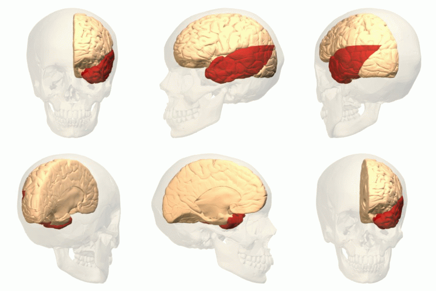
Temporal Lobe
The temporal lobe is one of the four Lobes of the brain, major lobes of the cerebral cortex in the brain of mammals. The temporal lobe is located beneath the lateral fissure on both cerebral hemispheres of the mammalian brain. The temporal lobe is involved in processing sensory input into derived meanings for the appropriate retention of visual memory, language comprehension, and emotion association. ''Temporal'' refers to the head's Temple (anatomy), temples. Structure The Temple (anatomy)#Etymology, temporal Lobe (anatomy), lobe consists of structures that are vital for declarative or long-term memory. Declarative memory, Declarative (denotative) or Explicit memory, explicit memory is conscious memory divided into semantic memory (facts) and episodic memory (events). Medial temporal lobe structures that are critical for long-term memory include the hippocampus, along with the surrounding Hippocampal formation, hippocampal region consisting of the Perirhinal cortex, perirhinal, ... [...More Info...] [...Related Items...] OR: [Wikipedia] [Google] [Baidu] |
Limbic System
The limbic system, also known as the paleomammalian cortex, is a set of brain structures located on both sides of the thalamus, immediately beneath the medial temporal lobe of the cerebrum primarily in the forebrain.Schacter, Daniel L. 2012. ''Psychology''.sec. 3.20 It supports a variety of functions including emotion, behavior, long-term memory, and olfaction. Emotional life is largely housed in the limbic system, and it critically aids the formation of memories. With a primordial structure, the limbic system is involved in lower order emotional processing of input from sensory systems and consists of the amygdaloid nuclear complex (amygdala), mammillary bodies, stria medullaris, central gray and dorsal and ventral nuclei of Gudden. This processed information is often relayed to a collection of structures from the telencephalon, diencephalon, and mesencephalon, including the prefrontal cortex, cingulate gyrus, limbic thalamus, hippocampus including the parahippocampal gyrus an ... [...More Info...] [...Related Items...] OR: [Wikipedia] [Google] [Baidu] |
Cytoarchitecture
Cytoarchitecture (Greek '' κύτος''= "cell" + '' ἀρχιτεκτονική''= "architecture"), also known as cytoarchitectonics, is the study of the cellular composition of the central nervous system's tissues under the microscope. Cytoarchitectonics is one of the ways to parse the brain, by obtaining sections of the brain using a microtome and staining them with chemical agents which reveal where different neurons are located. The study of the parcellation of ''nerve fibers'' (primarily axons) into layers forms the subject of myeloarchitectonics ( History of the cerebral cytoarchitecture Defining cerebral cytoarchitecture began with the advent of —the science of slicing a ...[...More Info...] [...Related Items...] OR: [Wikipedia] [Google] [Baidu] |
Allocortex
The allocortex or heterogenetic cortex, and neocortex are the two types of cerebral cortex in the brain. The allocortex is the much smaller area of cortex taking up just 10 %, the neocortex takes up the remaining 90 %. It is characterized by having just three or four cortical layers, in contrast with the six layers of the neocortex. There are three subtypes of allocortex: the paleocortex, the archicortex, and the periallocortex—a transitional zone between the neocortex and the allocortex. The specific regions of the brain usually described as belonging to the allocortex are the olfactory system, and the hippocampus. Allocortex is termed heterogenetic cortex, because during development it never has the six-layered architecture of homogenetic neocortex. It differs from hetero''typic'' cortex, a type of cerebral cortex, which during prenatal development, passes through a six-layered stage to have fewer layers, such as in Brodmann area 4 that lacks granule cells. Structure The all ... [...More Info...] [...Related Items...] OR: [Wikipedia] [Google] [Baidu] |


.png)

