|
Tooth Eruption
Tooth eruption is a process in tooth development in which the teeth enter the mouth and become visible. It is currently believed that the periodontal ligament plays an important role in tooth eruption. The first human teeth to appear, the deciduous (primary) teeth (also known as baby or milk teeth), erupt into the mouth from around 6 months until 2 years of age, in a process known as " teething". These teeth are the only ones in the mouth until a person is about 6 years old creating the primary dentition stage. At that time, the first permanent tooth erupts and begins a time in which there is a combination of primary and permanent teeth, known as the mixed dentition stage, which lasts until the last primary tooth is lost. Then, the remaining permanent teeth erupt into the mouth during the permanent dentition stage. Theories Although researchers agree that tooth eruption is a complex process, there is little agreement on the identity of the mechanism that controls eruption. There ... [...More Info...] [...Related Items...] OR: [Wikipedia] [Google] [Baidu] |
Developmental Biology
Developmental biology is the study of the process by which animals and plants grow and develop. Developmental biology also encompasses the biology of regeneration, asexual reproduction, metamorphosis, and the growth and differentiation of stem cells in the adult organism. Perspectives The main processes involved in the embryonic development of animals are: tissue patterning (via regional specification and patterned cell differentiation); tissue growth; and tissue morphogenesis. * Regional specification refers to the processes that create the spatial patterns in a ball or sheet of initially similar cells. This generally involves the action of cytoplasmic determinants, located within parts of the fertilized egg, and of inductive signals emitted from signaling centers in the embryo. The early stages of regional specification do not generate functional differentiated cells, but cell populations committed to developing to a specific region or part of the organism. These are ... [...More Info...] [...Related Items...] OR: [Wikipedia] [Google] [Baidu] |
Dentition
Dentition pertains to the development of teeth and their arrangement in the mouth. In particular, it is the characteristic arrangement, kind, and number of teeth in a given species at a given age. That is, the number, type, and morpho-physiology (that is, the relationship between the shape and form of the tooth in question and its inferred function) of the teeth of an animal. Animals whose teeth are all of the same type, such as most non-mammalian vertebrates, are said to have '' homodont'' dentition, whereas those whose teeth differ morphologically are said to have ''heterodont'' dentition. The dentition of animals with two successions of teeth (deciduous, permanent) is referred to as ''diphyodont'', while the dentition of animals with only one set of teeth throughout life is ''monophyodont''. The dentition of animals in which the teeth are continuously discarded and replaced throughout life is termed ''polyphyodont''. The dentition of animals in which the teeth are set in soc ... [...More Info...] [...Related Items...] OR: [Wikipedia] [Google] [Baidu] |
Maxillary Third Molar
A third molar, commonly called wisdom tooth, is one of the three molars per quadrant of the human dentition. It is the most posterior of the three. The age at which wisdom teeth come through ( erupt) is variable, but this generally occurs between late teens and early twenties. Most adults have four wisdom teeth, one in each of the four quadrants, but it is possible to have none, fewer, or more, in which case the extras are called supernumerary teeth. Wisdom teeth may get stuck ( impacted) against other teeth if there is not enough space for them to come through normally. Impacted wisdom teeth are still sometimes removed for orthodontic treatment, believing that they move the other teeth and cause crowding, though this is not held anymore as true. Impacted wisdom teeth may suffer from tooth decay if oral hygiene becomes more difficult. Wisdom teeth which are partially erupted through the gum may also cause inflammation and infection in the surrounding gum tissues, termed peric ... [...More Info...] [...Related Items...] OR: [Wikipedia] [Google] [Baidu] |
Maxillary Second Molar
The maxillary second molar is the tooth located distally (away from the midline of the face) from both the maxillary first molars of the mouth but mesial (toward the midline of the face) from both maxillary third molars. This is true only in permanent teeth. In deciduous (baby) teeth, the maxillary second molar is the last tooth in the mouth and does not have a third molar behind it. The function of this molar is similar to that of all molars in regard to grinding being the principal action during mastication, commonly known as chewing. There are usually four cusps on maxillary molars, two on the buccal (side nearest the cheek) and two palatal (side nearest the palate). There are great differences between the deciduous (baby) maxillary molars and those of the permanent maxillary molars, even though their function are similar. The permanent maxillary molars are not considered to have any teeth that precede it. Despite being named molars, the deciduous molars are followed b ... [...More Info...] [...Related Items...] OR: [Wikipedia] [Google] [Baidu] |
Maxillary Canine
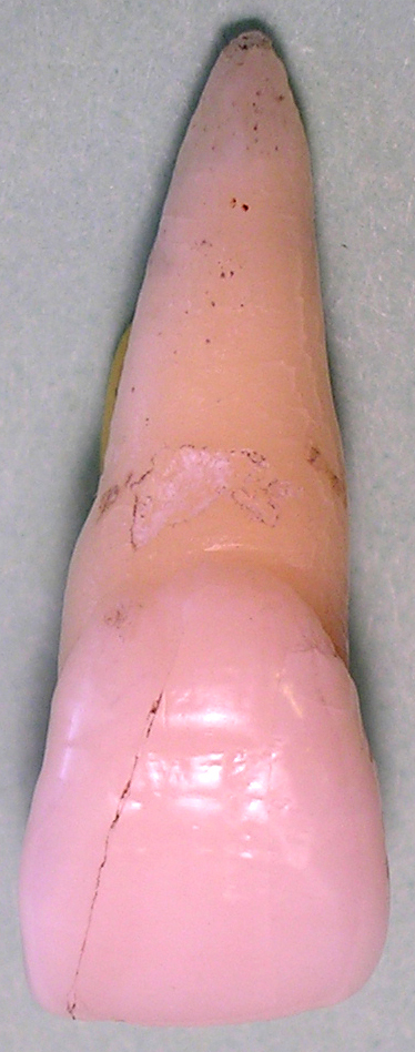
In human dentistry, the maxillary canine is the tooth located laterally (away from the midline of the face) from both maxillary lateral incisors of the mouth but mesial (toward the midline of the face) from both maxillary first premolars. Both the maxillary and mandibular canines are called the "cornerstone" of the mouth because they are all located three teeth away from the midline, and separate the premolars from the incisors. The location of the canines reflect their dual function as they complement both the premolars and incisors during mastication, commonly known as chewing. Nonetheless, the most common action of the canines is tearing of food. The canines often erupt in the upper gums several millimeters above the gum line. The canine teeth are able to withstand the tremendous lateral pressure caused by chewing. There is a single cusp on canines, and they resemble the prehensile teeth found in carnivorous animals such as the extinct saber-toothed cat. Though relatively th ... [...More Info...] [...Related Items...] OR: [Wikipedia] [Google] [Baidu] |
Maxillary Second Premolar
The maxillary second premolar is one of two teeth located in the upper maxilar, laterally (away from the midline of the face) from both the maxillary first premolars of the mouth but mesial (toward the midline of the face) from both maxillary first molars. The function of this premolar is similar to that of first molars in regard to grinding being the principal action during mastication, commonly known as chewing. There are two cusps on maxillary second premolars, but both of them are less sharp than those of the maxillary first premolars. There are no deciduous (baby) maxillary premolars. Instead, the teeth that precede the permanent maxillary premolars are the deciduous maxillary molars. In the universal system of notation, the permanent maxillary premolars are designated by a number. The right permanent maxillary second premolar is known as "4", and the left one is known as "13". In the Palmer notation Palmer notation (sometimes called the "Military System" and named ... [...More Info...] [...Related Items...] OR: [Wikipedia] [Google] [Baidu] |
Maxillary First Premolar
The maxillary first premolar is one of two teeth located in the upper jaw, laterally (away from the midline of the face) from both the maxillary canines of the mouth but mesial (toward the midline of the face) from both maxillary second premolars. The function of this premolar is similar to that of canines in regard to tearing being the principal action during mastication, commonly known as chewing. There are two cusps on maxillary first premolars, and the buccal (closest to the cheek) cusp is sharp enough to resemble the prehensile teeth found in carnivorous animals. There are no deciduous maxillary premolars. Around 10-11 years of age, the primary molars are shed and the permanent premolars erupt in their place. It takes about 3 years for the adult premolar and its root to fully calcify. Due to its long buccal root with narrow root canal and short palatal root with wide root canal, the upper 1st premolar is very prone to fracture during exodontia, hence, it is sometimes refer ... [...More Info...] [...Related Items...] OR: [Wikipedia] [Google] [Baidu] |
Maxillary Lateral Incisor
The maxillary lateral incisors are a pair of upper (maxillary) teeth that are located laterally (away from the midline of the face) from both maxillary central incisors of the mouth and medially (toward the midline of the face) from both maxillary canines. As with all incisors, their function is for shearing or cutting food during mastication, commonly known as chewing. There are generally no cusps on the teeth, but the rare condition known as talon cusps are most prevalent on the maxillary lateral incisors. The surface area of the tooth used in eating is called an incisal ridge or incisal edge. Though relatively the same, there are some minor differences between the deciduous (baby) maxillary lateral incisor and that of the permanent maxillary lateral incisor. The maxillary lateral incisors occlude in opposition to the mandibular lateral incisors. Notation In the universal system of notation, the deciduous maxillary lateral incisors are designated by a letter written in ... [...More Info...] [...Related Items...] OR: [Wikipedia] [Google] [Baidu] |
Maxillary Central Incisor
The maxillary central incisor is a human tooth in the front upper jaw, or maxilla, and is usually the most visible of all teeth in the mouth. It is located mesial (closer to the midline of the face) to the maxillary lateral incisor. As with all incisors, their function is for shearing or cutting food during mastication (chewing). There is typically a single cusp on each tooth, called an incisal ridge or incisal edge. Formation of these teeth begins at 14 weeks in utero for the deciduous (baby) set and 3–4 months of age for the permanent set. There are some minor differences between the deciduous maxillary central incisor and that of the permanent maxillary central incisor. The deciduous tooth appears in the mouth at 8–12 months of age and shed at 6–7 years, and is replaced by the permanent tooth around 7–8 years of age. The permanent tooth is larger and is longer than it is wide. The maxillary central incisors contact each other at the midline of the face. T ... [...More Info...] [...Related Items...] OR: [Wikipedia] [Google] [Baidu] |
Maxillary First Molar
The maxillary first molar is the human tooth located laterally (away from the midline of the face) from both the maxillary second premolars of the mouth but mesial (toward the midline of the face) from both maxillary second molars. The function of this molar is similar to that of all molars in regard to grinding being the principal action during mastication, commonly known as chewing. There are usually four cusps on maxillary molars, two on the buccal (side nearest the cheek) and two palatal (side nearest the palate). There may also be a fifth smaller cusp on the palatal side known as the Cusp of Carabelli. Normally, maxillary molars have four lobes, two buccal and two lingual, which are named in the same manner as the cusps that represent them ( mesiobuccal, distobuccal, mesiolingual, and distolingual lobes). Unlike the anterior teeth and premolars, molars do not exhibit facial developmental depressions. Evidence of lobe separation can be found in the central groove, whic ... [...More Info...] [...Related Items...] OR: [Wikipedia] [Google] [Baidu] |
Canine Tooth
In mammalian oral anatomy, the canine teeth, also called cuspids, dog teeth, or (in the context of the upper jaw) fangs, eye teeth, vampire teeth, or vampire fangs, are the relatively long, pointed teeth. They can appear more flattened however, causing them to resemble incisors and leading them to be called ''incisiform''. They developed and are used primarily for firmly holding food in order to tear it apart, and occasionally as weapons. They are often the largest teeth in a mammal's mouth. Individuals of most species that develop them normally have four, two in the upper jaw and two in the lower, separated within each jaw by incisors; humans and dogs are examples. In most species, canines are the anterior-most teeth in the maxillary bone. The four canines in humans are the two maxillary canines and the two mandibular canines. Details There are generally four canine teeth: two in the upper (maxillary) and two in the lower (mandibular) arch. A canine is placed laterall ... [...More Info...] [...Related Items...] OR: [Wikipedia] [Google] [Baidu] |
Incisor
Incisors (from Latin ''incidere'', "to cut") are the front teeth present in most mammals. They are located in the premaxilla above and on the mandible below. Humans have a total of eight (two on each side, top and bottom). Opossums have 18, whereas armadillos have none. Structure Adult humans normally have eight incisors, two of each type. The types of incisor are: * maxillary central incisor (upper jaw, closest to the center of the lips) * maxillary lateral incisor (upper jaw, beside the maxillary central incisor) * mandibular central incisor (lower jaw, closest to the center of the lips) * mandibular lateral incisor (lower jaw, beside the mandibular central incisor) Children with a full set of deciduous teeth (primary teeth) also have eight incisors, named the same way as in permanent teeth. Young children may have from zero to eight incisors depending on the stage of their tooth eruption and tooth development. Typically, the mandibular central incisors erupt first, ... [...More Info...] [...Related Items...] OR: [Wikipedia] [Google] [Baidu] |


