|
Maxillary Lateral Incisor
The maxillary lateral incisors are a pair of upper (maxillary) teeth that are located laterally (away from the midline of the face) from both maxillary central incisors of the mouth and medially (toward the midline of the face) from both maxillary canines. As with all incisors, their function is for shearing or cutting food during mastication, commonly known as chewing. There are generally no cusps on the teeth, but the rare condition known as talon cusps are most prevalent on the maxillary lateral incisors. The surface area of the tooth used in eating is called an incisal ridge or incisal edge. Though relatively the same, there are some minor differences between the deciduous (baby) maxillary lateral incisor and that of the permanent maxillary lateral incisor. The maxillary lateral incisors occlude in opposition to the mandibular lateral incisors. Notation In the universal system of notation, the deciduous maxillary lateral incisors are designated by a letter written in ... [...More Info...] [...Related Items...] OR: [Wikipedia] [Google] [Baidu] |
Maxilla
The maxilla (plural: ''maxillae'' ) in vertebrates is the upper fixed (not fixed in Neopterygii) bone of the jaw formed from the fusion of two maxillary bones. In humans, the upper jaw includes the hard palate in the front of the mouth. The two maxillary bones are fused at the intermaxillary suture, forming the anterior nasal spine. This is similar to the mandible (lower jaw), which is also a fusion of two mandibular bones at the mandibular symphysis. The mandible is the movable part of the jaw. Structure In humans, the maxilla consists of: * The body of the maxilla * Four processes ** the zygomatic process ** the frontal process of maxilla ** the alveolar process ** the palatine process * three surfaces – anterior, posterior, medial * the Infraorbital foramen * the maxillary sinus * the incisive foramen Articulations Each maxilla articulates with nine bones: * two of the cranium: the frontal and ethmoid * seven of the face: the nasal, zygomatic, lacrimal, ... [...More Info...] [...Related Items...] OR: [Wikipedia] [Google] [Baidu] |
Permanent Teeth
Permanent teeth or adult teeth are the second set of teeth formed in diphyodont mammals. In humans and old world simians, there are thirty-two permanent teeth, consisting of six maxillary and six mandibular molars, four maxillary and four mandibular premolars, two maxillary and two mandibular canines, four maxillary and four mandibular incisors. Timeline The first permanent tooth usually appears in the mouth In animal anatomy, the mouth, also known as the oral cavity, or in Latin cavum oris, is the opening through which many animals take in food and issue vocal sounds. It is also the cavity lying at the upper end of the alimentary canal, bounded on t ... at around six years of age, and the mouth will then be in a transition time with both primary (or deciduous dentition) teeth and permanent teeth during the mixed dentition period until the last primary tooth is lost or shed. The first of the permanent teeth to erupt are the permanent first molars, right behind the last ' ... [...More Info...] [...Related Items...] OR: [Wikipedia] [Google] [Baidu] |
Cementoenamel Junction
The cementoenamel junction, frequently abbreviated as the CEJ, is a slightly visible anatomical border identified on a tooth. It is the location where the enamel, which covers the anatomical crown of a tooth, and the cementum, which covers the anatomical root of a tooth, meet. Informally it is known as the neck of the tooth. The border created by these two dental tissues has much significance as it is usually the location where the gingiva The gums or gingiva (plural: ''gingivae'') consist of the mucosal tissue that lies over the mandible and maxilla inside the mouth. Gum health and disease can have an effect on general health. Structure The gums are part of the soft tissue l ... attaches to a healthy tooth by fibers called the gingival fibers. Active recession of the gingiva reveals the cementoenamel junction in the mouth and is usually a sign of an unhealthy condition. There exists a normal variation in the relationship of the cementum and the enamel at the cemento ... [...More Info...] [...Related Items...] OR: [Wikipedia] [Google] [Baidu] |
Maxillary Third Molar
A third molar, commonly called wisdom tooth, is one of the three molars per quadrant of the human dentition. It is the most posterior of the three. The age at which wisdom teeth come through ( erupt) is variable, but this generally occurs between late teens and early twenties. Most adults have four wisdom teeth, one in each of the four quadrants, but it is possible to have none, fewer, or more, in which case the extras are called supernumerary teeth. Wisdom teeth may get stuck ( impacted) against other teeth if there is not enough space for them to come through normally. Impacted wisdom teeth are still sometimes removed for orthodontic treatment, believing that they move the other teeth and cause crowding, though this is not held anymore as true. Impacted wisdom teeth may suffer from tooth decay if oral hygiene becomes more difficult. Wisdom teeth which are partially erupted through the gum may also cause inflammation and infection in the surrounding gum tissues, termed peric ... [...More Info...] [...Related Items...] OR: [Wikipedia] [Google] [Baidu] |
Palmer Notation
Palmer notation (sometimes called the "Military System" and named for 19th-century American dentist Dr. Corydon Palmer from Warren, Ohio) is a dental notation (tooth numbering system). Despite the adoption of the FDI World Dental Federation notation (ISO 3950) in most of the world and by the World Health Organization, the Palmer notation continued to be the overwhelmingly preferred method used by orthodontists, dental students and practitioners in the United Kingdom as of 1998. The notation was originally termed the Zsigmondy system after Hungarian dentist Adolf Zsigmondy, who developed the idea in 1861 using a Zsigmondy cross to record quadrants of tooth positions. Adult teeth were numbered 1 to 8, and the child primary dentition (also called deciduous, milk or baby teeth) were depicted with a quadrant grid using Roman numerals I, II, III, IV, V to number the teeth from the midline. Palmer changed this to A, B, C, D, E, which made it less confusing and less prone to errors in in ... [...More Info...] [...Related Items...] OR: [Wikipedia] [Google] [Baidu] |
Maxillary Lateral Incisors01-01-06
{{disambig ...
Maxillary means "related to the maxilla (upper jaw bone)". Terms containing "maxillary" include: * Maxillary artery *Maxillary nerve * Maxillary prominence *Maxillary sinus The pyramid-shaped maxillary sinus (or antrum of Nathaniel Highmore (surgeon), Highmore) is the largest of the paranasal sinuses, and drains into the middle meatus of the nose through the osteomeatal complex.Human Anatomy, Jacobs, Elsevier, 2008, ... [...More Info...] [...Related Items...] OR: [Wikipedia] [Google] [Baidu] |
FDI World Dental Federation Notation
FDI World Dental Federation notation (also "FDI notation" or "ISO 3950 notation") is the world's most commonly used dental notation (tooth numbering system). It is designated by the International Organization for Standardization as standard ISO 3950 "Dentistry — Designation system for teeth and areas of the oral cavity". The system is developed by the FDI World Dental Federation. It is also used by the World Health Organization, and is used in most countries of the world except the United States (which uses the UNS). Orientation of the chart is traditionally "dentist's view", i.e. patient's right corresponds to notation chart left. The designations "left" and "right" on the chart below correspond to the patient's left and right. Table of codes Codes, names, and usual number of roots: (see chart of teeth at Universal Numbering System) *11 21 51 61 maxillary central incisor 1 *41 31 81 71 mandibular central incisor 1 *12 22 52 62 maxillary lateral incisor 1 *42 ... [...More Info...] [...Related Items...] OR: [Wikipedia] [Google] [Baidu] |
Universal Numbering System (dental)
The Universal Numbering System, sometimes called the "American System", is a dental notation system commonly used in the United States. Most of the rest of the world uses the FDI World Dental Federation notation, accepted as an international standard by the International Standards Organization as ISO 3950. However, dentists in the United Kingdom commonly still use the older Palmer notation despite the difficulty in representing its graphical components in computerized (non-handwritten) records. Left and right Dental charts are normally arranged from the viewpoint of a dental practitioner facing a patient. The patient's right side appears on the left side of the chart, and the patient's left side appears on the right side of the chart. The labels "right" and "left" on the charts in this article correspond to the patient's right and left, respectively. Universal numbering system Although it is named the "universal numbering system", it is also called the "American system" a ... [...More Info...] [...Related Items...] OR: [Wikipedia] [Google] [Baidu] |
Mandibular Lateral Incisor
The mandibular lateral incisor is the tooth located distally (away from the midline of the face) from both mandibular central incisors of the mouth and mesially (toward the midline of the face) from both mandibular canines. As with all incisors, their function is for shearing or cutting food during mastication, commonly known as chewing. There are no cusps on the teeth. Instead, the surface area of the tooth used in eating is called an incisal ridge or incisal edge. Though relatively the same, there are some minor differences between the deciduous (baby) mandibular lateral incisor and that of the permanent mandibular lateral incisor. In the universal system of notation, the deciduous mandibular lateral incisors are designated by a letter written in uppercase. The right deciduous mandibular lateral incisor is known as "Q", and the left one is known as "N". The international notation has a different system of notation. Thus, the right deciduous mandibular lateral incisor ... [...More Info...] [...Related Items...] OR: [Wikipedia] [Google] [Baidu] |
Occlusion (dentistry)
Occlusion, in a dental context, means simply the contact between teeth. More technically, it is the relationship between the maxillary (upper) and mandibular (lower) teeth when they approach each other, as occurs during chewing or at rest. Static occlusion refers to contact between teeth when the jaw is closed and stationary, while dynamic occlusion refers to occlusal contacts made when the jaw is moving. The masticatory system also involves the periodontium, the TMJ (and other skeletal components) and the neuromusculature, therefore the tooth contacts should not be looked at in isolation, but in relation to the overall masticatory system. Anatomy of Masticatory System One cannot fully understand occlusion without an in depth understanding of the anatomy including that of the teeth, TMJ, musculature surrounding this and the skeletal components. The Dentition and Surrounding Structures The human dentition consists of 32 permanent teeth and these are distributed betwee ... [...More Info...] [...Related Items...] OR: [Wikipedia] [Google] [Baidu] |
Deciduous Teeth
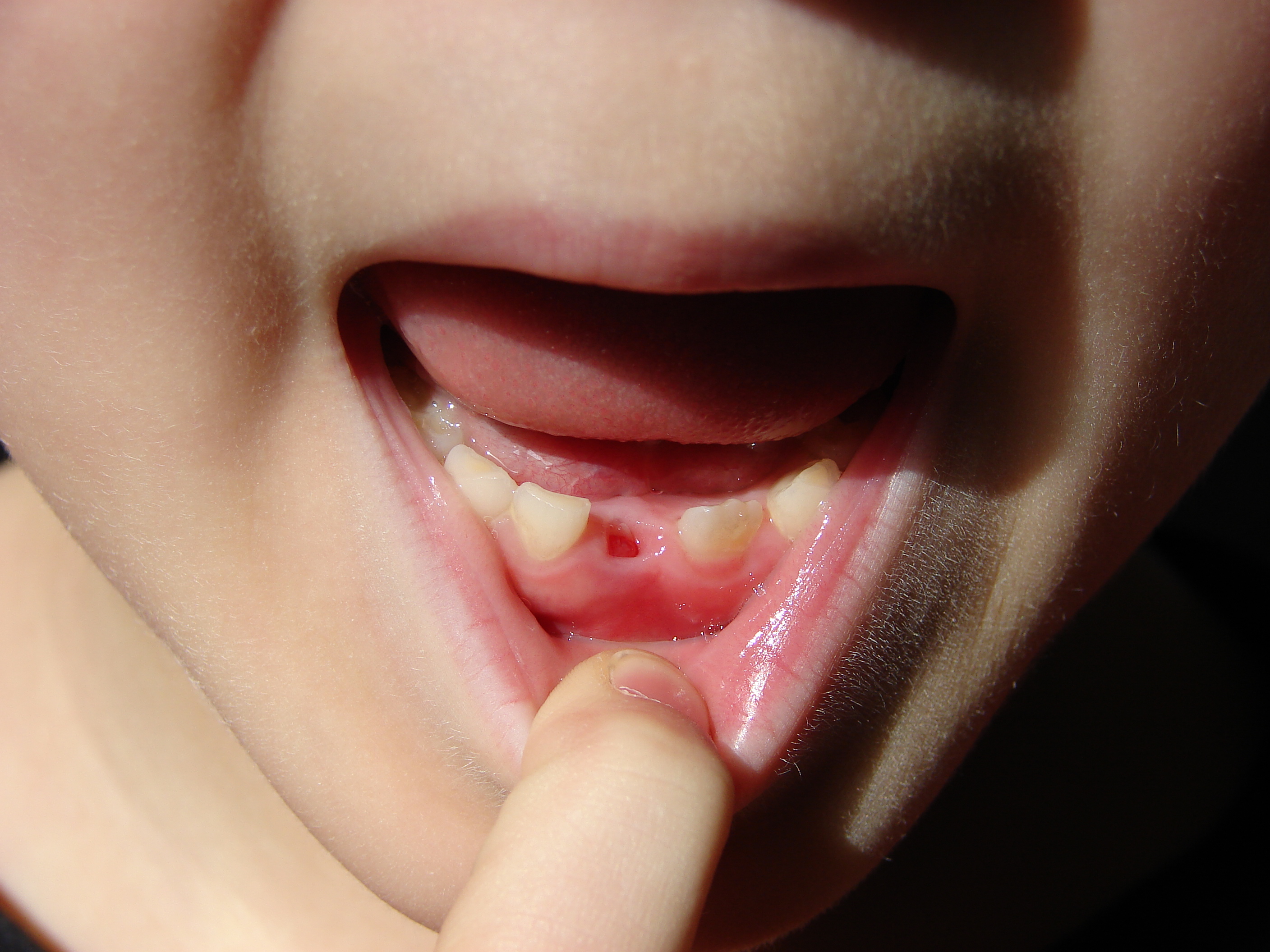
Deciduous teeth or primary teeth, also informally known as baby teeth, milk teeth, or temporary teeth,Illustrated Dental Embryology, Histology, and Anatomy, Bath-Balogh and Fehrenbach, Elsevier, 2011, page 255 are the first set of teeth in the growth and development of humans and other diphyodonts, which include most mammals but not elephants, kangaroos, or manatees which are polyphyodonts. Deciduous teeth tooth development, develop during the embryonic stage of development and tooth eruption, erupt (break through the gums and become visible in the mouth) during infancy. They are usually lost and replaced by permanent teeth, but in the absence of their permanent replacements, they can remain functional for many years into adulthood. Development Formation Primary teeth start to form during the embryonic phase of human development (biology), human life. The development of primary teeth starts at the sixth week of tooth development as the dental lamina. This process starts at t ... [...More Info...] [...Related Items...] OR: [Wikipedia] [Google] [Baidu] |
Tooth
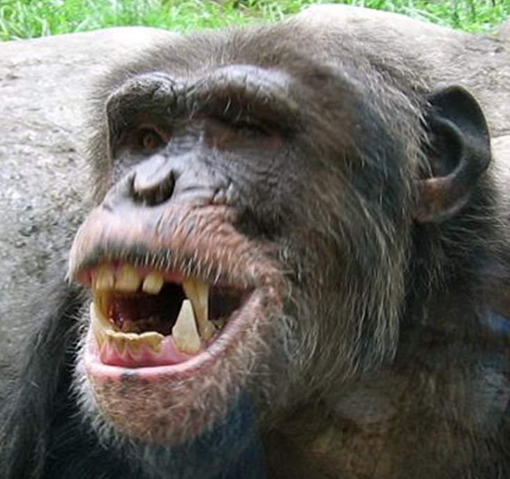
A tooth ( : teeth) is a hard, calcified structure found in the jaws (or mouths) of many vertebrates and used to break down food. Some animals, particularly carnivores and omnivores, also use teeth to help with capturing or wounding prey, tearing food, for defensive purposes, to intimidate other animals often including their own, or to carry prey or their young. The roots of teeth are covered by gums. Teeth are not made of bone, but rather of multiple tissues of varying density and hardness that originate from the embryonic germ layer, the ectoderm. The general structure of teeth is similar across the vertebrates, although there is considerable variation in their form and position. The teeth of mammals have deep roots, and this pattern is also found in some fish, and in crocodilians. In most teleost fish, however, the teeth are attached to the outer surface of the bone, while in lizards they are attached to the inner surface of the jaw by one side. In cartilaginous fi ... [...More Info...] [...Related Items...] OR: [Wikipedia] [Google] [Baidu] |


