|
Testicular Vein
The testicular vein (or spermatic vein), the male gonadal vein, carries deoxygenated blood from its corresponding testis to the inferior vena cava or one of its tributaries. It is the male equivalent of the ovarian vein, and is the venous counterpart of the testicular artery. Structure It is a paired vein, with one supplying each testis: * the right testicular vein generally joins the inferior vena cava; * the left testicular vein, unlike the right one, joins the left renal vein instead of the inferior vena cava. The veins emerge from the back of the testis, and receive tributaries from the epididymis. They unite and form a convoluted plexus, called the pampiniform plexus, which constitutes the greater mass of the spermatic cord; the vessels composing this plexus are very numerous, and ascend along the cord, in front of the ductus deferens. Below the subcutaneous inguinal ring, they unite to form three or four veins, which pass along the inguinal canal, and, entering the abdome ... [...More Info...] [...Related Items...] OR: [Wikipedia] [Google] [Baidu] |
Pampiniform Plexus
The pampiniform plexus (from Latin ''pampinus'', a tendril, + ''forma'', form) is a venous plexus – a network of many small veins found in the human male spermatic cord, and the suspensory ligament of the testicle, suspensory ligament of the ovary. In the male, it is formed by the union of multiple testicular veins from the back of the testis and tributaries from the epididymis. In the male The veins of the plexus ascend along the spermatic cord in front of the vas deferens. Below the superficial inguinal ring they unite to form three or four veins, which pass along the inguinal canal, and, entering the abdomen through the deep inguinal ring, coalesce to form two veins. These again unite to form a single vein, the testicular vein, which opens on the right side into the inferior vena cava, at an acute angle, and on the left side into the left renal vein, at a right angle. The pampiniform plexus forms the chief mass of the cord. In addition to its function in venous return from t ... [...More Info...] [...Related Items...] OR: [Wikipedia] [Google] [Baidu] |
Inguinal Canal
The inguinal canals are the two passages in the anterior abdominal wall of humans and animals which in males convey the spermatic cords and in females the round ligament of the uterus. The inguinal canals are larger and more prominent in males. There is one inguinal canal on each side of the midline. Structure The inguinal canals are situated just above the medial half of the inguinal ligament. In both sexes the canals transmit the ilioinguinal nerves. The canals are approximately 3.75 to 4 cm long. , angled anteroinferiorly and medially. In males, its diameter is normally 2 cm (±1 cm in standard deviation) at the deep inguinal ring.The diameter has been estimated to be ±2.2cm ±1.08cm in Africans, and 2.1 cm ±0.41cm in Europeans. A first-order approximation is to visualize each canal as a cylinder. Walls To help define the boundaries, these canals are often further approximated as boxes with six sides. Not including the two rings, the remaining four sides are usually ca ... [...More Info...] [...Related Items...] OR: [Wikipedia] [Google] [Baidu] |
European Journal Of Vascular And Endovascular Surgery
The ''European Journal of Vascular and Endovascular Surgery'' is a monthly peer-reviewed journal publishing articles describing endovascular methods and their critical evaluation. According to the ''Journal Citation Reports'', the journal has a 2019 impact factor The impact factor (IF) or journal impact factor (JIF) of an academic journal is a scientometric index calculated by Clarivate that reflects the yearly mean number of citations of articles published in the last two years in a given journal, as i ... of 5.328. References {{Reflist English-language journals ... [...More Info...] [...Related Items...] OR: [Wikipedia] [Google] [Baidu] |
Nutcracker Syndrome
The nutcracker syndrome (NCS) results most commonly from the compression of the left renal vein (LRV) between the abdominal aorta (AA) and superior mesenteric artery (SMA), although other variants exist. The name derives from the fact that, in the sagittal plane and/or transverse plane, the SMA and AA (with some imagination) appear to be a nutcracker crushing a nut (the renal vein). Furthermore, the venous return from the left gonadal vein returning to the left renal vein is blocked, thus causing testicular pain (colloquially referred to as "nut pain"). There is a wide spectrum of clinical presentations and diagnostic criteria are not well defined, which frequently results in delayed or incorrect diagnosis. The first clinical report of Nutcracker phenomenon appeared in 1950. This condition is not to be confused with superior mesenteric artery syndrome, which is the compression of the third portion of the duodenum by the SMA and the AA. Signs and symptoms The signs and symptoms of N ... [...More Info...] [...Related Items...] OR: [Wikipedia] [Google] [Baidu] |
Superior Mesenteric Artery
In human anatomy, the superior mesenteric artery (SMA) is an artery which arises from the anterior surface of the abdominal aorta, just inferior to the origin of the celiac trunk, and supplies blood to the intestine from the lower part of the duodenum through two-thirds of the transverse colon, as well as the pancreas. Structure It arises anterior to lower border of vertebra L1 in an adult. It is usually 1 cm lower than the celiac trunk. It initially travels in an anterior/inferior direction, passing behind/under the neck of the pancreas and the splenic vein. Located under this portion of the superior mesenteric artery, between it and the aorta, are the following: * left renal vein - travels between the left kidney and the inferior vena cava (can be compressed between the SMA and the abdominal aorta at this location, leading to nutcracker syndrome). * the third part of the duodenum, a segment of the small intestines (can be compressed by the SMA at this location, lea ... [...More Info...] [...Related Items...] OR: [Wikipedia] [Google] [Baidu] |
Abdominal Aorta
In human anatomy, the abdominal aorta is the largest artery in the abdominal cavity. As part of the aorta, it is a direct continuation of the descending aorta (of the thorax). Structure The abdominal aorta begins at the level of the thoracic diaphragm, diaphragm, crossing it via the aortic hiatus, technically behind the diaphragm, at the vertebral level of T12. It travels down the posterior wall of the abdomen, anterior to the vertebral column. It thus follows the curvature of the lumbar vertebrae, that is, convex anteriorly. The peak of this convexity is at the level of the third lumbar vertebra (L3). It runs parallel to the inferior vena cava, which is located just to the right of the abdominal aorta, and becomes smaller in diameter as it gives off branches. This is thought to be due to the large size of its principal branches. At the 11th rib, the diameter is 122mm long and 55mm wide and this is because of the constant pressure. The abdominal aorta is clinically divided int ... [...More Info...] [...Related Items...] OR: [Wikipedia] [Google] [Baidu] |
Ligature (medicine)
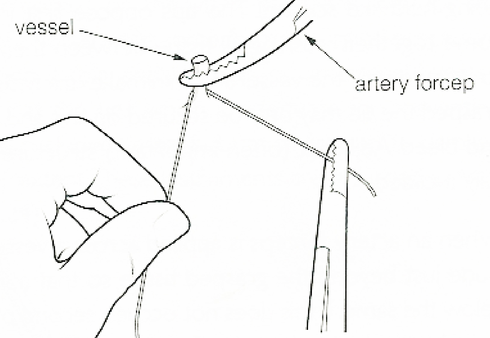
In surgery or medical procedure, a ligature consists of a piece of thread ( suture) tied around an anatomical structure, usually a blood vessel or another hollow structure (e.g. urethra) to shut it off. History The principle of ligation is attributed to Hippocrates and Galen. In ancient Rome, ligatures were used to treat hemorrhoids. The concept of a ligature was reintroduced some 1,500 years later by Ambroise Paré, and finally it found its modern use in 1870–80, made popular by Jules-Émile Péan. Procedure With a blood vessel the surgeon will clamp the vessel perpendicular to the axis of the artery or vein with a hemostat, then secure it by ligating it; i.e. using a piece of suture around it before dividing the structure and releasing the hemostat. It is different from a tourniquet in that the tourniquet will not be secured by knots and it can therefore be released/tightened at will. Ligature is one of the remedies to treat skin tag, or acrochorda. It is done by tying str ... [...More Info...] [...Related Items...] OR: [Wikipedia] [Google] [Baidu] |
Varicocele
varicocele is an abnormal enlargement of the pampiniform venous plexus in the scrotum. This plexus of veins drains blood from the testicles back to the heart. The vessels originate in the abdomen and course down through the inguinal canal as part of the spermatic cord on their way to the testis. Varicoceles occur in around 15% to 20% of all men. The incidence of varicocele increase with age. Signs and symptoms Varicocele might be noticed as soft lumps, usually above the testicle and mostly on the left side of the scrotum. Right-sided and bilateral varicocele does also occur. Men with varicocele can feel symptoms of pain or heaviness in their scrotum. Large varicoceles present as plexus of veins and may be described as a "bag of worms". Varicocele is sometimes discovered when investigating the cause of male infertility. Cause There are three main theories as to the anatomical cause; the first has to do with the geometry of the veins, wherein the vein on the left side connects to ... [...More Info...] [...Related Items...] OR: [Wikipedia] [Google] [Baidu] |
Vein
Veins are blood vessels in humans and most other animals that carry blood towards the heart. Most veins carry deoxygenated blood from the tissues back to the heart; exceptions are the pulmonary and umbilical veins, both of which carry oxygenated blood to the heart. In contrast to veins, arteries carry blood away from the heart. Veins are less muscular than arteries and are often closer to the skin. There are valves (called ''pocket valves'') in most veins to prevent backflow. Structure Veins are present throughout the body as tubes that carry blood back to the heart. Veins are classified in a number of ways, including superficial vs. deep, pulmonary vs. systemic, and large vs. small. * Superficial veins are those closer to the surface of the body, and have no corresponding arteries. *Deep veins are deeper in the body and have corresponding arteries. *Perforator veins drain from the superficial to the deep veins. These are usually referred to in the lower limbs and feet. *Communic ... [...More Info...] [...Related Items...] OR: [Wikipedia] [Google] [Baidu] |
Iliac Colon
The iliac colon is the portion of the descending colon which is situated within the left iliac fossa (between the crest of the left ilium, and the pelvic brim). It is about 12 to 15 cm long. It curves inferior-ward and medial-ward anterior of the iliacus and psoas muscles. It ends by transitioning into the sigmoid colon at the superior aperture of the lesser pelvis. It is covered by peritoneum The peritoneum is the serous membrane forming the lining of the abdominal cavity or coelom in amniotes and some invertebrates, such as annelids. It covers most of the intra-abdominal (or coelomic) organs, and is composed of a layer of meso ... only on its anterior and lateral surfaces. Additional images File:Gray1165.png, Female pelvis and its contents, seen from above and in front. References Large intestine {{digestive-stub ... [...More Info...] [...Related Items...] OR: [Wikipedia] [Google] [Baidu] |
Internal Spermatic Artery
The testicular artery (the male gonadal artery, also called the internal spermatic arteries in older texts) is a branch of the abdominal aorta that supplies blood to the testis. It is a paired artery, with one for each of the testes. It is the male equivalent of the ovarian artery. Because the testis is found in a different location than that of its female equivalent, it has a different course than the ovarian artery. They are two slender vessels of considerable length, and arise from the front of the aorta a little below the renal arteries. Each passes obliquely downward and lateralward behind the peritoneum, resting on the Psoas major, the right lying in front of the inferior vena cava and behind the middle colic and ileocolic arteries and the terminal part of the ileum, the left behind the left colic and sigmoid arteries and the iliac colon. Each crosses obliquely over the ureter and the lower part of the external iliac artery to reach the abdominal inguinal ring, through ... [...More Info...] [...Related Items...] OR: [Wikipedia] [Google] [Baidu] |
Peritoneum
The peritoneum is the serous membrane forming the lining of the abdominal cavity or coelom in amniotes and some invertebrates, such as annelids. It covers most of the intra-abdominal (or coelomic) organs, and is composed of a layer of mesothelium supported by a thin layer of connective tissue. This peritoneal lining of the cavity supports many of the abdominal organs and serves as a conduit for their blood vessels, lymphatic vessels, and nerves. The abdominal cavity (the space bounded by the vertebrae, abdominal muscles, diaphragm, and pelvic floor) is different from the intraperitoneal space (located within the abdominal cavity but wrapped in peritoneum). The structures within the intraperitoneal space are called "intraperitoneal" (e.g., the stomach and intestines), the structures in the abdominal cavity that are located behind the intraperitoneal space are called "retroperitoneal" (e.g., the kidneys), and those structures below the intraperitoneal space are called "subp ... [...More Info...] [...Related Items...] OR: [Wikipedia] [Google] [Baidu] |

.gif)


