|
Iliac Colon
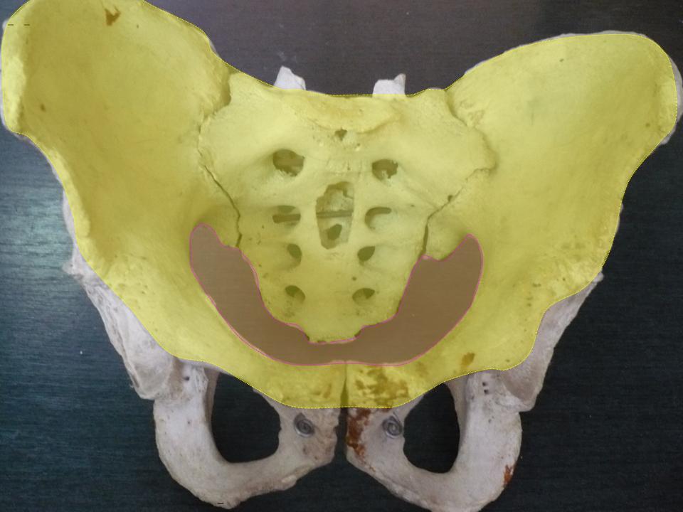
The iliac colon is the portion of the descending colon which is situated within the left iliac fossa (between the crest of the left ilium, and the pelvic brim). It is about 12 to 15 cm long. It curves inferior-ward and medial-ward anterior of the iliacus and psoas muscles. It ends by transitioning into the sigmoid colon at the superior aperture of the lesser pelvis. It is covered by peritoneum The peritoneum is the serous membrane forming the lining of the abdominal cavity or coelom in amniotes and some invertebrates, such as annelids. It covers most of the intra-abdominal (or coelomic) organs, and is composed of a layer of mesot ... only on its anterior and lateral surfaces. Additional images File:Gray1165.png, Female pelvis and its contents, seen from above and in front. References Large intestine {{digestive-stub ... [...More Info...] [...Related Items...] OR: [Wikipedia] [Google] [Baidu] |
Descending Colon
In the anatomy of humans and homologous primates, the descending colon is the part of the colon extending from the left colic flexure to the level of the iliac crest (whereupon it transitions into the sigmoid colon). The function of the descending colon in the digestive system is to store the remains of digested food that will be emptied into the rectum. The descending colon is on the left side of the body (barring any malformations). The term left colon is hypernymous to ''descending colon'' in precise use; many casual mentions of the left colon chiefly concern the descending colon. Structure The descending colon extends from the left colic flexure at the upper left part of the abdomen inferior-ward through the left hypochondrium and lumbar regions, along the outer border of the left kidney, ending at the level of the iliac crest at the lower left part of the abdomen, being contunued thenceforth as the sigmoid colon. It usually retroperitoneal (being lined by peritoneu ... [...More Info...] [...Related Items...] OR: [Wikipedia] [Google] [Baidu] |
Iliac Fossa
The iliac fossa is a large, smooth, concave surface on the internal surface of the ilium (part of the three fused bones making the hip bone). Structure The iliac fossa is bounded above by the iliac crest, and below by the arcuate line. It is bordered in front and behind by the anterior and posterior borders of the ilium. The iliac fossa gives origin to the iliacus muscle. The obturator nerve passes around the iliac fossa. It is perforated at its inner part by a nutrient canal. Below it there is a smooth, rounded border, the arcuate line, which runs anterior, inferior, and medial. When the "left" or "right" adjective is used (e.g "right iliac fossa"), the iliac fossa usually means one of the inguinal regions of the nine regions of the abdomen The abdomen (colloquially called the belly, tummy, midriff, tucky or stomach) is the part of the body between the thorax (chest) and pelvis, in humans and in other vertebrates. The abdomen is the front part of the abdominal segmen ... [...More Info...] [...Related Items...] OR: [Wikipedia] [Google] [Baidu] |
Ilium (bone)
The ilium () (plural ilia) is the uppermost and largest part of the hip bone, and appears in most vertebrates including mammals and birds, but not bony fish. All reptiles have an ilium except snakes, although some snake species have a tiny bone which is considered to be an ilium. The ilium of the human is divisible into two parts, the body and the wing; the separation is indicated on the top surface by a curved line, the arcuate line, and on the external surface by the margin of the acetabulum. The name comes from the Latin (''ile'', ''ilis''), meaning "groin" or "flank". Structure The ilium consists of the body and wing. Together with the ischium and pubis, to which the ilium is connected, these form the pelvic bone, with only a faint line indicating the place of union. The body ( la, corpus) forms less than two-fifths of the acetabulum; and also forms part of the acetabular fossa. The internal surface of the body is part of the wall of the lesser pelvis and gives or ... [...More Info...] [...Related Items...] OR: [Wikipedia] [Google] [Baidu] |
Pelvic Brim
The pelvic brim is the edge of the pelvic inlet. It is an approximately Mickey Mouse head-shaped line passing through the prominence of the sacrum, the arcuate and pectineal lines, and the upper margin of the pubic symphysis. Structure The pelvic brim is an approximately Mickey Mouse head-shaped line passing through the prominence of the sacrum, the arcuate and pectineal lines, and the upper margin of the pubic symphysis. The pelvic brim is obtusely pointed in front, diverging on either side, and encroached upon behind by the projection forward of the promontory of the sacrum. The oblique plane passing approximately through the pelvic brim divides the internal part of the pelvis (pelvic cavity) into the false or greater pelvis and the true or lesser pelvis. The false pelvis, which is above that plane, is sometimes considered to be a part of the abdominal cavity, rather than a part of the pelvic cavity. In this case, the pelvic cavity coincides with the true pelvis, which ... [...More Info...] [...Related Items...] OR: [Wikipedia] [Google] [Baidu] |
Iliacus
The iliacus is a flat, triangular muscle which fills the iliac fossa. It forms the lateral portion of iliopsoas, providing flexion of the thigh and lower limb at the acetabulofemoral joint. Structure The iliacus arises from the iliac fossa on the interior side of the hip bone, and also from the region of the anterior inferior iliac spine (AIIS). It joins the psoas major to form the iliopsoas. It proceeds across the iliopubic eminence through the muscular lacuna to its insertion on the lesser trochanter of the femur. Its fibers are often inserted in front of those of the psoas major and extend distally over the lesser trochanter.Platzer (2004), p 234 Nerve supply The iliopsoas is innervated by the femoral nerve and direct branches from the lumbar plexus.''Thieme Atlas of Anatomy'' (2006), p 422 Function In open-chain exercises, as part of the iliopsoas, the iliacus is important for lifting (flexing) the femur forward (e.g. front scale). In closed-chain exercises, the iliopsoas ... [...More Info...] [...Related Items...] OR: [Wikipedia] [Google] [Baidu] |
Psoas Major Muscle
The psoas major ( or ; from grc, ψόᾱ, psóā, muscles of the loins) is a long fusiform muscle located in the lateral lumbar region between the vertebral column and the brim of the lesser pelvis. It joins the iliacus muscle to form the iliopsoas. In animals, this muscle is equivalent to the tenderloin. Structure The psoas major is divided into a superficial and a deep part. The deep part originates from the transverse processes of lumbar vertebrae L1–L5. The superficial part originates from the lateral surfaces of the last thoracic vertebra, lumbar vertebrae L1–L4, and the neighboring intervertebral discs. The lumbar plexus lies between the two layers. Together, the iliacus muscle and the psoas major form the iliopsoas, which is surrounded by the iliac fascia. The iliopsoas runs across the iliopubic eminence through the muscular lacuna to its insertion on the lesser trochanter of the femur. The iliopectineal bursa separates the tendon of the iliopsoas muscl ... [...More Info...] [...Related Items...] OR: [Wikipedia] [Google] [Baidu] |
Sigmoid Colon
The sigmoid colon (or pelvic colon) is the part of the large intestine that is closest to the rectum and anus. It forms a loop that averages about in length. The loop is typically shaped like a Greek letter sigma (ς) or Latin letter S (thus ''sigma'' + '' -oid''). This part of the colon normally lies within the pelvis, but due to its freedom of movement it is liable to be displaced into the abdominal cavity. Structure The sigmoid colon begins at the superior aperture of the lesser pelvis, where it is continuous with the iliac colon, and passes transversely across the front of the sacrum to the right side of the pelvis. It then curves on itself and turns toward the left to reach the middle line at the level of the third piece of the sacrum, where it bends downward and ends in the rectum. Its function is to expel solid and gaseous waste from the gastrointestinal tract. The curving path it takes toward the anus allows it to store gas in the superior arched portion, enabling ... [...More Info...] [...Related Items...] OR: [Wikipedia] [Google] [Baidu] |
Superior Aperture
The pelvic inlet or superior aperture of the pelvis is a planar surface which defines the boundary between the pelvic cavity and the abdominal cavity (or, according to some authors, between two parts of the pelvic cavity, called lesser pelvis and greater pelvis). It is a major target of measurements of pelvimetry. Its position and orientation relative to the skeleton of the pelvis is anatomically defined by its edge, the pelvic brim. The pelvic brim is an approximately apple-shaped line passing through the prominence of the sacrum, the arcuate and pectineal lines, and the upper margin of the pubic symphysis. Occasionally, the terms pelvic inlet and pelvic brim are used interchangeably. Boundaries The edge of the pelvic inlet (pelvic brim) is formed as follows: Diameters The diameters or conjugates of the pelvis are measured at the pelvic inlet and outlet and as oblique diameters. Two diameters may be measured from the outside of the body using a pelvimeter Additiona ... [...More Info...] [...Related Items...] OR: [Wikipedia] [Google] [Baidu] |
Lesser Pelvis
The pelvic cavity is a body cavity that is bounded by the bones of the pelvis. Its oblique roof is the pelvic inlet (the superior opening of the pelvis). Its lower boundary is the pelvic floor. The pelvic cavity primarily contains the reproductive organs, urinary bladder, distal ureters, proximal urethra, terminal sigmoid colon, rectum, and anal canal. In females, the uterus, Fallopian tubes, ovaries and upper vagina occupy the area between the other viscera. The rectum is located at the back of the pelvis, in the curve of the sacrum and coccyx; the bladder is in front, behind the pubic symphysis. The pelvic cavity also contains major arteries, veins, muscles, and nerves. These structures coexist in a crowded space, and disorders of one pelvic component may impact upon another; for example, constipation may overload the rectum and compress the urinary bladder, or childbirth might damage the pudendal nerves and later lead to anal weakness. Structure The pelvis ... [...More Info...] [...Related Items...] OR: [Wikipedia] [Google] [Baidu] |
Peritoneum
The peritoneum is the serous membrane forming the lining of the abdominal cavity or coelom in amniotes and some invertebrates, such as annelids. It covers most of the intra-abdominal (or coelomic) organs, and is composed of a layer of mesothelium supported by a thin layer of connective tissue. This peritoneal lining of the cavity supports many of the abdominal organs and serves as a conduit for their blood vessels, lymphatic vessels, and nerves. The abdominal cavity (the space bounded by the vertebrae, abdominal muscles, diaphragm, and pelvic floor) is different from the intraperitoneal space (located within the abdominal cavity but wrapped in peritoneum). The structures within the intraperitoneal space are called "intraperitoneal" (e.g., the stomach and intestines), the structures in the abdominal cavity that are located behind the intraperitoneal space are called " retroperitoneal" (e.g., the kidneys), and those structures below the intraperitoneal space are called "su ... [...More Info...] [...Related Items...] OR: [Wikipedia] [Google] [Baidu] |
