|
Tubal Tonsils
The tubal tonsil, also known as Gerlach tonsil, is one of the four main tonsil groups forming Waldeyer's tonsillar ring. Structure Each tubal tonsil is located posterior to the opening of the Eustachian tube on the lateral wall of the nasopharynx. It is one of the four main tonsil groups forming Waldeyer's tonsillar ring. This ring also includes the palatine tonsils, the lingual tonsils, and the adenoid. Clinical significance The tubal tonsil may be affected by tonsillitis. However, this usually affects only the palatine tonsils. History The tubal tonsil may also be known as the Gerlach tonsil. It is very close to the torus tubarius The base of the cartilaginous portion of the Eustachian tube (auditory tube) lies directly under the mucous membrane of the nasal part of the pharynx, where it forms an elevation or cushion, the torus tubarius or torus of the auditory tube, behind ..., which is why this tonsil is sometimes also called the ''tonsil of (the) torus tubarius''.En ... [...More Info...] [...Related Items...] OR: [Wikipedia] [Google] [Baidu] |
Tonsil
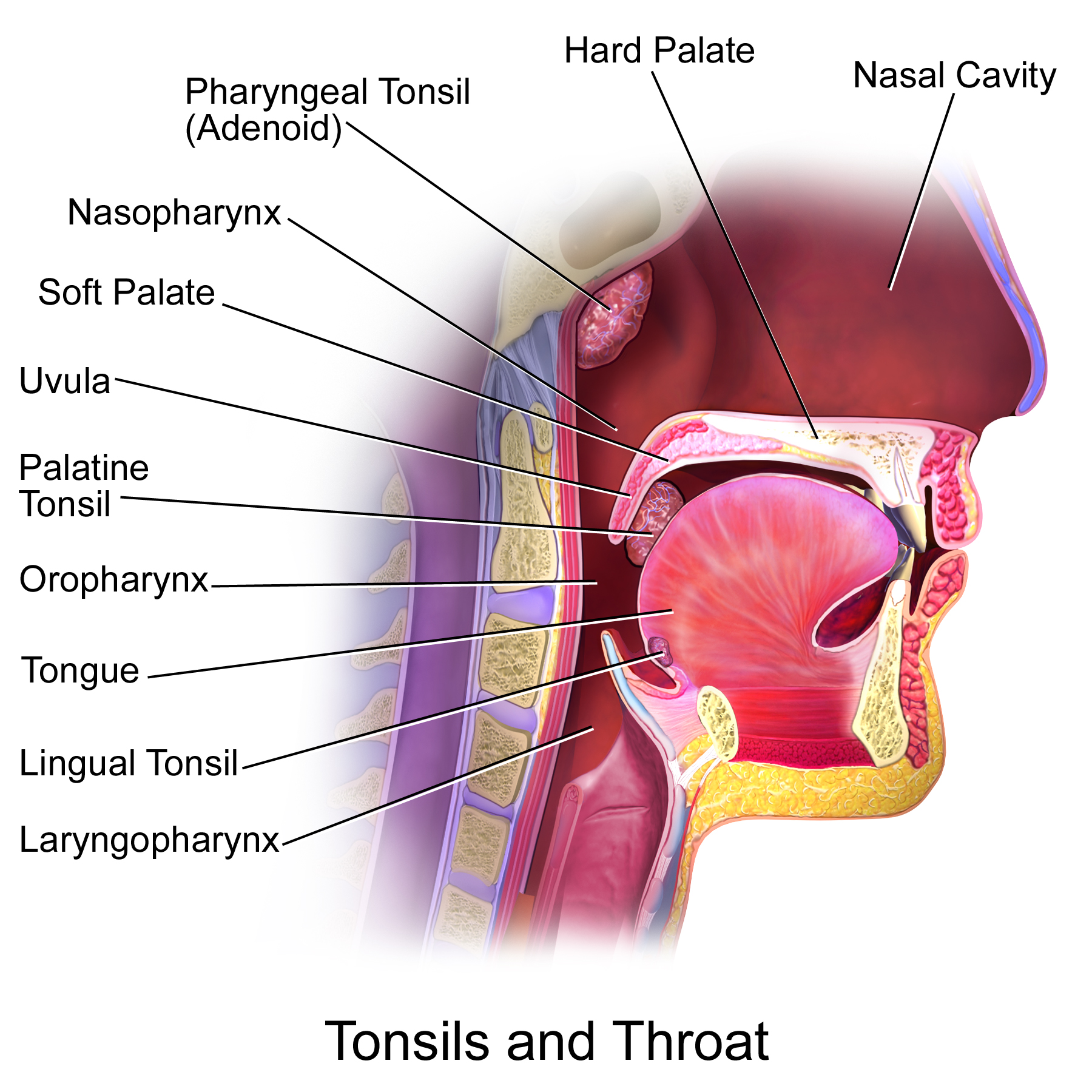
The tonsils are a set of lymphoid organs facing into the aerodigestive tract, which is known as Waldeyer's tonsillar ring and consists of the adenoid tonsil, two tubal tonsils, two palatine tonsils, and the lingual tonsils. These organs play an important role in the immune system. When used unqualified, the term most commonly refers specifically to the palatine tonsils, which are two lymphoid organs situated at either side of the back of the human throat. The palatine tonsils and the adenoid tonsil are organs consisting of lymphoepithelial tissue located near the oropharynx and nasopharynx (parts of the throat). Structure Humans are born with four types of tonsils: the pharyngeal tonsil, two tubal tonsils, two palatine tonsils and the lingual tonsils. Development The palatine tonsils tend to reach their largest size in puberty, and they gradually undergo atrophy thereafter. However, they are largest relative to the diameter of the throat in young children. In adults, each pal ... [...More Info...] [...Related Items...] OR: [Wikipedia] [Google] [Baidu] |
Waldeyer's Tonsillar Ring
Waldeyer's tonsillar ring (pharyngeal lymphoid ring, Waldeyer's lymphatic ring, or tonsillar ring) is a ringed arrangement of lymphoid organs in the pharynx. Waldeyer's ring surrounds the naso- and oropharynx, with some of its tonsillar tissue located above and some below the soft palate (and to the back of the mouth cavity). Structure The ring consists of the (from top to bottom): * 1 pharyngeal tonsil (or "adenoid"), located on the roof of the nasopharynx, under the sphenoid bone. * 2 tubal tonsils on each side, where each auditory tube opens into the nasopharynx * 2 palatine tonsils (commonly called "the tonsils") located in the oropharynx *lingual tonsils, a collection of lymphatic tissue located on the back part of the tongue Terminology Some authors speak of two pharyngeal tonsils/two adenoids. These authors simply look at the left and right halves of the pharyngeal tonsil as two tonsils. Many authors also speak of lingual tonsils (in the plural), because this accumulation o ... [...More Info...] [...Related Items...] OR: [Wikipedia] [Google] [Baidu] |
Eustachian Tube
In anatomy, the Eustachian tube, also known as the auditory tube or pharyngotympanic tube, is a tube that links the nasopharynx to the middle ear, of which it is also a part. In adult humans, the Eustachian tube is approximately long and in diameter. It is named after the sixteenth-century Italian anatomist Bartolomeo Eustachi. In humans and other tetrapods, both the middle ear and the ear canal are normally filled with air. Unlike the air of the ear canal, however, the air of the middle ear is not in direct contact with the atmosphere outside the body; thus, a pressure difference can develop between the atmospheric pressure of the ear canal and the middle ear. Normally, the Eustachian tube is collapsed, but it gapes open with swallowing and with positive pressure, allowing the middle ear's pressure to adjust to the atmospheric pressure. When taking off in an aircraft, the ambient air pressure goes from higher (on the ground) to lower (in the sky). The air in the middle ear ... [...More Info...] [...Related Items...] OR: [Wikipedia] [Google] [Baidu] |
Nasopharynx
The pharynx (plural: pharynges) is the part of the throat behind the mouth and nasal cavity, and above the oesophagus and trachea (the tubes going down to the stomach and the lungs). It is found in vertebrates and invertebrates, though its structure varies across species. The pharynx carries food and air to the esophagus and larynx respectively. The flap of cartilage called the epiglottis stops food from entering the larynx. In humans, the pharynx is part of the digestive system and the conducting zone of the respiratory system. (The conducting zone—which also includes the nostrils of the nose, the larynx, trachea, bronchi, and bronchioles—filters, warms and moistens air and conducts it into the lungs). The human pharynx is conventionally divided into three sections: the nasopharynx, oropharynx, and laryngopharynx. It is also important in vocalization. In humans, two sets of pharyngeal muscles form the pharynx and determine the shape of its lumen. They are arranged as an in ... [...More Info...] [...Related Items...] OR: [Wikipedia] [Google] [Baidu] |
Elsevier
Elsevier () is a Dutch academic publishing company specializing in scientific, technical, and medical content. Its products include journals such as ''The Lancet'', ''Cell'', the ScienceDirect collection of electronic journals, '' Trends'', the '' Current Opinion'' series, the online citation database Scopus, the SciVal tool for measuring research performance, the ClinicalKey search engine for clinicians, and the ClinicalPath evidence-based cancer care service. Elsevier's products and services also include digital tools for data management, instruction, research analytics and assessment. Elsevier is part of the RELX Group (known until 2015 as Reed Elsevier), a publicly traded company. According to RELX reports, in 2021 Elsevier published more than 600,000 articles annually in over 2,700 journals; as of 2018 its archives contained over 17 million documents and 40,000 e-books, with over one billion annual downloads. Researchers have criticized Elsevier for its high profit marg ... [...More Info...] [...Related Items...] OR: [Wikipedia] [Google] [Baidu] |
Palatine Tonsil
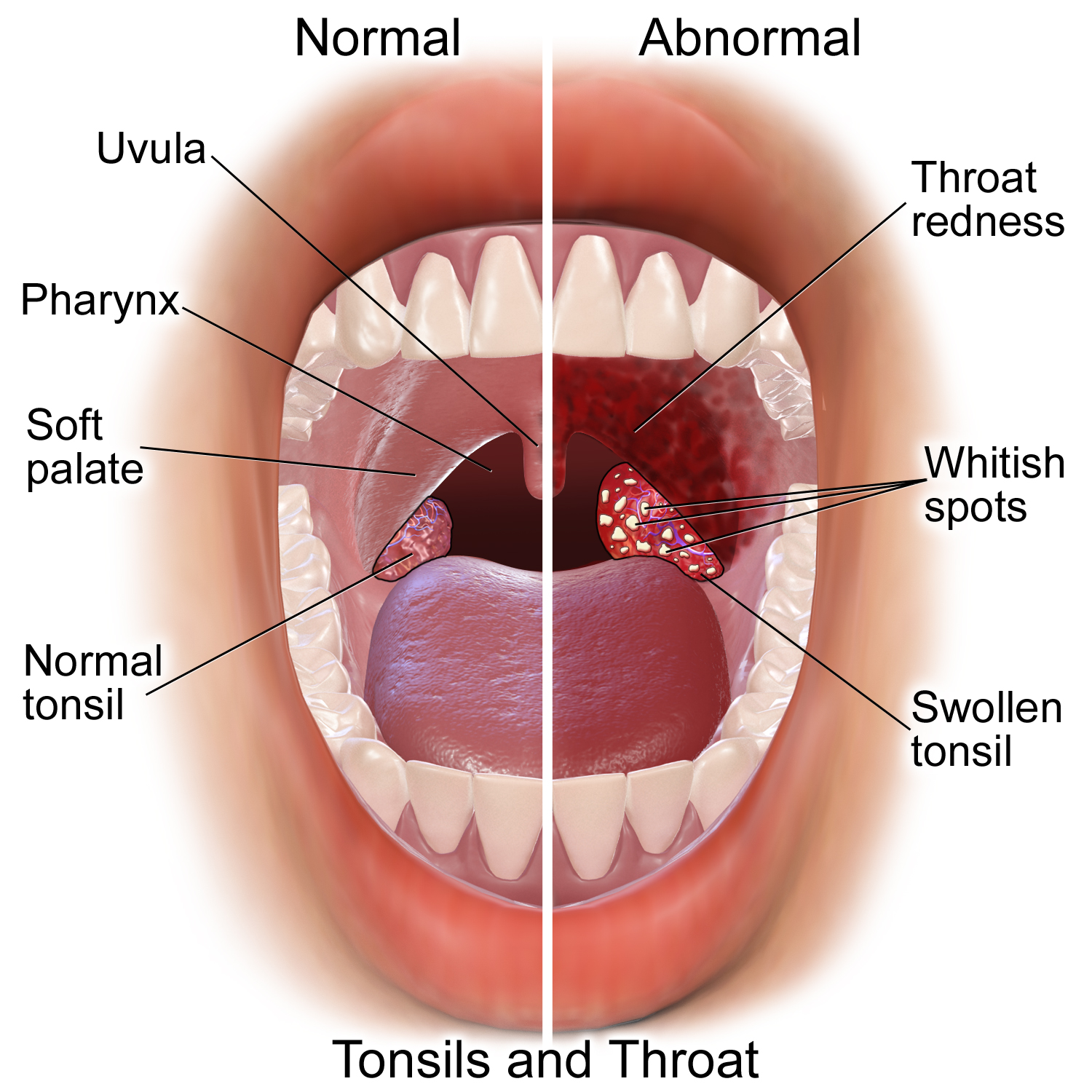
Palatine tonsils, commonly called the tonsils and occasionally called the faucial tonsils, are tonsils located on the left and right sides at the back of the throat, which can often be seen as flesh-colored, pinkish lumps. Tonsils only present as "white lumps" if they are inflamed or infected with symptoms of exudates (pus drainage) and severe swelling. Tonsillitis is an inflammation of the tonsils and will often, but not necessarily, cause a sore throat and fever. In Chronic (medicine), chronic cases tonsillectomy may be indicated. Structure The palatine tonsils are located in the isthmus of the fauces, between the palatoglossal arch and the palatopharyngeal arch of the soft palate. The palatine tonsil is one of the mucosa-associated lymphoid tissues (MALT), located at the entrance to the upper respiratory and gastrointestinal tracts to protect the body from the entry of exogenous material through mucosal sites. In consequence it is a site of, and potential focus for, infection ... [...More Info...] [...Related Items...] OR: [Wikipedia] [Google] [Baidu] |
Lingual Tonsils
The lingual tonsils are a collection of lymphatic tissue located in the lamina propria of the root of the tongue. This lymphatic tissue consists of the lymphatic nodules rich in cells of the immune system (immunocytes). The immunocytes initiate the immune response when the lingual tonsils get in contact with invading microorganisms (pathogenic bacteria, viruses or parasites). Structure Microanatomy Lingual tonsils are covered externally by stratified squamous nonkeratinized epithelium that invaginates inward forming crypts. Beneath the epithelium is a layer of lymphoid nodules containing lymphocytes. Mucous glands located at the root of tongue are drained through several ducts into the crypt of lingual tonsils. Secretions of these mucous glands keep the crypt clean and free of any debris. Blood supply Lingual tonsils are located on posterior aspect of tongue which is supplied through: * Lingual artery, branch of external carotid artery * Tonsillar branch of facial artery *Asc ... [...More Info...] [...Related Items...] OR: [Wikipedia] [Google] [Baidu] |
Adenoid
In anatomy, the adenoid, also known as the pharyngeal tonsil or nasopharyngeal tonsil, is the superior-most of the tonsils. It is a mass of lymphatic tissue located behind the nasal cavity, in the roof of the nasopharynx, where the nose blends into the throat. In children, it normally forms a soft mound in the roof and back wall of the nasopharynx, just above and behind the uvula. The term ''adenoid'' is also used to represent adenoid hypertrophy, the abnormal growth of the pharyngeal tonsils. Structure The adenoid is a mass of lymphatic tissue located behind the nasal cavity, in the roof of the nasopharynx, where the nose blends into the throat. The adenoid, unlike the palatine tonsils, has pseudostratified epithelium. The adenoids are part of the so-called Waldeyer ring of lymphoid tissue which also includes the palatine tonsils, the lingual tonsils and the tubal tonsils. Development Adenoids develop from a subepithelial infiltration of lymphocytes after the 16th week o ... [...More Info...] [...Related Items...] OR: [Wikipedia] [Google] [Baidu] |
Tonsillitis
Tonsillitis is inflammation of the tonsils in the upper part of the throat. It can be acute or chronic. Acute tonsillitis typically has a rapid onset. Symptoms may include sore throat, fever, enlargement of the tonsils, trouble swallowing, and enlarged lymph nodes around the neck. Complications include peritonsillar abscess. Tonsillitis is most commonly caused by a viral infection and about 5% to 40% of cases are caused by a bacterial infection.Lang 2009p. 2083./ref> When caused by the bacterium group A streptococcus, it is classed as streptococcal tonsillitis also referred to as ''strep throat''. Rarely bacteria such as '' Neisseria gonorrhoeae'', '' Corynebacterium diphtheriae'', or ''Haemophilus influenzae'' may be the cause. Typically the infection is spread between people through the air. A scoring system, such as the Centor score, may help separate possible causes. Confirmation may be by a throat swab or rapid strep test. Treatment efforts involve improving symptoms and ... [...More Info...] [...Related Items...] OR: [Wikipedia] [Google] [Baidu] |
Palatine Tonsil
Palatine tonsils, commonly called the tonsils and occasionally called the faucial tonsils, are tonsils located on the left and right sides at the back of the throat, which can often be seen as flesh-colored, pinkish lumps. Tonsils only present as "white lumps" if they are inflamed or infected with symptoms of exudates (pus drainage) and severe swelling. Tonsillitis is an inflammation of the tonsils and will often, but not necessarily, cause a sore throat and fever. In Chronic (medicine), chronic cases tonsillectomy may be indicated. Structure The palatine tonsils are located in the isthmus of the fauces, between the palatoglossal arch and the palatopharyngeal arch of the soft palate. The palatine tonsil is one of the mucosa-associated lymphoid tissues (MALT), located at the entrance to the upper respiratory and gastrointestinal tracts to protect the body from the entry of exogenous material through mucosal sites. In consequence it is a site of, and potential focus for, infection ... [...More Info...] [...Related Items...] OR: [Wikipedia] [Google] [Baidu] |
Torus Tubarius
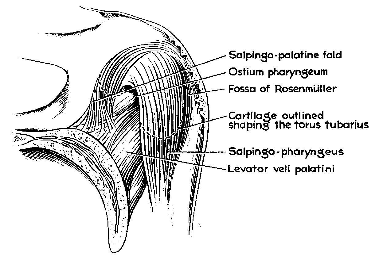
The base of the cartilaginous portion of the Eustachian tube (auditory tube) lies directly under the mucous membrane of the nasal part of the pharynx, where it forms an elevation or cushion, the torus tubarius or torus of the auditory tube, behind the pharyngeal orifice of the tube. The torus tubarius is very close to the tubal tonsil, which is sometimes also called the ''tonsil of (the) torus tubarius''. Two folds run posteriorly and anteriorly: *posteriorly, the vertical fold of mucous membrane, the salpingopharyngeal fold, stretches from the lower part of the torus tubarius; it contains the Salpingopharyngeus muscle which originates from the superior border of the medial lamina of the cartilage of the auditory tube, and passes downward and blends with the posterior fasciculus of the palatopharyngeus muscle. *anteriorly, the second and smaller fold, the salpingopalatine fold, smaller than the salpingopharyngeal fold, contains some fibers of muscle, called ''salpingopalatine musc ... [...More Info...] [...Related Items...] OR: [Wikipedia] [Google] [Baidu] |
Lymphatics Of The Head And Neck
The lymphatic vessels (or lymph vessels or lymphatics) are thin-walled vessels (tubes), structured like blood vessels, that carry lymph. As part of the lymphatic system, lymph vessels are complementary to the cardiovascular system. Lymph vessels are lined by endothelial cells, and have a thin layer of smooth muscle, and adventitia that binds the lymph vessels to the surrounding tissue. Lymph vessels are devoted to the propulsion of the lymph from the lymph capillaries, which are mainly concerned with the absorption of interstitial fluid from the tissues. Lymph capillaries are slightly bigger than their counterpart capillaries of the vascular system. Lymph vessels that carry lymph to a lymph node are called afferent lymph vessels, and those that carry it from a lymph node are called efferent lymph vessels, from where the lymph may travel to another lymph node, may be returned to a vein, or may travel to a larger lymph duct. Lymph ducts drain the lymph into one of the subclavi ... [...More Info...] [...Related Items...] OR: [Wikipedia] [Google] [Baidu] |


.jpg)


