|
Tonotopy
In physiology, tonotopy (from Greek tono = frequency and topos = place) is the spatial arrangement of where sounds of different frequency are processed in the brain. Tones close to each other in terms of frequency are represented in topologically neighbouring regions in the brain. Tonotopic maps are a particular case of topographic organization, similar to retinotopy in the visual system. Tonotopy in the auditory system begins at the cochlea, the small snail-like structure in the inner ear that sends information about sound to the brain. Different regions of the basilar membrane in the organ of Corti, the sound-sensitive portion of the cochlea, vibrate at different sinusoidal frequencies due to variations in thickness and width along the length of the membrane. Nerves that transmit information from different regions of the basilar membrane therefore encode frequency tonotopically. This tonotopy then projects through the vestibulocochlear nerve and associated midbrain structures ... [...More Info...] [...Related Items...] OR: [Wikipedia] [Google] [Baidu] |
Topographic Map (neuroanatomy)
A topographic map is the ordered projection of a sensory surface, like the retina or the skin, or an effector system, like the musculature, to one or more structures of the central nervous system. Topographic maps can be found in all sensory systems and in many motor systems. Visual system The visual system refers to the part of the central nervous system that allows an organism to see. It interprets information from visible light to build a representation of the world. The ganglion cells of the retina project in an orderly fashion to the lateral geniculate nucleus of the thalamus and from there to the primary visual cortex(V1); adjacent spots on the retina are represented by adjacent neurons in the lateral geniculate nucleus and the primary visual cortex. The term for this pattern of projection is ''topography''. There are many types of topographic maps in the visual cortices, including retinotopic maps, occular dominance maps and orientation maps. Retinotopic maps are the eas ... [...More Info...] [...Related Items...] OR: [Wikipedia] [Google] [Baidu] |
Retinotopy
Retinotopy (from Greek τόπος, place) is the mapping of visual input from the retina to neurons, particularly those neurons within the visual stream. For clarity, 'retinotopy' can be replaced with 'retinal mapping', and 'retinotopic' with 'retinally mapped'. Visual field maps (retinotopic maps) are found in many amphibian and mammalian species, though the specific size, number, and spatial arrangement of these maps can differ considerably. Sensory topographies can be found throughout the brain and are critical to the understanding of one's external environment. Moreover, the study of sensory topographies and retinotopy in particular has furthered our understanding of how neurons encode and organize sensory signals. Retinal mapping of the visual field is maintained through various points of the visual pathway including but not limited to the retina, the dorsal lateral geniculate nucleus, the optic tectum, the primary visual cortex (V1), and higher visual areas (V2-V4). Retin ... [...More Info...] [...Related Items...] OR: [Wikipedia] [Google] [Baidu] |
Medial Geniculate Nucleus
The medial geniculate nucleus (MGN) or medial geniculate body (MGB) is part of the auditory thalamus and represents the thalamic relay between the inferior colliculus (IC) and the auditory cortex (AC). It is made up of a number of sub-nuclei that are distinguished by their neuronal morphology and density, by their afferent and efferent connections, and by the coding properties of their neurons. It is thought that the MGN influences the direction and maintenance of attention. Divisions The MGN has three major divisions; ventral (VMGN), dorsal (DMGN) and medial (MMGN). Whilst the VMGN is specific to auditory information processing, the DMGN and MMGN also receive information from non-auditory pathways. Ventral subnucleus Cell types There are two main cell types in the ventral subnucleus of the medial geniculate body (VMGN): * Thalamocortical relay cells (or principal neurons): The dendritic Dendrite derives from the Greek word "dendron" meaning ( "tree-like"), and may refer to: ... [...More Info...] [...Related Items...] OR: [Wikipedia] [Google] [Baidu] |
Wave
In physics, mathematics, and related fields, a wave is a propagating dynamic disturbance (change from equilibrium) of one or more quantities. Waves can be periodic, in which case those quantities oscillate repeatedly about an equilibrium (resting) value at some frequency. When the entire waveform moves in one direction, it is said to be a ''traveling wave''; by contrast, a pair of superimposed periodic waves traveling in opposite directions makes a '' standing wave''. In a standing wave, the amplitude of vibration has nulls at some positions where the wave amplitude appears smaller or even zero. Waves are often described by a '' wave equation'' (standing wave field of two opposite waves) or a one-way wave equation for single wave propagation in a defined direction. Two types of waves are most commonly studied in classical physics. In a '' mechanical wave'', stress and strain fields oscillate about a mechanical equilibrium. A mechanical wave is a local deformation (str ... [...More Info...] [...Related Items...] OR: [Wikipedia] [Google] [Baidu] |
Place Theory (hearing)
Place theory is a theory of hearing that states that our perception of sound depends on where each component frequency produces vibrations along the basilar membrane. By this theory, the pitch of a sound, such as a human voice or a musical tone, is determined by the places where the membrane vibrates, based on frequencies corresponding to the tonotopic organization of the primary auditory neurons. More generally, schemes that base attributes of auditory perception on the neural firing rate as a function of place are known as rate–place schemes. The main alternative to the place theory is the temporal theory, also known as timing theory. These theories are closely linked with the volley principle or volley theory, a mechanism by which groups of neurons can encode the timing of a sound waveform. In all cases, neural firing patterns in time determine the perception of pitch. The combination known as the place–volley theory uses both mechanisms in combination, primarily cod ... [...More Info...] [...Related Items...] OR: [Wikipedia] [Google] [Baidu] |
Interaural Time Difference
The interaural time difference (or ITD) when concerning humans or animals, is the difference in arrival time of a sound between two ears. It is important in the localization of sounds, as it provides a cue to the direction or angle of the sound source from the head. If a signal arrives at the head from one side, the signal has further to travel to reach the far ear than the near ear. This pathlength difference results in a time difference between the sound's arrivals at the ears, which is detected and aids the process of identifying the direction of sound source. When a signal is produced in the horizontal plane, its angle in relation to the head is referred to as its azimuth, with 0 degrees (0°) azimuth being directly in front of the listener, 90° to the right, and 180° being directly behind. Different methods for measuring ITDs *For an abrupt stimulus such as a click, onset ITDs are measured. An onset ITD is the time difference between the onset of the signal reaching two ea ... [...More Info...] [...Related Items...] OR: [Wikipedia] [Google] [Baidu] |
Auditory Cortex
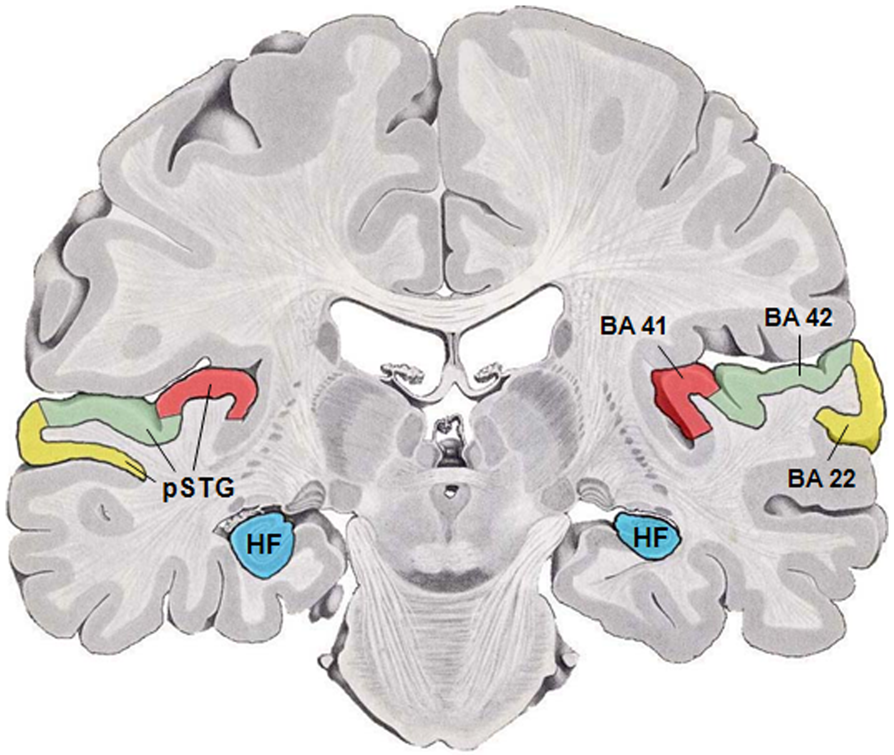
The auditory cortex is the part of the temporal lobe that processes auditory information in humans and many other vertebrates. It is a part of the auditory system, performing basic and higher functions in hearing, such as possible relations to language switching.Cf. Pickles, James O. (2012). ''An Introduction to the Physiology of Hearing'' (4th ed.). Bingley, UK: Emerald Group Publishing Limited, p. 238. It is located bilaterally, roughly at the upper sides of the temporal lobes – in humans, curving down and onto the medial surface, on the superior temporal plane, within the lateral sulcus and comprising parts of the transverse temporal gyri, and the superior temporal gyrus, including the planum polare and planum temporale (roughly Brodmann areas 41 and 42, and partially 22). The auditory cortex takes part in the spectrotemporal, meaning involving time and frequency, analysis of the inputs passed on from the ear. The cortex then filters and passes on the information to ... [...More Info...] [...Related Items...] OR: [Wikipedia] [Google] [Baidu] |
Transverse Temporal Gyrus
The transverse temporal gyri, also called Heschl's gyri () or Heschl's convolutions, are gyri found in the area of primary auditory cortex buried within the lateral sulcus of the human brain, occupying Brodmann areas 41 and 42. Transverse temporal gyri are superior to and separated from the planum temporale (cortex involved in language production) by Heschl's sulcus. Transverse temporal gyri are found in varying numbers in both the right and left hemispheres of the brain and one study found that this number is not related to the hemisphere or dominance of hemisphere studied in subjects. Transverse temporal gyri can be viewed in the sagittal plane as either an omega shape (if one gyrus is present) or a heart shape (if two gyri and a sulcus are present). Transverse temporal gyri are the first cortical structures to process incoming auditory information. Anatomically, the transverse temporal gyri are distinct in that they run mediolaterally (toward the center of the brain), rather t ... [...More Info...] [...Related Items...] OR: [Wikipedia] [Google] [Baidu] |
Functional Magnetic Resonance Imaging
Functional magnetic resonance imaging or functional MRI (fMRI) measures brain activity by detecting changes associated with blood flow. This technique relies on the fact that cerebral blood flow and neuronal activation are coupled. When an area of the brain is in use, blood flow to that region also increases. The primary form of fMRI uses the blood-oxygen-level dependent (BOLD) contrast, discovered by Seiji Ogawa in 1990. This is a type of specialized brain and body scan used to map neural activity in the brain or spinal cord of humans or other animals by imaging the change in blood flow ( hemodynamic response) related to energy use by brain cells. Since the early 1990s, fMRI has come to dominate brain mapping research because it does not involve the use of injections, surgery, the ingestion of substances, or exposure to ionizing radiation. This measure is frequently corrupted by noise from various sources; hence, statistical procedures are used to extract the underlying sig ... [...More Info...] [...Related Items...] OR: [Wikipedia] [Google] [Baidu] |
Positron Emission Tomography
Positron emission tomography (PET) is a functional imaging technique that uses radioactive substances known as radiotracers to visualize and measure changes in metabolic processes, and in other physiological activities including blood flow, regional chemical composition, and absorption. Different tracers are used for various imaging purposes, depending on the target process within the body. For example: * Fluorodeoxyglucose ( 18F">sup>18FDG or FDG) is commonly used to detect cancer; * 18Fodium fluoride">sup>18Fodium fluoride (Na18F) is widely used for detecting bone formation; * Oxygen-15 (15O) is sometimes used to measure blood flow. PET is a common imaging technique, a medical scintillography technique used in nuclear medicine. A radiopharmaceutical – a radioisotope attached to a drug – is injected into the body as a radioactive tracer, tracer. When the radiopharmaceutical undergoes beta plus decay, a positron is emitted, and when the positron interacts with an or ... [...More Info...] [...Related Items...] OR: [Wikipedia] [Google] [Baidu] |
Auditory Cortex
The auditory cortex is the part of the temporal lobe that processes auditory information in humans and many other vertebrates. It is a part of the auditory system, performing basic and higher functions in hearing, such as possible relations to language switching.Cf. Pickles, James O. (2012). ''An Introduction to the Physiology of Hearing'' (4th ed.). Bingley, UK: Emerald Group Publishing Limited, p. 238. It is located bilaterally, roughly at the upper sides of the temporal lobes – in humans, curving down and onto the medial surface, on the superior temporal plane, within the lateral sulcus and comprising parts of the transverse temporal gyri, and the superior temporal gyrus, including the planum polare and planum temporale (roughly Brodmann areas 41 and 42, and partially 22). The auditory cortex takes part in the spectrotemporal, meaning involving time and frequency, analysis of the inputs passed on from the ear. The cortex then filters and passes on the information to ... [...More Info...] [...Related Items...] OR: [Wikipedia] [Google] [Baidu] |


_entre_izquierdo_(inferior)_y_correcto_(superior)_orejas.jpg)


