|
Superior Nasal Concha
The superior nasal concha is a small, curved plate of bone representing a medial bony process of the labyrinth of the ethmoid bone. The superior nasal concha forms the roof of the superior nasal meatus. Anatomy Anatomical relations The superior nasal concha is situated posterosuperiorly to the middle nasal concha. It forms the superior boundary of the superior nasal meatus. Superior to the superior nasal concha is the sphenoethmoidal recess where the sphenoid sinus communicates with the nasal cavity; the sphenoethmoidal recess is interposed between the superior nasal concha, and (the anterior aspect of) the body of sphenoid bone. See also * Nasal concha In anatomy, a nasal concha (), plural conchae (), also called a nasal turbinate or turbinal, is a long, narrow, curled shelf of bone that protrudes into the breathing passage of the nose in humans and various animals. The conchae are shaped like ... Additional images File:Gray152.png, Ethmoid bone from the right si ... [...More Info...] [...Related Items...] OR: [Wikipedia] [Google] [Baidu] |
Ethmoid Bone
The ethmoid bone (; from grc, ἡθμός, hēthmós, sieve) is an unpaired bone in the skull that separates the nasal cavity from the brain. It is located at the roof of the nose, between the two orbits. The cubical bone is lightweight due to a spongy construction. The ethmoid bone is one of the bones that make up the orbit of the eye. Structure The ethmoid bone is an anterior cranial bone located between the eyes. It contributes to the medial wall of the orbit, the nasal cavity, and the nasal septum. The ethmoid has three parts: cribriform plate, ethmoidal labyrinth, and perpendicular plate. The cribriform plate forms the roof of the nasal cavity and also contributes to formation of the anterior cranial fossa, the ethmoidal labyrinth consists of a large mass on either side of the perpendicular plate, and the perpendicular plate forms the superior two-thirds of the nasal septum. Between the orbital plate and the nasal conchae are the ethmoidal sinuses or ethmoidal air cell ... [...More Info...] [...Related Items...] OR: [Wikipedia] [Google] [Baidu] |
Labyrinth Of Ethmoid
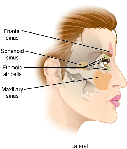
The ethmoidal labyrinth or lateral mass of the ethmoid bone consists of a number of thin-walled cellular cavities, the ethmoid air cells, arranged in three groups, anterior, middle, and posterior, and interposed between two vertical plates of bone; the lateral plate forms part of the orbit, the medial plate forms part of the nasal cavity. In the disarticulated bone many of these cells are opened into, but when the bones are articulated, they are closed in at every part, except where they open into the nasal cavity. Surfaces The upper surface of the labyrinth presents a number of half-broken cells, the walls of which are completed, in the articulated skull, by the edges of the ethmoidal notch of the frontal bone. Crossing this surface are two grooves, converted into two openings by articulation with the frontal; they are the anterior and posterior ethmoidal foramina, and open on the inner wall of the orbit. The posterior surface presents large irregular cellular cavities, which a ... [...More Info...] [...Related Items...] OR: [Wikipedia] [Google] [Baidu] |
Superior Nasal Meatus
In anatomy, the term nasal meatus can refer to any of the three meatuses (passages) through the skulls nasal cavity: the superior meatus (''meatus nasi superior''), middle meatus (''meatus nasi medius''), and inferior meatus (''meatus nasi inferior''). The nasal meatuses are located beneath each of the corresponding nasal conchae. In the case where a fourth, supreme nasal concha is present, there is a fourth supreme nasal meatus. Structure The superior meatus is the smallest of the three. It is a narrow cavity located obliquely below the superior concha. This meatus is short, lies above and extends from the middle part of the middle concha below. From behind, the sphenopalatine foramen opens into the cavity of the superior meatus and the meatus communicates with the posterior ethmoidal cells. Above and at the back of the superior concha is the sphenoethmoidal recess which the sphenoidal sinus opens into. The superior meatus occupies the middle third of the nasal cavity’s ... [...More Info...] [...Related Items...] OR: [Wikipedia] [Google] [Baidu] |
Middle Nasal Concha
The medial surface of the labyrinth of ethmoid consists of a thin lamella, which descends from the under surface of the cribriform plate, and ends below in a free, convoluted margin, the middle nasal concha (middle nasal turbinate). It is rough, and marked above by numerous grooves, directed nearly vertically downward from the cribriform plate; they lodge branches of the olfactory nerves, which are distributed to the mucous membrane covering the superior nasal concha. Additional images File:Illu nose nasal cavities.jpg, Nose and nasal cavities File:Gray152.png, Ethmoid bone from the right side. File:Gray196.png, Roof, floor, and lateral wall of left nasal cavity. File:Gray780.png, The sphenopalatine ganglion and its branches. File:Gray859.png, Coronal section of nasal cavities. File:Gray994.png, Sagittal section of nose, mouth, pharynx, and larynx. File:Nasenmuscheln1.JPG, Nasal conchae See also * Nasal concha References External links * * upstate.edu - Fronta ... [...More Info...] [...Related Items...] OR: [Wikipedia] [Google] [Baidu] |
Sphenoethmoidal Recess
The sphenoethmoidal recess is a small space in the nasal cavity into which the sphenoidal sinus and posterior ethmoid sinus open. It lies posterior and superior to the superior concha The superior nasal concha is a small, curved plate of bone representing a medial bony process of the labyrinth of the ethmoid bone. The superior nasal concha forms the roof of the superior nasal meatus. Anatomy Anatomical relations The superi .... The sphenoethmoidal recess drains the posterior ethmoid air cells and sphenoid sinuses into the superior meatus of the nasal cavity. References External links * - "The turbinates have been cut and removed to illustrate the meatus and openings into them." Nose {{respiratory-stub ... [...More Info...] [...Related Items...] OR: [Wikipedia] [Google] [Baidu] |
Sphenoid Sinus
The sphenoid sinus is a paired paranasal sinus occurring within the within the body of the sphenoid bone. It represents one pair of the four paired paranasal sinuses.Illustrated Anatomy of the Head and Neck, Fehrenbach and Herring, Elsevier, 2012, page 64 The pair of sphenoid sinuses are separated in the middle by a septum of sphenoid sinuses. Each sphenoid sinus communicates with the nasal cavity via the opening of sphenoidal sinus. The two sphenoid sinuses vary in size and shape, and are usually asymmetrical. Anatomy On average, a sphenoid sinus measures 2.2 cm vertical height, 2 cm in transverse breadth; and 2.2 cm antero-posterior depth. Each spehoid sinus is contained within the body of sphenoid bone, being situated just inferior to the sella turcica. The two sphenoid sinuses are separated medially by the septum of sphenoidal sinuses (which is usually asymmetrical). An opening of sphenoidal sinus forms a passage between each sphenoidal sinus, and the n ... [...More Info...] [...Related Items...] OR: [Wikipedia] [Google] [Baidu] |
Nasal Cavity
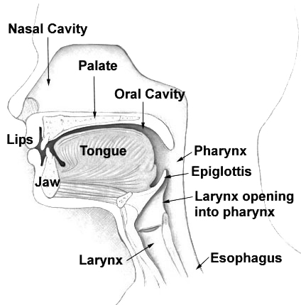
The nasal cavity is a large, air-filled space above and behind the nose in the middle of the face. The nasal septum divides the cavity into two cavities, also known as fossae. Each cavity is the continuation of one of the two nostrils. The nasal cavity is the uppermost part of the respiratory system and provides the nasal passage for inhaled air from the nostrils to the nasopharynx and rest of the respiratory tract. The paranasal sinuses surround and drain into the nasal cavity. Structure The term "nasal cavity" can refer to each of the two cavities of the nose, or to the two sides combined. The lateral wall of each nasal cavity mainly consists of the maxilla. However, there is a deficiency that is compensated for by the perpendicular plate of the palatine bone, the medial pterygoid plate, the labyrinth of ethmoid and the inferior concha. The paranasal sinuses are connected to the nasal cavity through small orifices called ostia. Most of these ostia communicate with ... [...More Info...] [...Related Items...] OR: [Wikipedia] [Google] [Baidu] |
Body Of Sphenoid Bone
The body of the sphenoid bone, more or less cubical in shape, is hollowed out in its interior to form two large cavities, the sphenoidal sinuses, which are separated from each other by a septum. Superior surface The superior surface of the body ig. 1presents in front a prominent spine, the ethmoidal spine, for articulation with the cribriform plate of the ethmoid bone; behind this is a smooth surface slightly raised in the middle line, and grooved on either side for the olfactory lobes of the brain. This surface is bounded behind by a ridge, which forms the anterior border of a narrow, transverse groove, the prechiasmatic groove, above and behind which lies the optic chiasma; the groove ends on either side in the optic foramen, which transmits the optic nerve and ophthalmic artery into the orbital cavity. Behind the chiasmatic groove is an elevation, the tuberculum sellae; and behind this is a deep depression, the saddle-shaped sella turcica (Turkish seat), the deepest ... [...More Info...] [...Related Items...] OR: [Wikipedia] [Google] [Baidu] |
Turbinate
In anatomy, a nasal concha (), plural conchae (), also called a nasal turbinate or turbinal, is a long, narrow, curled shelf of bone that protrudes into the breathing passage of the nose in humans and various animals. The conchae are shaped like an elongated seashell, which gave them their name (Latin ''concha'' from Greek ''κόγχη''). A concha is any of the scrolled spongy bones of the nasal passages in vertebrates.''Anatomy of the Human Body'' Gray, Henry (1918) The Nasal Cavity. In humans, the conchae divide the nasal airway into four groove-like air passages, and are responsible for forcing inhaled air to flow in a steady, regular pattern around the largest possible of [...More Info...] [...Related Items...] OR: [Wikipedia] [Google] [Baidu] |


