|
Subscapularis
The subscapularis is a large triangular muscle which fills the subscapular fossa and inserts into the lesser tubercle of the humerus and the front of the capsule of the shoulder-joint. Structure It arises from its medial two-thirds and Some fibers arise from tendinous laminae, which intersect the muscle and are attached to ridges on the bone; others from an aponeurosis, which separates the muscle from the teres major and the long head of the triceps brachii. The fibers pass laterally and coalesce into a tendon that is inserted into the lesser tubercle of the humerus and the anterior part of the shoulder-joint capsule. Tendinous fibers extend to the greater tubercle with insertions into the bicipital groove. Relations The tendon of the muscle is separated from the neck of the scapula by a large bursa, which communicates with the cavity of the shoulder-joint through an aperture in the capsule. The subscapularis is separated from the serratus anterior books.google.com/books? ... [...More Info...] [...Related Items...] OR: [Wikipedia] [Google] [Baidu] |
Rotator Cuff
The rotator cuff is a group of muscles and their tendons that act to stabilize the human shoulder and allow for its extensive range of motion. Of the seven scapulohumeral muscles, four make up the rotator cuff. The four muscles are the supraspinatus muscle, the infraspinatus muscle, teres minor muscle, and the subscapularis muscle. Structure Muscles composing rotator cuff The supraspinatus muscle spreads out in a horizontal band to insert on the superior facet of the greater tubercle of the humerus. The greater tubercle projects as the most lateral structure of the humeral head. Medial to this, in turn, is the lesser tubercle of the humeral head. The subscapularis muscle origin is divided from the remainder of the rotator cuff origins as it is deep to the scapula. The four tendons of these muscles converge to form the rotator cuff tendon. These tendinous insertions along with the articular capsule, the coracohumeral ligament, and the glenohumeral ligament complex, ... [...More Info...] [...Related Items...] OR: [Wikipedia] [Google] [Baidu] |
Lower Subscapular Nerve
The lower subscapular nerve, also known as the inferior subscapular nerve, is the third branch of the posterior cord of the brachial plexus. It innervates the inferior portion of the subscapularis muscle and the teres major muscle. Structure The lower subscapular nerve contains axons from the ventral rami of the C5 and C6 cervical spinal nerves. It is the third branch of the posterior cord of the brachial plexus. It gives branches to 2 muscles: * subscapularis muscle. It usually gives 4 branches to innervate the subscapularis, and can give up to 8 branches. * teres major muscle. Function The lower subscapular nerve innervates the subscapularis muscle and the teres major muscle. These muscles medially rotate and adduct the humerus The humerus (; ) is a long bone in the arm that runs from the shoulder to the elbow. It connects the scapula and the two bones of the lower arm, the radius and ulna, and consists of three sections. The humeral upper extremity consists of a roun ... [...More Info...] [...Related Items...] OR: [Wikipedia] [Google] [Baidu] |
Cervical Spinal Nerve 6
The cervical spinal nerve 6 (C6) is a spinal nerve of the cervical segment. It originates from the spinal column from above the cervical vertebra 6 (C6). The C6 nerve root shares a common branch from C5, and has a role in innervating many muscles of the rotator cuff and distal arm, including: *Subclavius *Supraspinatus *Infraspinatus * Biceps Brachii * Brachialis * Deltoid *Teres Minor *Brachioradialis *Serratus Anterior *Subscapularis *Pectoralis Major *Coracobrachialis *Teres Major * Supinator *Extensor Carpi Radialis Brevis * Extensor Carpi Radialis Longus * Latissimus Dorsi Damage to the C6 motor neuron, by way of impingement, ischemia, trauma, or degeneration of nerve tissue, can cause denervation Denervation is any loss of nerve supply regardless of the cause. If the nerves lost to denervation are part of the neuronal communication to a specific function in the body then altered or a loss of physiological functioning can occur. Denervati ... of one or more of the asso ... [...More Info...] [...Related Items...] OR: [Wikipedia] [Google] [Baidu] |
Shoulder-joint
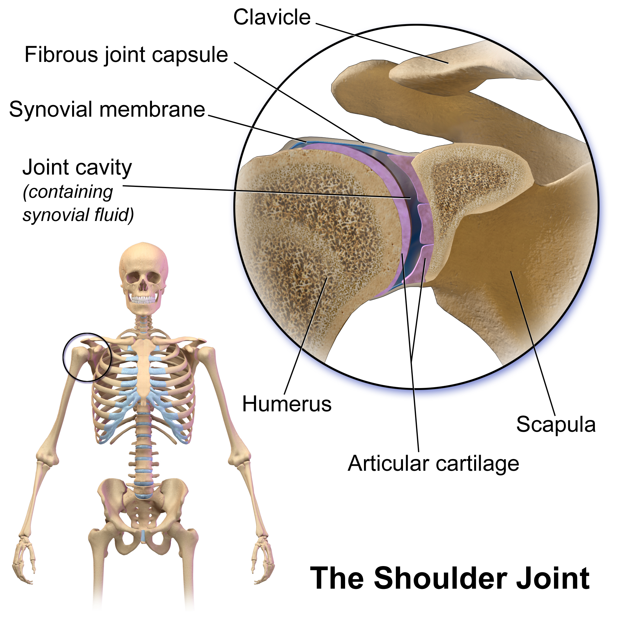
The shoulder joint (or glenohumeral joint from Greek ''glene'', eyeball, + -''oid'', 'form of', + Latin ''humerus'', shoulder) is structurally classified as a synovial ball-and-socket joint and functionally as a diarthrosis and multiaxial joint. It involves an articulation between the glenoid fossa of the scapula (shoulder blade) and the head of the humerus (upper arm bone). Due to the very loose joint capsule that gives a limited interface of the humerus and scapula, it is the most mobile joint of the human body. Structure The shoulder joint is a ball-and-socket joint between the scapula and the humerus. The socket of the glenoid fossa of the scapula is itself quite shallow, but it is made deeper by the addition of the glenoid labrum. The glenoid labrum is a ring of cartilaginous fibre attached to the circumference of the cavity. This ring is continuous with the tendon of the biceps brachii above. Spaces Significant joint spaces are: * The normal glenohumeral space is 4–5 ... [...More Info...] [...Related Items...] OR: [Wikipedia] [Google] [Baidu] |
Head Of The Humerus
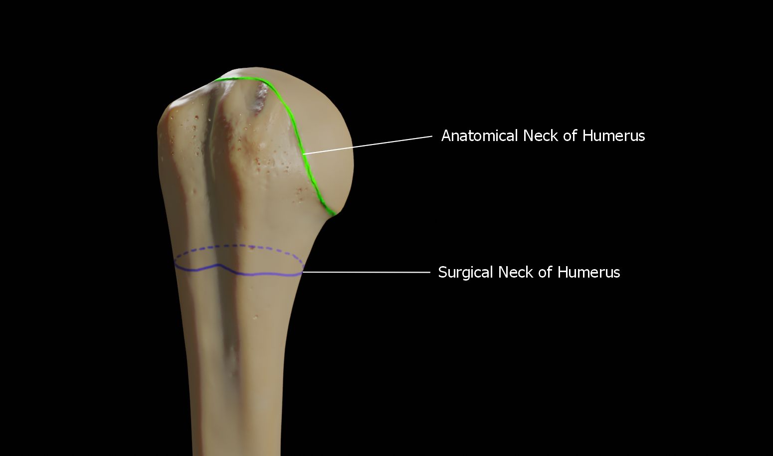
The humerus (; ) is a long bone in the arm that runs from the shoulder to the elbow. It connects the scapula and the two bones of the lower arm, the radius and ulna, and consists of three sections. The humeral upper extremity consists of a rounded head, a narrow neck, and two short processes (tubercles, sometimes called tuberosities). The body is cylindrical in its upper portion, and more prismatic below. The lower extremity consists of 2 epicondyles, 2 processes (trochlea & capitulum), and 3 fossae (radial fossa, coronoid fossa, and olecranon fossa). As well as its true anatomical neck, the constriction below the greater and lesser tubercles of the humerus is referred to as its surgical neck due to its tendency to fracture, thus often becoming the focus of surgeons. Etymology The word "humerus" is derived from la, humerus, umerus meaning upper arm, shoulder, and is linguistically related to Gothic ''ams'' shoulder and Greek ''ōmos''. Structure Upper extremity The upper or pro ... [...More Info...] [...Related Items...] OR: [Wikipedia] [Google] [Baidu] |
Upper Subscapular Nerve
The upper (superior) subscapular nerve is the first branch of the posterior cord of the brachial plexus. The upper subscapular nerve contains axons from the ventral rami of the C5 and C6 cervical spinal nerves. It innervates the superior portion of the subscapularis muscle. The inferior portion of the subscapularis is innervated by the lower subscapular nerve. Structure The axons which form the upper subscapular nerve travel from the ventral rami of C5 and C6. They join at the upper trunk and move through its posterior division to form the posterior cord, along with the other two posterior divisions of the middle and lower trunks. The axons then branch from the posterior cord and form the upper subscapular nerve. Function The upper subscapular nerve innervates the superior portion of the subscapularis muscle The subscapularis is a large triangular muscle which fills the subscapular fossa and inserts into the lesser tubercle of the humerus and the front of the capsul ... [...More Info...] [...Related Items...] OR: [Wikipedia] [Google] [Baidu] |
Glenohumeral Joint
The shoulder joint (or glenohumeral joint from Greek ''glene'', eyeball, + -''oid'', 'form of', + Latin ''humerus'', shoulder) is structurally classified as a synovial ball-and-socket joint and functionally as a diarthrosis and multiaxial joint. It involves an articulation between the glenoid fossa of the scapula (shoulder blade) and the head of the humerus (upper arm bone). Due to the very loose joint capsule that gives a limited interface of the humerus and scapula, it is the most mobile joint of the human body. Structure The shoulder joint is a ball-and-socket joint between the scapula and the humerus. The socket of the glenoid fossa of the scapula is itself quite shallow, but it is made deeper by the addition of the glenoid labrum. The glenoid labrum is a ring of cartilaginous fibre attached to the circumference of the cavity. This ring is continuous with the tendon of the biceps brachii above. Spaces Significant joint spaces are: * The normal glenohumeral space is 4– ... [...More Info...] [...Related Items...] OR: [Wikipedia] [Google] [Baidu] |
Subscapular Fossa
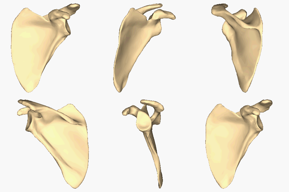
The scapula (plural scapulae or scapulas), also known as the shoulder blade, is the bone that connects the humerus (upper arm bone) with the clavicle (collar bone). Like their connected bones, the scapulae are paired, with each scapula on either side of the body being roughly a mirror image of the other. The name derives from the Classical Latin word for trowel or small shovel, which it was thought to resemble. In compound terms, the prefix omo- is used for the shoulder blade in medical terminology. This prefix is derived from ὦμος (ōmos), the Ancient Greek word for shoulder, and is cognate with the Latin , which in Latin signifies either the shoulder or the upper arm bone. The scapula forms the back of the shoulder girdle. In humans, it is a flat bone, roughly triangular in shape, placed on a posterolateral aspect of the thoracic cage. Structure The scapula is a thick, flat bone lying on the thoracic wall that provides an attachment for three groups of muscles: intrin ... [...More Info...] [...Related Items...] OR: [Wikipedia] [Google] [Baidu] |
Upper Subscapular Nerve
The upper (superior) subscapular nerve is the first branch of the posterior cord of the brachial plexus. The upper subscapular nerve contains axons from the ventral rami of the C5 and C6 cervical spinal nerves. It innervates the superior portion of the subscapularis muscle. The inferior portion of the subscapularis is innervated by the lower subscapular nerve. Structure The axons which form the upper subscapular nerve travel from the ventral rami of C5 and C6. They join at the upper trunk and move through its posterior division to form the posterior cord, along with the other two posterior divisions of the middle and lower trunks. The axons then branch from the posterior cord and form the upper subscapular nerve. Function The upper subscapular nerve innervates the superior portion of the subscapularis muscle The subscapularis is a large triangular muscle which fills the subscapular fossa and inserts into the lesser tubercle of the humerus and the front of the capsul ... [...More Info...] [...Related Items...] OR: [Wikipedia] [Google] [Baidu] |
Humerus
The humerus (; ) is a long bone in the arm that runs from the shoulder to the elbow. It connects the scapula and the two bones of the lower arm, the radius and ulna, and consists of three sections. The humeral upper extremity consists of a rounded head, a narrow neck, and two short processes (tubercles, sometimes called tuberosities). The body is cylindrical in its upper portion, and more prismatic below. The lower extremity consists of 2 epicondyles, 2 processes (trochlea & capitulum), and 3 fossae (radial fossa, coronoid fossa, and olecranon fossa). As well as its true anatomical neck, the constriction below the greater and lesser tubercles of the humerus is referred to as its surgical neck due to its tendency to fracture, thus often becoming the focus of surgeons. Etymology The word "humerus" is derived from la, humerus, umerus meaning upper arm, shoulder, and is linguistically related to Gothic ''ams'' shoulder and Greek ''ōmos''. Structure Upper extremity The upper or pr ... [...More Info...] [...Related Items...] OR: [Wikipedia] [Google] [Baidu] |
Cervical Spinal Nerve 5
The cervical spinal nerve 5 (C5) is a spinal nerve of the cervical segment. Nervous System -- Groups of Nerves It originates from the spinal column from above the (C5). It contributes to the phrenic nerve, , and before joining |


