|
Stomatitis Nicotina
Stomatitis nicotina is a diffuse white patch on the hard palate, usually caused by tobacco smoking, usually pipe or cigar smoking. It is painless, and it is caused by a response of the palatal oral mucosa to chronic heat. A more pronounced appearance can occur with reverse smoking, sometimes distinguished from stomatitis nicotina by the term reverse smoker's stomatitis. While stomatitis nicotina that is caused by heat is not a premalignant condition (i.e. it does not carry an increased risk of transformation to oral cancer), the condition that is caused by reverse smoking is premalignant. Signs and symptoms The palate may appear gray or white and contain many papules or nodules that are slightly elevated with red dots in their center. These red dots represent the ducts of minor salivary glands which have become inflamed by heat. The condition is painless. If a denture is normally worn while smoking, then the mucosa underneath the denture appears unaffected by the condition. In seve ... [...More Info...] [...Related Items...] OR: [Wikipedia] [Google] [Baidu] |
Hard Palate
The hard palate is a thin horizontal bony plate made up of two bones of the facial skeleton, located in the roof of the mouth. The bones are the palatine process of the maxilla and the horizontal plate of palatine bone. The hard palate spans the alveolar process, alveolar arch formed by the alveolar process that holds the upper teeth (when these are developed). Structure The hard palate is formed by the palatine process of the maxilla and horizontal plate of palatine bone. It forms a partition between the nasal passages and the mouth. On the anterior portion of the hard palate are the plicae, irregular ridges in the mucous membrane that help facilitate the movement of food backward towards the larynx. This partition is continued deeper into the mouth by a fleshy extension called the soft palate. On the ventral surface of hard palate, some projections or transverse ridges are present which are called as palatine rugae. Function The hard palate is important for feeding and sp ... [...More Info...] [...Related Items...] OR: [Wikipedia] [Google] [Baidu] |
Erythroplakia
Erythroplakia is a clinical term to describe any erythematous (red) area on a mucous membrane, that cannot be attributed to any other pathology. The term erythroplasia was coined by Louis Queyrat to describe a precancerous red lesion of the penis. This gave rise to the term erythoplasia of Queyrat. Depending upon the context, this term may refer specifically to carcinoma in situ of the glans penis or vulva appearing as a red patch, or may be used as a synonym of erythroplasia on other mucous membrane or transitional sites. It mainly affects the glans penis (the head of the penis), although uncommonly it may present on the mucous membranes of the larynx, and rarely, the mouth, or the anus. Erythroplakia is analogous to the term leukoplakia which describes white patches. Together, these are the 2 traditionally accepted types of premalignant lesion in the mouth, When a lesion contains both red and white areas, the term "speckled leukoplakia" or "eyrthroleukoplakia" is used. Altho ... [...More Info...] [...Related Items...] OR: [Wikipedia] [Google] [Baidu] |
Dysplasia
Dysplasia is any of various types of abnormal growth or development of cells (microscopic scale) or organs (macroscopic scale), and the abnormal histology or anatomical structure(s) resulting from such growth. Dysplasias on a mainly microscopic scale include epithelial dysplasia and fibrous dysplasia of bone. Dysplasias on a mainly macroscopic scale include hip dysplasia (human), hip dysplasia, myelodysplastic syndrome, and multicystic dysplastic kidney. In one of the modern histopathology, histopathological senses of the term, dysplasia is sometimes differentiated from other categories of tissue change including hyperplasia, metaplasia, and neoplasia, and dysplasias are thus generally not cancerous. An exception is that the myelodysplasias include a range of benign tumor, benign, precancerous condition, precancerous, and cancerous forms. Various other dysplasias tend to be precancerous. The word's meanings thus cover a spectrum of histopathological variations. Microscopic sca ... [...More Info...] [...Related Items...] OR: [Wikipedia] [Google] [Baidu] |
Cell (biology)
The cell is the basic structural and functional unit of life forms. Every cell consists of a cytoplasm enclosed within a membrane, and contains many biomolecules such as proteins, DNA and RNA, as well as many small molecules of nutrients and metabolites.Cell Movements and the Shaping of the Vertebrate Body in Chapter 21 of Molecular Biology of the Cell '' fourth edition, edited by Bruce Alberts (2002) published by Garland Science. The Alberts text discusses how the "cellular building blocks" move to shape developing embryos. It is also common to describe small molecules such as ... [...More Info...] [...Related Items...] OR: [Wikipedia] [Google] [Baidu] |
Keratin
Keratin () is one of a family of structural fibrous proteins also known as ''scleroproteins''. Alpha-keratin (α-keratin) is a type of keratin found in vertebrates. It is the key structural material making up scales, hair, nails, feathers, horns, claws, hooves, and the outer layer of skin among vertebrates. Keratin also protects epithelial cells from damage or stress. Keratin is extremely insoluble in water and organic solvents. Keratin monomers assemble into bundles to form intermediate filaments, which are tough and form strong unmineralized epidermal appendages found in reptiles, birds, amphibians, and mammals. Excessive keratinization participate in fortification of certain tissues such as in horns of cattle and rhinos, and armadillos' osteoderm. The only other biological matter known to approximate the toughness of keratinized tissue is chitin. Keratin comes in two types, the primitive, softer forms found in all vertebrates and harder, derived forms found only amon ... [...More Info...] [...Related Items...] OR: [Wikipedia] [Google] [Baidu] |
Neutrophil
Neutrophils (also known as neutrocytes or heterophils) are the most abundant type of granulocytes and make up 40% to 70% of all white blood cells in humans. They form an essential part of the innate immune system, with their functions varying in different animals. They are formed from stem cells in the bone marrow and Cellular differentiation, differentiated into #Subpopulations, subpopulations of neutrophil-killers and neutrophil-cagers. They are short-lived and highly mobile, as they can enter parts of tissue where other cells/molecules cannot. Neutrophils may be subdivided into segmented neutrophils and banded neutrophils (or Band cell, bands). They form part of the polymorphonuclear cells family (PMNs) together with basophils and eosinophils. The name ''neutrophil'' derives from staining characteristics on hematoxylin and eosin (H&E stain, H&E) histology, histological or cell biology, cytological preparations. Whereas basophilic white blood cells stain dark blue and eosinoph ... [...More Info...] [...Related Items...] OR: [Wikipedia] [Google] [Baidu] |
Hyperplasia
Hyperplasia (from ancient Greek ὑπέρ ''huper'' 'over' + πλάσις ''plasis'' 'formation'), or hypergenesis, is an enlargement of an organ or tissue caused by an increase in the amount of organic tissue that results from cell proliferation. It may lead to the gross enlargement of an organ, and the term is sometimes confused with benign neoplasia or benign tumor. Hyperplasia is a common preneoplastic response to stimulus. Microscopically, cells resemble normal cells but are increased in numbers. Sometimes cells may also be increased in size (hypertrophy). Hyperplasia is different from hypertrophy in that the adaptive cell change in hypertrophy is an increase in the ''size'' of cells, whereas hyperplasia involves an increase in the ''number'' of cells. Causes Hyperplasia may be due to any number of causes, including proliferation of basal layer of epidermis to compensate skin loss, chronic inflammatory response, hormonal dysfunctions, or compensation for damage o ... [...More Info...] [...Related Items...] OR: [Wikipedia] [Google] [Baidu] |
Squamous Metaplasia
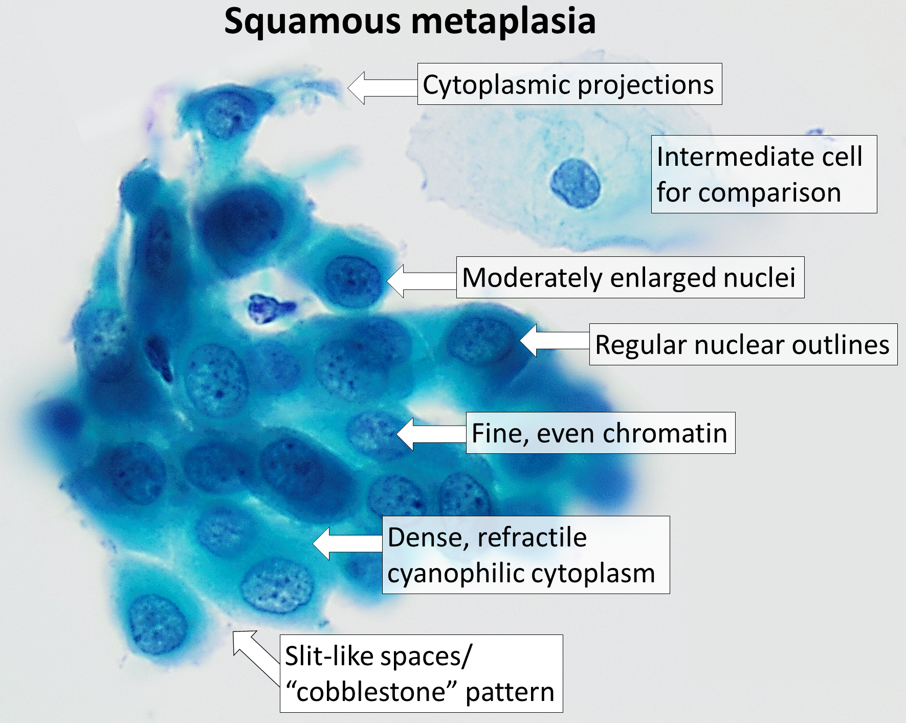
Squamous metaplasia is a benign non-cancerous change (metaplasia) of surfacing lining cells (epithelium) to a squamous morphology. Location Common sites for squamous metaplasia include the bladder and cervix. Smokers often exhibit squamous metaplasia in the linings of their airways. These changes don't signify a specific disease, but rather usually represent the body's response to stress or irritation. Vitamin A deficiency or overdose can also lead to squamous metaplasia. Uterine cervix In regard to the cervix, squamous metaplasia can sometimes be found in the endocervix, as it is composed of simple columnar epithelium, whereas the ectocervix is composed of stratified squamous non-keratinized epithelium.Kumar, Vinay; Abbas, Abul K.; Fausto, Nelson; & Mitchell, Richard N. (2007) ''Robbins Basic Pathology'' (8th ed.). Saunders Elsevier. pp. 716-720 Significance Squamous metaplasia may be seen in the context of benign lesions (e.g., atypical polypoid adenomyoma), chronic irri ... [...More Info...] [...Related Items...] OR: [Wikipedia] [Google] [Baidu] |
Hyperkeratosis
Hyperkeratosis is thickening of the stratum corneum (the outermost layer of the epidermis, or skin), often associated with the presence of an abnormal quantity of keratin,Kumar, Vinay; Fausto, Nelso; Abbas, Abul (2004) ''Robbins & Cotran Pathologic Basis of Disease'' (7th ed.). Saunders. Page 1230. . and also usually accompanied by an increase in the granular layer. As the corneum layer normally varies greatly in thickness in different sites, some experience is needed to assess minor degrees of hyperkeratosis. It can be caused by vitamin A deficiency or chronic exposure to arsenic. Hyperkeratosis can also be caused by B-Raf inhibitor drugs such as Vemurafenib and Dabrafenib.Niezgoda, Anna; Niezgoda, Piotr; Czajkowski, Rafal (2015) ''Novel Approaches to Treatment of Advanced Melanoma: A Review of Targeted Therapy and Immunotherapy'' BioMed Research International It can be treated with urea-containing creams, which dissolve the intercellular matrix of the cells of the stratum co ... [...More Info...] [...Related Items...] OR: [Wikipedia] [Google] [Baidu] |
Oral Lichen Planus
Lichen planus (LP) is a chronic inflammatory and immune-mediated disease that affects the skin, nails, hair, and mucous membranes. It is not an actual lichen, and is only named that because it looks like one. It is characterized by polygonal, flat-topped, violaceous papules and plaques with overlying, reticulated, fine white scale ( Wickham's striae), commonly affecting dorsal hands, flexural wrists and forearms, trunk, anterior lower legs and oral mucosa. The hue may be gray-brown in people with darker skin. Although there is a broad clinical range of LP manifestations, the skin and oral cavity remain as the major sites of involvement. The cause is unknown, but it is thought to be the result of an autoimmune process with an unknown initial trigger. There is no cure, but many different medications and procedures have been used in efforts to control the symptoms. The term lichenoid reaction (lichenoid eruption or lichenoid lesion) refers to a lesion of similar or identical histop ... [...More Info...] [...Related Items...] OR: [Wikipedia] [Google] [Baidu] |
Oral Candidiasis
Oral candidiasis, also known as oral thrush among other names, is candidiasis that occurs in the mouth. That is, oral candidiasis is a mycosis (yeast/fungal infection) of ''Candida'' species on the mucous membranes of the mouth. ''Candida albicans'' is the most commonly implicated organism in this condition. ''C. albicans'' is carried in the mouths of about 50% of the world's population as a normal component of the oral microbiota. This candidal carriage state is not considered a disease, but when ''Candida'' species become pathogenic and invade host tissues, oral candidiasis can occur. This change usually constitutes an opportunistic infection by normally harmless micro-organisms because of local (i.e., mucosal) or systemic factors altering host immunity. Classification Oral candidiasis is a mycosis (fungal infection). Traditionally, oral candidiasis is classified using the Lehner system, originally described in the 1960s, into acute and chronic forms (see table). Some of the ... [...More Info...] [...Related Items...] OR: [Wikipedia] [Google] [Baidu] |
Discoid Lupus Erythematosus
Discoid lupus erythematosus is the most common type of chronic cutaneous lupus (CCLE), an autoimmune skin condition on the lupus erythematosus spectrum of illnesses. It presents with red, painful, inflamed and coin-shaped patches of skin with a scaly and crusty appearance, most often on the scalp, cheeks, and ears. Hair loss may occur if the lesions are on the scalp.James, William; Berger, Timothy; Elston, Dirk (2005). ''Andrews' Diseases of the Skin: Clinical Dermatology''. (10th ed.) Saunders. Chapter 8. . The lesions can then develop severe scarring, and the centre areas may appear lighter in color with a rim darker than the normal skin. These lesions can last for years without treatment. Patients with systemic lupus erythematous develop discoid lupus lesions with some frequency. However, patients who present initially with discoid lupus infrequently develop systemic lupus. Discoid lupus can be divided into localized, generalized, and childhood discoid lupus. The lesions are d ... [...More Info...] [...Related Items...] OR: [Wikipedia] [Google] [Baidu] |


