|
Splanchnocranium
The splanchnocranium (or visceral skeleton) is the portion of the cranium that is derived from pharyngeal arches. ''Splanchno'' indicates to the gut because the face forms around the mouth, which is an end of the gut. The splanchnocranium consists of cartilage and endochondral bone. In mammals, the splanchnocranium comprises the three ear ossicles (i.e., incus, malleus, and stapes), as well as the alisphenoid, the styloid process, the hyoid apparatus, and the thyroid cartilage. In other tetrapods, such as amphibians and reptiles, homologous bones to those of mammals, such as the quadrate, articular, columella, and entoglossus are part of the splanchnocranium. See also * Dermatocranium * Endocranium The endocranium in comparative anatomy is a part of the skull base in vertebrates and it represents the basal, inner part of the cranium. The term is also applied to the outer layer of the dura mater in human anatomy. Structure Structurally, t ... References {{anatomy-stub ... [...More Info...] [...Related Items...] OR: [Wikipedia] [Google] [Baidu] |
Dermatocranium
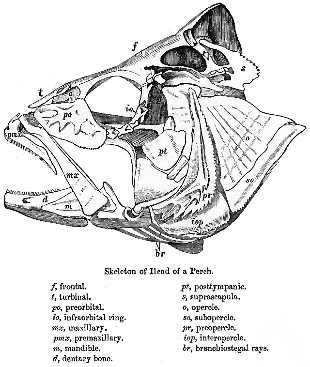
The dermatocranium is the portion of the cranium that is composed of dermal bone, as opposed to the endocranium and splanchnocranium, which are composed of endochondral bone. The dermatocranium comprises the skull roof, the facial skeleton (usually excluding the dentary In anatomy, the mandible, lower jaw or jawbone is the largest, strongest and lowest bone in the human facial skeleton. It forms the lower jaw and holds the lower tooth, teeth in place. The mandible sits beneath the maxilla. It is the only movabl ...), and—in fishes—the opercular bones. References Human anatomy Vertebrate anatomy {{musculoskeletal-stub ... [...More Info...] [...Related Items...] OR: [Wikipedia] [Google] [Baidu] |
Cranium
The skull is a bone protective cavity for the brain. The skull is composed of four types of bone i.e., cranial bones, facial bones, ear ossicles and hyoid bone. However two parts are more prominent: the cranium and the mandible. In humans, these two parts are the neurocranium and the viscerocranium ( facial skeleton) that includes the mandible as its largest bone. The skull forms the anterior-most portion of the skeleton and is a product of cephalisation—housing the brain, and several sensory structures such as the eyes, ears, nose, and mouth. In humans these sensory structures are part of the facial skeleton. Functions of the skull include protection of the brain, fixing the distance between the eyes to allow stereoscopic vision, and fixing the position of the ears to enable sound localisation of the direction and distance of sounds. In some animals, such as horned ungulates (mammals with hooves), the skull also has a defensive function by providing the mount (on the fron ... [...More Info...] [...Related Items...] OR: [Wikipedia] [Google] [Baidu] |
Thyroid Cartilage
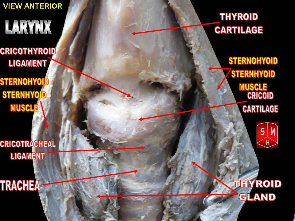
The thyroid cartilage is the largest of the nine cartilages that make up the ''laryngeal skeleton'', the cartilage structure in and around the trachea that contains the larynx. It does not completely encircle the larynx (only the cricoid cartilage encircles it). Structure The thyroid cartilage is a hyaline cartilage structure that sits in front of the larynx and above the thyroid gland. The cartilage is composed of two halves, which meet in the middle at a peak called the laryngeal prominence, also called the Adam's apple. In the midline above the prominence is the superior thyroid notch. A counterpart notch at the bottom of the cartilage is called the inferior thyroid notch. The two halves of the cartilage that make out the outer surfaces extend obliquely to cover the sides of the trachea. The posterior edge of each half articulates with the cricoid cartilage inferiorly at a joint called the cricothyroid joint. The most posterior part of the cartilage also has two projection ... [...More Info...] [...Related Items...] OR: [Wikipedia] [Google] [Baidu] |
Endocranium
The endocranium in comparative anatomy is a part of the skull base in vertebrates and it represents the basal, inner part of the cranium. The term is also applied to the outer layer of the dura mater in human anatomy. Structure Structurally, the endocranium consists of a boxlike shape, open at the top. The posterior margin exhibit the '' foramen magnum'', an opening for the spinal cord. The floor of the endocranium has several paired openings for the cranial nerves, and the anterior margin holds a spongy construction, allowing for the external nasal nerves to pass through. Romer, A.S. & T.S. Parsons. 1977. ''The Vertebrate Body.'' 5th ed. Saunders, Philadelphia. (6th ed. 1985) All bones of the structure derive from the cranial neural crest during fetal development. Endocranial elements in humans In humans and other mammals, the endocranium forms during fetal development as a cartilaginous neurocranium, that ossifies from several centers. Several of these bones merge, and in t ... [...More Info...] [...Related Items...] OR: [Wikipedia] [Google] [Baidu] |
Entoglossus
The entoglossus is an elongated bony process in lizards and birds that projects from the body of the hyoid apparatus into the tongue The tongue is a muscular organ (anatomy), organ in the mouth of a typical tetrapod. It manipulates food for mastication and swallowing as part of the digestive system, digestive process, and is the primary organ of taste. The tongue's upper surfa .... References Vertebrate anatomy {{Vertebrate anatomy-stub ... [...More Info...] [...Related Items...] OR: [Wikipedia] [Google] [Baidu] |
Columella (auditory System)
In the auditory system, the columella contributes to hearing in amphibians, reptiles and birds. The columella form thin, bony structures in the interior of the skull and serve the purpose of transmitting sounds from the eardrum. It is an evolutionary homolog of the stapes, one of the auditory ossicles in mammals. In many species, the extracolumella is a cartilaginous structure that grows in association with the columella. During development, the columella is derived from the dorsal end of the hyoid arch. Evolution The evolution of the columella is closely related to the evolution of the jaw joint. It is an ancestral homolog of the stapes, and is derived from the hyomandibular bone of fishes. As the columella is derived from the hyomandibula, many of its functional relationships remain the same. The columella resides in the air-filled tympanic cavity of the middle ear. The footplate, or proximal end of the columella, rests in the oval window. Sound is conducted through the ov ... [...More Info...] [...Related Items...] OR: [Wikipedia] [Google] [Baidu] |
Articular
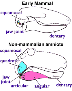
The articular bone is part of the lower jaw of most vertebrates, including most jawed fish, amphibians, birds and various kinds of reptiles, as well as ancestral mammals. Anatomy In most vertebrates, the articular bone is connected to two other lower jaw bones, the suprangular and the angular. Developmentally, it originates from the embryonic mandibular cartilage. The most caudal portion of the mandibular cartilage ossifies to form the articular bone, while the remainder of the mandibular cartilage either remains cartilaginous or disappears. In snakes In snakes, the articular, surangular, and prearticular bones have fused to form the compound bone. The mandible is suspended from the quadrate bone and articulates at this compound bone. Function In amphibians and reptiles In most tetrapods, the articular bone forms the lower portion of the jaw joint. The upper jaw articulates at the quadrate bone. In mammals In mammals, the articular bone evolves to form the malle ... [...More Info...] [...Related Items...] OR: [Wikipedia] [Google] [Baidu] |
Quadrate Bone
The quadrate bone is a skull bone in most tetrapods, including amphibians, sauropsids (reptiles, birds), and early synapsids. In most tetrapods, the quadrate bone connects to the quadratojugal and squamosal bones in the skull, and forms upper part of the jaw joint. The lower jaw articulates at the articular bone, located at the rear end of the lower jaw. The quadrate bone forms the lower jaw articulation in all classes except mammals. Evolutionarily, it is derived from the hindmost part of the primitive cartilaginous upper jaw. Function in reptiles In certain extinct reptiles, the variation and stability of the morphology of the quadrate bone has helped paleontologists in the species-level taxonomy and identification of mosasaur squamates and spinosaurine dinosaurs. In some lizards and dinosaurs, the quadrate is articulated at both ends and movable. In snakes, the quadrate bone has become elongated and very mobile, and contributes greatly to their ability to swallow very ... [...More Info...] [...Related Items...] OR: [Wikipedia] [Google] [Baidu] |
Reptile
Reptiles, as most commonly defined are the animals in the class Reptilia ( ), a paraphyletic grouping comprising all sauropsids except birds. Living reptiles comprise turtles, crocodilians, squamates (lizards and snakes) and rhynchocephalians (tuatara). As of March 2022, the Reptile Database includes about 11,700 species. In the traditional Linnaean classification system, birds are considered a separate class to reptiles. However, crocodilians are more closely related to birds than they are to other living reptiles, and so modern cladistic classification systems include birds within Reptilia, redefining the term as a clade. Other cladistic definitions abandon the term reptile altogether in favor of the clade Sauropsida, which refers to all amniotes more closely related to modern reptiles than to mammals. The study of the traditional reptile orders, historically combined with that of modern amphibians, is called herpetology. The earliest known proto-reptiles originated around ... [...More Info...] [...Related Items...] OR: [Wikipedia] [Google] [Baidu] |
Amphibian
Amphibians are tetrapod, four-limbed and ectothermic vertebrates of the Class (biology), class Amphibia. All living amphibians belong to the group Lissamphibia. They inhabit a wide variety of habitats, with most species living within terrestrial animal, terrestrial, fossorial, arboreal or freshwater aquatic ecosystems. Thus amphibians typically start out as larvae living in water, but some species have developed behavioural adaptations to bypass this. The young generally undergo metamorphosis from larva with gills to an adult air-breathing form with lungs. Amphibians use their skin as a secondary respiratory surface and some small terrestrial salamanders and frogs lack lungs and rely entirely on their skin. They are superficially similar to reptiles like lizards but, along with mammals and birds, reptiles are amniotes and do not require water bodies in which to breed. With their complex reproductive needs and permeable skins, amphibians are often ecological indicators; in re ... [...More Info...] [...Related Items...] OR: [Wikipedia] [Google] [Baidu] |
Tetrapod
Tetrapods (; ) are four-limbed vertebrate animals constituting the superclass Tetrapoda (). It includes extant and extinct amphibians, sauropsids ( reptiles, including dinosaurs and therefore birds) and synapsids (pelycosaurs, extinct therapsids and all extant mammals). Tetrapods evolved from a clade of primitive semiaquatic animals known as the Tetrapodomorpha which, in turn, evolved from ancient lobe-finned fish (sarcopterygians) around 390 million years ago in the Middle Devonian period; their forms were transitional between lobe-finned fishes and true four-limbed tetrapods. Limbed vertebrates (tetrapods in the broad sense of the word) are first known from Middle Devonian trackways, and body fossils became common near the end of the Late Devonian but these were all aquatic. The first crown-tetrapods (last common ancestors of extant tetrapods capable of terrestrial locomotion) appeared by the very early Carboniferous, 350 million years ago. The specific aquatic ancestors ... [...More Info...] [...Related Items...] OR: [Wikipedia] [Google] [Baidu] |
Hyoid Apparatus
The hyoid apparatus is the collective term used in veterinary anatomy for the bones which suspend the tongue and larynx. It consists of pairs of stylohyoid, thyrohyoid, epihyoid and ceratohyoid bones, and a single basihyoid bone. The hyoid apparatus resembles the shape of a trapeze, or a bent letter "H". The basihyoid bone lies within the muscle at the base of the tongue. In humans, the single hyoid bone The hyoid bone (lingual bone or tongue-bone) () is a horseshoe-shaped bone situated in the anterior midline of the neck between the chin and the thyroid cartilage. At rest, it lies between the base of the mandible and the third cervical vertebr ... is an equivalent of the hyoid apparatus. References Bird anatomy Mammal anatomy Human head and neck {{animal-anatomy-stub ... [...More Info...] [...Related Items...] OR: [Wikipedia] [Google] [Baidu] |


.png)
_(14771939372).jpg)