|
Sex Cord–gonadal Stromal Tumour
Sex cord–gonadal stromal tumour is a group of tumours derived from the stromal component of the ovary and testis, which comprises the granulosa, thecal cells and fibrocytes. In contrast, the epithelial cells originate from the outer epithelial lining surrounding the gonad while the germ cell tumors arise from the precursor cells of the gametes, hence the name germ cell. In humans, this group accounts for 8% of ovarian cancers and under 5% of testicular cancers. Their diagnosis is histological: only a biopsy of the tumour can make an exact diagnosis. They are often suspected of being malignant prior to operation, being solid ovarian tumours that tend to occur most commonly in post menopausal women. This group of tumours is significantly less common than testicular germ cell tumours in men, and slightly less common than ovarian germ cell tumours in women (see Ovarian cancer). Types Tumour types in order of prevalence * Granulosa cell tumour. This tumour produces granulosa ... [...More Info...] [...Related Items...] OR: [Wikipedia] [Google] [Baidu] |
Micrograph
A micrograph or photomicrograph is a photograph or digital image taken through a microscope or similar device to show a magnified image of an object. This is opposed to a macrograph or photomacrograph, an image which is also taken on a microscope but is only slightly magnified, usually less than 10 times. Micrography is the practice or art of using microscopes to make photographs. A micrograph contains extensive details of microstructure. A wealth of information can be obtained from a simple micrograph like behavior of the material under different conditions, the phases found in the system, failure analysis, grain size estimation, elemental analysis and so on. Micrographs are widely used in all fields of microscopy. Types Photomicrograph A light micrograph or photomicrograph is a micrograph prepared using an optical microscope, a process referred to as ''photomicroscopy''. At a basic level, photomicroscopy may be performed simply by connecting a camera to a microscope, th ... [...More Info...] [...Related Items...] OR: [Wikipedia] [Google] [Baidu] |
Testicle
A testicle or testis (plural testes) is the male reproductive gland or gonad in all bilaterians, including humans. It is homologous to the female ovary. The functions of the testes are to produce both sperm and androgens, primarily testosterone. Testosterone release is controlled by the anterior pituitary luteinizing hormone, whereas sperm production is controlled both by the anterior pituitary follicle-stimulating hormone and gonadal testosterone. Structure Appearance Males have two testicles of similar size contained within the scrotum, which is an extension of the abdominal wall. Scrotal asymmetry, in which one testicle extends farther down into the scrotum than the other, is common. This is because of the differences in the vasculature's anatomy. For 85% of men, the right testis hangs lower than the left one. Measurement and volume The volume of the testicle can be estimated by palpating it and comparing it to ellipsoids of known sizes. Another method is to use caliper ... [...More Info...] [...Related Items...] OR: [Wikipedia] [Google] [Baidu] |
Theca Of Follicle
The theca folliculi comprise a layer of the ovarian follicles. They appear as the follicles become secondary follicles. The theca are divided into two layers, the theca interna and the theca externa. Theca cells are a group of endocrine cells in the ovary made up of connective tissue surrounding the follicle. They have many diverse functions, including promoting folliculogenesis and recruitment of a single follicle during ovulation. Theca cells and granulosa cells together form the stroma of the ovary. Androgen synthesis Theca cells are responsible for synthesizing androgens, providing signal transduction between granulosa cells and oocytes during development by the establishment of a vascular system, providing nutrients, and providing structure and support to the follicle as it matures. Theca cells are responsible for the production of androstenedione, and indirectly the production of 17β estradiol, also called E2, by supplying the neighboring granulosa cells with andr ... [...More Info...] [...Related Items...] OR: [Wikipedia] [Google] [Baidu] |
Testicle
A testicle or testis (plural testes) is the male reproductive gland or gonad in all bilaterians, including humans. It is homologous to the female ovary. The functions of the testes are to produce both sperm and androgens, primarily testosterone. Testosterone release is controlled by the anterior pituitary luteinizing hormone, whereas sperm production is controlled both by the anterior pituitary follicle-stimulating hormone and gonadal testosterone. Structure Appearance Males have two testicles of similar size contained within the scrotum, which is an extension of the abdominal wall. Scrotal asymmetry, in which one testicle extends farther down into the scrotum than the other, is common. This is because of the differences in the vasculature's anatomy. For 85% of men, the right testis hangs lower than the left one. Measurement and volume The volume of the testicle can be estimated by palpating it and comparing it to ellipsoids of known sizes. Another method is to use caliper ... [...More Info...] [...Related Items...] OR: [Wikipedia] [Google] [Baidu] |
Sertoli Cell
Sertoli cells are a type of sustentacular "nurse" cell found in human testes which contribute to the process of spermatogenesis (the production of sperm) as a structural component of the seminiferous tubules. They are activated by follicle-stimulating hormone (FSH) secreted by the adenohypophysis and express FSH receptor on their membranes. History Sertoli cells are named after Enrico Sertoli, an Italian physiologist who discovered them while studying medicine at the University of Pavia, Italy. He published a description of his eponymous cell in 1865. The cell was discovered by Sertoli with a Belthle microscope which had been purchased in 1862. In the 1865 publication, his first description used the terms "tree-like cell" or "stringy cell"; most importantly, he referred to these as "mother cells". Other scientists later used Enrico's family name to label these cells in publications, beginning in 1888. As of 2006, two textbooks that are devoted specifically to the Sertoli cell have ... [...More Info...] [...Related Items...] OR: [Wikipedia] [Google] [Baidu] |
Hysterectomy
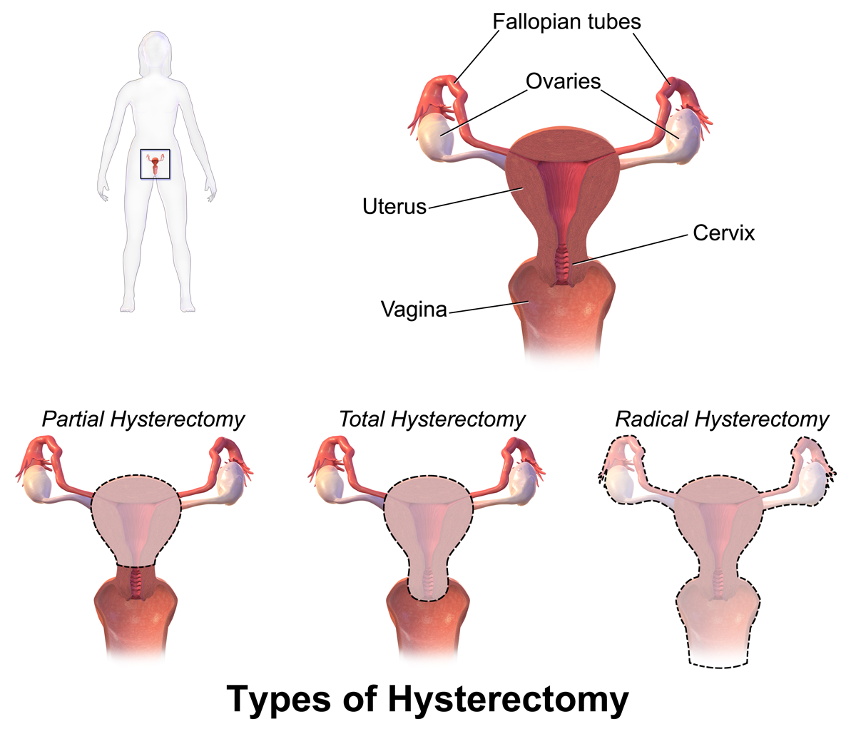
Hysterectomy is the surgical removal of the uterus. It may also involve removal of the cervix, ovaries (oophorectomy), Fallopian tubes (salpingectomy), and other surrounding structures. Usually performed by a gynecologist, a hysterectomy may be total (removing the body, fundus, and cervix of the uterus; often called "complete") or partial (removal of the uterine body while leaving the cervix intact; also called "supracervical"). Removal of the uterus renders the patient unable to bear children (as does removal of ovaries and fallopian tubes) and has surgical risks as well as long-term effects, so the surgery is normally recommended only when other treatment options are not available or have failed. It is the second most commonly performed gynecological surgical procedure, after cesarean section, in the United States. Nearly 68 percent were performed for conditions such as endometriosis, irregular bleeding, and uterine fibroids. It is expected that the frequency of hysterectom ... [...More Info...] [...Related Items...] OR: [Wikipedia] [Google] [Baidu] |
Granulosa Cells
A granulosa cell or follicular cell is a somatic cell of the sex cord that is closely associated with the developing female gamete (called an oocyte or egg) in the ovary of mammals. Structure and function In the primordial ovarian follicle, and later in follicle development (folliculogenesis), granulosa cells advance to form a multilayered cumulus oophorus surrounding the oocyte in the preovulatory or antral (or Graafian) follicle. The major functions of granulosa cells include the production of sex steroids, as well as myriad growth factors thought to interact with the oocyte during its development. The sex steroid production begins with follicle-stimulating hormone (FSH) from the anterior pituitary, stimulating granulosa cells to convert androgens (coming from the thecal cells) to estradiol by aromatase during the follicular phase of the menstrual cycle. However, after ovulation the granulosa cells turn into granulosa lutein cells that produce progesterone. The progesterone may ... [...More Info...] [...Related Items...] OR: [Wikipedia] [Google] [Baidu] |
Sertoli–Leydig Cell Tumour
Sertoli–Leydig cell tumour is a group of tumors composed of variable proportions of Sertoli cells, Leydig cells, and in the case of intermediate and poorly differentiated neoplasms, primitive gonadal stroma and sometimes heterologous elements.WHO, 2003 Sertoli–Leydig cell tumour (a sex-cord stromal tumor), is a testosterone-secreting ovarian tumor and is a member of the sex cord-stromal tumour group of ovarian and testicular cancers. The tumour occurs in early adulthood (not seen in newborn), is rare, comprising less than 1% of testicular tumours. While the tumour can occur at any age, it occurs most often in young adults. Recent studies have shown that many cases of Sertoli–Leydig cell tumor of the ovary are caused by germline mutations in the ''DICER1'' gene. These hereditary cases tend to be younger, often have a multinodular thyroid goiter and there may be a personal or family history of other rare tumors such as pleuropulmonary blastoma, Wilms tumor and cervical rhabdom ... [...More Info...] [...Related Items...] OR: [Wikipedia] [Google] [Baidu] |
Leydig Cell Tumour
Leydig cell tumour, also Leydig cell tumor (US spelling), (testicular) interstitial cell tumour and (testicular) interstitial cell tumor (US spelling), is a member of the sex cord-stromal tumour group of ovarian and testicular cancers. It arises from Leydig cells. While the tumour can occur at any age, it occurs most often in young adults. A Sertoli–Leydig cell tumour is a combination of a Leydig cell tumour and a Sertoli cell tumour from Sertoli cells. Presentation The majority of Leydig cell tumors are found in males, usually at 5–10 years of age or in middle adulthood (30–60 years). Children typically present with precocious puberty. Due to excess testosterone secreted by the tumour, one-third of female patients present with a recent history of progressive masculinization. Masculinization is preceded by anovulation, oligomenorrhea, amenorrhea and ''defeminization''. Additional signs include acne and hirsutism, voice deepening, clitoromegaly, temporal hair recession, a ... [...More Info...] [...Related Items...] OR: [Wikipedia] [Google] [Baidu] |
Fibroma
Fibromas are benign tumors that are composed of fibrous or connective tissue. They can grow in all organs, arising from mesenchyme tissue. The term "fibroblastic" or "fibromatous" is used to describe tumors of the fibrous connective tissue. When the term ''fibroma'' is used without modifier, it is usually considered benign, with the term fibrosarcoma reserved for malignant tumors. Types Hard fibroma The hard fibroma (fibroma durum) consists of many fibres and few cells, e.g. in skin it is called dermatofibroma (fibroma simplex or nodulus cutaneous). A special form is the keloid, which derives from hyperplastic growth of scars. Soft fibroma The soft fibroma (fibroma molle) or fibroma with a shaft (acrochordon, skin tag, fibroma pendulans) consist of many loosely connected cells and less fibroid tissue. It mostly appears at the neck, armpits or groin. The photo shows a soft fibroma of the eyelid. Other types of fibroma The fibroma cavernosum or angiofibroma, consists of ... [...More Info...] [...Related Items...] OR: [Wikipedia] [Google] [Baidu] |
Thecoma
Thecomas or theca cell tumors are benign Ovarian cancer, ovarian neoplasms composed only of theca cells. Histogenetically they are classified as sex cord-stromal tumours. They are typically estrogen-producing and they occur in older women (mean age 59; 84% after menopause). (They can, however, appear before menopause.) 60% of patients present with abnormal uterine bleeding, and 20% have endometrial carcinoma. Pathologic features Grossly, the tumour is solid and yellow. Grossly and microscopically, it consists of the ovarian cortex. Microscopically, the tumour cells have abundant lipid-filled cytoplasm. References External links Gynaecological neoplasia {{oncology-stub ... [...More Info...] [...Related Items...] OR: [Wikipedia] [Google] [Baidu] |
Stromal Cell
Stromal cells, or mesenchymal stromal cells, are differentiating cells found in abundance within bone marrow but can also be seen all around the body. Stromal cells can become connective tissue cells of any organ, for example in the uterine mucosa (endometrium), prostate, bone marrow, lymph node and the ovary. They are cells that support the function of the parenchymal cells of that organ. The most common stromal cells include fibroblasts and pericytes. The term ''stromal'' comes from Latin , "bed covering", and Ancient Greek , , "bed". Stromal cells are an important part of the body's immune response and modulate inflammation through multiple pathways. They also aid in differentiation of hematopoietic cells and forming necessary blood elements. The interaction between stromal cells and tumor cells is known to play a major role in cancer growth and progression. In addition, by regulating local cytokine networks (e.g. M-CSF, LIF), bone marrow stromal cells have been described to be ... [...More Info...] [...Related Items...] OR: [Wikipedia] [Google] [Baidu] |


