 |
Scramblase
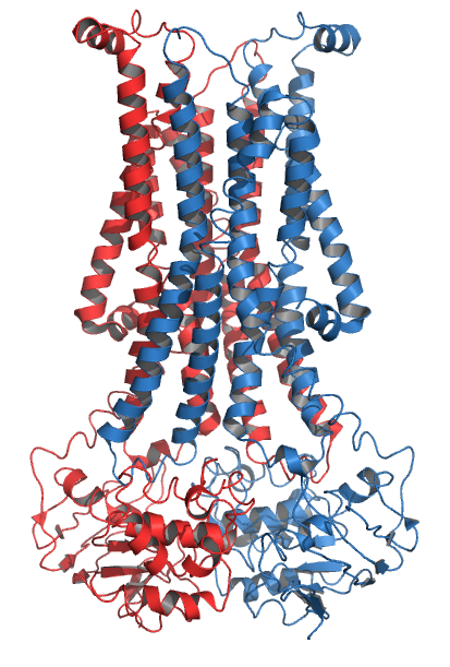
Scramblase is a protein responsible for the translocation of phospholipids between the two monolayers of a lipid bilayer of a cell membrane. In humans, phospholipid scramblases (PLSCRs) constitute a family of five homologous proteins that are named as hPLSCR1–hPLSCR5. Scramblases are not members of the general family of transmembrane lipid transporters known as flippases. Scramblases are distinct from flippases and floppases. Scramblases, flippases, and floppases are three different types of enzymatic groups of phospholipid transportation enzymes. The inner-leaflet, facing the inside of the cell, contains negatively charged amino-phospholipids and phosphatidylethanolamine. The outer-leaflet, facing the outside environment, contains phosphatidylcholine and sphingomyelin. Scramblase is an enzyme, present in the cell membrane, that can transport (''scramble'') the negatively charged phospholipids from the inner-leaflet to the outer-leaflet, and vice versa. Expression Wher ... [...More Info...] [...Related Items...] OR: [Wikipedia] [Google] [Baidu] |
 |
Flippase
Flippases (rarely spelled flipases) are transmembrane lipid transporter proteins located in the membrane which belong to ABC transporter or P4-type ATPase families. They are responsible for aiding the movement of phospholipid molecules between the two leaflets that compose a cell's membrane (transverse diffusion, also known as a "flip-flop" transition). The possibility of active maintenance of an asymmetric distribution of molecules in the phospholipid bilayer was predicted in the early 1970s by Mark Bretscher. Although phospholipids diffuse rapidly in the plane of the membrane, their polar head groups cannot pass easily through the hydrophobic center of the bilayer, limiting their diffusion in this dimension. Some flippases - often instead called scramblases - are energy-independent and bidirectional, causing reversible equilibration of phospholipid between the two sides of the membrane, whereas others are energy-dependent and unidirectional, using energy from ATP hydrolysis to ... [...More Info...] [...Related Items...] OR: [Wikipedia] [Google] [Baidu] |
|
PLSCR1
Phospholipid scramblase 1 (PL scramblase 1) is an enzyme that in humans is encoded by the ''PLSCR1'' gene. Interactions PLSCR1 has been shown to interact with: * CPSF6, * Epidermal growth factor receptor, * NEU4, * SHC1 SHC-transforming protein 1 is a protein that in humans is encoded by the ''SHC1'' gene. SHC has been found to be important in the regulation of apoptosis and drug resistance in mammalian cells. SCOP classifies the 3D structure as belonging to t ..., * SLPI, and * TFG. See also * Scramblase References Further reading * * * * * * * * * * * * * * * * * * {{gene-3-stub ... [...More Info...] [...Related Items...] OR: [Wikipedia] [Google] [Baidu] |
|
|
Tubby Protein
The tubby protein is encoded by the ''TUB'' gene. It is an upstream cell signaling protein common to multicellular eukaryotes. The first ''tubby'' gene was identified in mice, and proteins that are homologous to tubby are known as "tubby-like proteins" (TULPs). They share a common and characteristic tertiary structure that consists of a beta barrel packed around an alpha helix in the central pore. The gene derives its name from its role in metabolism; mice with a mutated tubby gene develop delayed-onset obesity, sensorineural hearing loss and retinal degeneration. Structure Tubby proteins are classified as α+β proteins and have a 12- beta stranded barrel surrounding a central alpha helix. Tubby proteins can bind the small cell signaling molecule phosphatidylinositol, which is typically localized to the cell membrane. A similar structural fold to the Tubby like proteins has been identified in the Scramblase family of proteins. Function Tubby proteins have been implicated as ... [...More Info...] [...Related Items...] OR: [Wikipedia] [Google] [Baidu] |
|
|
PLSCR3
Phospholipid scramblase 3 is an enzyme that in humans is encoded by the ''PLSCR3'' gene (abbreviated to PLS3 in this section). Like the other phospholipid scramblase family members (PLS1, PLS2, PLS4), PLS3 is a type II plasma membrane protein that is rich in proline and integral in apoptosis, or programmed cell death. The regulation of apoptosis is critical for both cell development and tissue homeostasis Although phospholipid scramblase is thought to exist in all eukaryotic cells, PLS3 is a protein that is novel to the mitochondria. This is very important because mitochondria are central in the apoptotic cell pathway. This newly found member of the scramblase family is "responsible for phospholipid translocation between two lipid compartments," the inner mitochondrial membrane and the outer membrane. Further experimental evidence suggests that the mechanism and effectors of PLS3's enzymatic activity are rather nuanced. Effect on mitochondrial cardiolipin Cardiolipin is a mi ... [...More Info...] [...Related Items...] OR: [Wikipedia] [Google] [Baidu] |
|
|
PLSCR4
Phospholipid scramblase 4, also known as Ca2+-dependent phospholipid scramblase 4, is a protein that is encoded in humans by the ''PLSCR4'' gene. See also * Scramblase Scramblase is a protein responsible for the translocation of phospholipids between the two monolayers of a lipid bilayer of a cell membrane. In humans, phospholipid scramblases (PLSCRs) constitute a family of five homologous proteins tha ... References Further reading * * * * * * * * * {{gene-3-stub ... [...More Info...] [...Related Items...] OR: [Wikipedia] [Google] [Baidu] |
|
 |
Cell Nucleus
The cell nucleus (pl. nuclei; from Latin or , meaning ''kernel'' or ''seed'') is a membrane-bound organelle found in eukaryotic cells. Eukaryotic cells usually have a single nucleus, but a few cell types, such as mammalian red blood cells, have no nuclei, and a few others including osteoclasts have many. The main structures making up the nucleus are the nuclear envelope, a double membrane that encloses the entire organelle and isolates its contents from the cellular cytoplasm; and the nuclear matrix, a network within the nucleus that adds mechanical support. The cell nucleus contains nearly all of the cell's genome. Nuclear DNA is often organized into multiple chromosomes – long stands of DNA dotted with various proteins, such as histones, that protect and organize the DNA. The genes within these chromosomes are structured in such a way to promote cell function. The nucleus maintains the integrity of genes and controls the activities of the cell by regulating g ... [...More Info...] [...Related Items...] OR: [Wikipedia] [Google] [Baidu] |
|
1Y2A
{{Letter-NumberCombDisambig ...
1Y or 1-Y may refer to: *UH-1Y; see H-1 upgrade program **Bell UH-1Y Venom *SSH 1Y (WA); see Washington State Route 532 *1Y-J, a model of Toyota Y engine *1Y, IATA code for Sun Air (Sudan) *1Y, IATA code for Electronic Data Systems * 1-Y classification in the U.S. Selective Service System, no longer in use See also *Year *Y1 (other) Y1 has several uses including: * Boeing Y1, the anticipated replacement for the company's existing Boeing 737 airliner * Great Northern Y-1, an electric locomotive used by the Great Northern Railway. * Y1 adrenocortical cell, a mouse cell line * Y1 ... [...More Info...] [...Related Items...] OR: [Wikipedia] [Google] [Baidu] |
|
 |
Erythrocyte
Red blood cells (RBCs), also referred to as red cells, red blood corpuscles (in humans or other animals not having nucleus in red blood cells), haematids, erythroid cells or erythrocytes (from Greek ''erythros'' for "red" and ''kytos'' for "hollow vessel", with ''-cyte'' translated as "cell" in modern usage), are the most common type of blood cell and the vertebrate's principal means of delivering oxygen (O2) to the body tissues—via blood flow through the circulatory system. RBCs take up oxygen in the lungs, or in fish the gills, and release it into tissues while squeezing through the body's capillaries. The cytoplasm of a red blood cell is rich in hemoglobin, an iron-containing biomolecule that can bind oxygen and is responsible for the red color of the cells and the blood. Each human red blood cell contains approximately 270 million hemoglobin molecules. The cell membrane is composed of proteins and lipids, and this structure provides properties essential for physio ... [...More Info...] [...Related Items...] OR: [Wikipedia] [Google] [Baidu] |
 |
Sickle Cell Disease
Sickle cell disease (SCD) is a group of blood disorders typically inherited from a person's parents. The most common type is known as sickle cell anaemia. It results in an abnormality in the oxygen-carrying protein haemoglobin found in red blood cells. This leads to a rigid, sickle-like shape under certain circumstances. Problems in sickle cell disease typically begin around 5 to 6 months of age. A number of health problems may develop, such as attacks of pain (known as a sickle cell crisis), anemia, swelling in the hands and feet, bacterial infections and stroke. Long-term pain may develop as people get older. The average life expectancy in the developed world is 40 to 60 years. Sickle cell disease occurs when a person inherits two abnormal copies of the β-globin gene (''HBB'') that makes haemoglobin, one from each parent. This gene occurs in chromosome 11. Several subtypes exist, depending on the exact mutation in each haemoglobin gene. An attack can be set off by te ... [...More Info...] [...Related Items...] OR: [Wikipedia] [Google] [Baidu] |
 |
Alpha Helix
The alpha helix (α-helix) is a common motif in the secondary structure of proteins and is a right hand-helix conformation in which every backbone N−H group hydrogen bonds to the backbone C=O group of the amino acid located four residues earlier along the protein sequence. The alpha helix is also called a classic Pauling–Corey–Branson α-helix. The name 3.613-helix is also used for this type of helix, denoting the average number of residues per helical turn, with 13 atoms being involved in the ring formed by the hydrogen bond. Among types of local structure in proteins, the α-helix is the most extreme and the most predictable from sequence, as well as the most prevalent. Discovery In the early 1930s, William Astbury showed that there were drastic changes in the X-ray fiber diffraction of moist wool or hair fibers upon significant stretching. The data suggested that the unstretched fibers had a coiled molecular structure with a characteristic repeat of ≈. Astbu ... [...More Info...] [...Related Items...] OR: [Wikipedia] [Google] [Baidu] |
 |
Redox
Redox (reduction–oxidation, , ) is a type of chemical reaction in which the oxidation states of substrate (chemistry), substrate change. Oxidation is the loss of Electron, electrons or an increase in the oxidation state, while reduction is the gain of electrons or a decrease in the oxidation state. There are two classes of redox reactions: * ''Electron-transfer'' – Only one (usually) electron flows from the reducing agent to the oxidant. This type of redox reaction is often discussed in terms of redox couples and electrode potentials. * ''Atom transfer'' – An atom transfers from one substrate to another. For example, in the rusting of iron, the oxidation state of iron atoms increases as the iron converts to an oxide, and simultaneously the oxidation state of oxygen decreases as it accepts electrons released by the iron. Although oxidation reactions are commonly associated with the formation of oxides, other chemical species can serve the same function. In hydrogen ... [...More Info...] [...Related Items...] OR: [Wikipedia] [Google] [Baidu] |
 |
Proline
Proline (symbol Pro or P) is an organic acid classed as a proteinogenic amino acid (used in the biosynthesis of proteins), although it does not contain the amino group but is rather a secondary amine. The secondary amine nitrogen is in the protonated form (NH2+) under biological conditions, while the carboxyl group is in the deprotonated −COO− form. The "side chain" from the α carbon connects to the nitrogen forming a pyrrolidine loop, classifying it as a aliphatic amino acid. It is non-essential in humans, meaning the body can synthesize it from the non-essential amino acid L-glutamate. It is encoded by all the codons starting with CC (CCU, CCC, CCA, and CCG). Proline is the only proteinogenic secondary amino acid which is a secondary amine, as the nitrogen atom is attached both to the α-carbon and to a chain of three carbons that together form a five-membered ring. History and etymology Proline was first isolated in 1900 by Richard Willstätter who obtained t ... [...More Info...] [...Related Items...] OR: [Wikipedia] [Google] [Baidu] |