|
Scapular Circumflex Artery
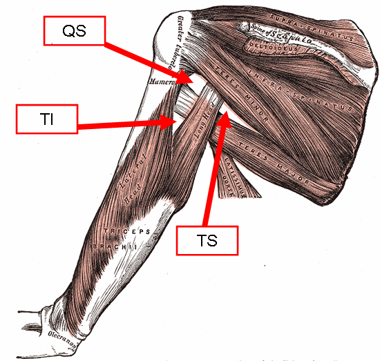
The circumflex scapular artery (scapular circumflex artery, dorsalis scapulae artery) is a branch of the subscapular artery and part of the scapular anastomoses. It curves around the axillary border of the scapula, traveling through the anatomical "Triangular space" made up of the Teres minor superiorly, the Teres major inferiorly, and the long head of the Triceps laterally. It enters the infraspinatous fossa under cover of the Teres minor, and anastomoses with the transverse scapular artery (suprascapular) and the descending branch of the transverse cervical (a.k.a. dorsal scapular artery). Branches In its course it gives off two branches: * one (infrascapular) enters the subscapular fossa beneath the Subscapularis, which it supplies, anastomosing with the transverse scapular artery and the descending branch of the transverse cervical. * the other is continued along the axillary border of the scapula, between the Teres major and minor, and at the dorsal surface of the infer ... [...More Info...] [...Related Items...] OR: [Wikipedia] [Google] [Baidu] |
Subscapular Artery
The subscapular artery, the largest branch of the axillary artery, arises from the third part of the axillary artery at the lower border of the subscapularis muscle, which it follows to the inferior angle of the scapula, where it anastomoses with the lateral thoracic and intercostal arteries, and with the descending branch of the dorsal scapular artery (a.k.a. deep branch of the transverse cervical artery if it arises from the cervical trunk), and ends in the neighboring muscles. About 4 cm from its origin it gives off two branches, first the scapular circumflex artery and then the thoracodorsal artery. From the thoracodorsal artery it supplies latissimus dorsi, while the scapular circumflex artery participates in the scapular anastamosis. It terminates in an anastomosis with the dorsal scapular artery The transverse cervical artery (transverse artery of neck or transversa colli artery) is an artery in the neck and a branch of the thyrocervical trunk, running at a h ... [...More Info...] [...Related Items...] OR: [Wikipedia] [Google] [Baidu] |
Artery
An artery (plural arteries) () is a blood vessel in humans and most animals that takes blood away from the heart to one or more parts of the body (tissues, lungs, brain etc.). Most arteries carry oxygenated blood; the two exceptions are the pulmonary and the umbilical arteries, which carry deoxygenated blood to the organs that oxygenate it (lungs and placenta, respectively). The effective arterial blood volume is that extracellular fluid which fills the arterial system. The arteries are part of the circulatory system, that is responsible for the delivery of oxygen and nutrients to all cells, as well as the removal of carbon dioxide and waste products, the maintenance of optimum blood pH, and the circulation of proteins and cells of the immune system. Arteries contrast with veins, which carry blood back towards the heart. Structure The anatomy of arteries can be separated into gross anatomy, at the macroscopic level, and microanatomy, which must be studied with a microscop ... [...More Info...] [...Related Items...] OR: [Wikipedia] [Google] [Baidu] |
Triangular Space
The triangular space (also known as the medial triangular space, upper triangular space, medial axillary space or foramen omotricipitale) is one of the three spaces found at the axillary space. The other two spaces are the quadrangular space and the triangular interval. Boundaries It has the following boundaries: * Inferior: the superior border of the teres major; * Lateral: the long head of the triceps; * Superior: Teres minor or Subscapularis For the superior border, some sources list the teres minor, while others list the subscapularis. Contents It contains the scapular circumflex vessels. Unlike the quadrangular space or the triangular interval, no major nerve passes through the triangular space. See also * Quadrangular space * Triangular interval The triangular interval (also known as the lateral triangular space, lower triangular space, and triceps hiatus) is a space found in the axilla. It is one of the three intermuscular spaces found in the axillary space. The ... [...More Info...] [...Related Items...] OR: [Wikipedia] [Google] [Baidu] |
Teres Minor
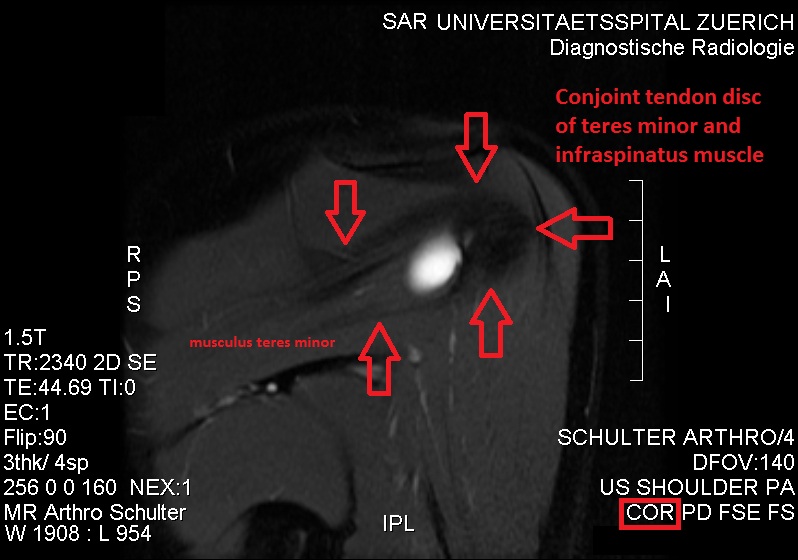
The teres minor (Latin ''teres'' meaning 'rounded') is a narrow, elongated muscle of the rotator cuff. The muscle originates from the lateral border and adjacent posterior surface of the corresponding right or left scapula and inserts at both the greater tubercle of the humerus and the posterior surface of the joint capsule. The primary function of the teres minor is to modulate the action of the deltoid, preventing the humeral head from sliding upward as the arm is abducted. It also functions to rotate the humerus laterally. The teres minor is innervated by the axillary nerve. Structure It arises from the dorsal surface of the axillary border of the scapula for the upper two-thirds of its extent, and from two aponeurotic laminae, one of which separates it from the infraspinatus muscle, the other from the teres major muscle. Its fibers run obliquely upwards and laterally; the upper ones end in a tendon which is inserted into the lowest of the three impressions on the greater tub ... [...More Info...] [...Related Items...] OR: [Wikipedia] [Google] [Baidu] |
Teres Major
The teres major muscle is a muscle of the upper limb. It attaches to the scapula and the humerus and is one of the seven scapulohumeral muscles. It is a thick but somewhat flattened muscle. The teres major muscle (from Latin ''teres'', meaning "rounded") is positioned above the latissimus dorsi muscle and assists in the Anatomical terms of motion#Flexion and extension, extension and Anatomical terms of motion#Rotation, medial rotation of the humerus. This muscle is commonly confused as a rotator cuff muscle, but it is not because it does not attach to the capsule of the shoulder joint, unlike the teres minor muscle for example. Structure The teres major muscle originates on the dorsal surface of the Scapula, inferior angle and the lower part of the Scapula, lateral border of the scapula. The fibers of teres major insert into the medial lip of the Bicipital groove, intertubercular sulcus of the humerus. Relations The tendon, at its insertion, lies behind that of the latissimus ... [...More Info...] [...Related Items...] OR: [Wikipedia] [Google] [Baidu] |
Triceps
The triceps, or triceps brachii (Latin for "three-headed muscle of the arm"), is a large muscle on the back of the upper limb of many vertebrates. It consists of 3 parts: the medial, lateral, and long head. It is the muscle principally responsible for extension of the elbow joint (straightening of the arm). Structure The long head arises from the infraglenoid tubercle of the scapula. It extends distally anterior to the teres minor and posterior to the teres major. The medial head arises proximally in the humerus, just inferior to the groove of the radial nerve; from the dorsal (back) surface of the humerus; from the medial intermuscular septum; and its distal part also arises from the lateral intermuscular septum. The medial head is mostly covered by the lateral and long heads, and is only visible distally on the humerus. The lateral head arises from the dorsal surface of the humerus, lateral and proximal to the groove of the radial nerve, from the greater tubercle down to ... [...More Info...] [...Related Items...] OR: [Wikipedia] [Google] [Baidu] |
Infraspinatous Fossa
The infraspinous fossa (infraspinatus fossa or infraspinatous fossa) of the scapula is much larger than the supraspinatous fossa; toward its vertebral margin a shallow concavity is seen at its upper part; its center presents a prominent convexity, while near the axillary border is a deep groove which runs from the upper toward the lower part. The medial two-thirds of the fossa give origin to the Infraspinatus; the lateral third is covered by this muscle. Additional images File:Infraspinatus fossa of left scapula- animation.gif, Left scapula. Infraspinatous fossa shown in red. File:Infraspinatus fossa - animation.gif, Animation. Infraspinatous fossa shown in red. File:Infraspinatous fossa of scapula01.png, Still image. File:Gray203.png, Left scapula. Dorsal surface. ("Infra-spinatous" fossa visible at bottom right.) File:Scapula post.jpg, Left scapula. Dorsal surface. Infraspinatous fossa not labeled, but visible at center. File:Infraspinatus muscle back2.png, Infraspin ... [...More Info...] [...Related Items...] OR: [Wikipedia] [Google] [Baidu] |
Anastomoses
An anastomosis (, plural anastomoses) is a connection or opening between two things (especially cavities or passages) that are normally diverging or branching, such as between blood vessels, leaf veins, or streams. Such a connection may be normal (such as the foramen ovale in a fetus's heart) or abnormal (such as the patent foramen ovale in an adult's heart); it may be acquired (such as an arteriovenous fistula) or innate (such as the arteriovenous shunt of a metarteriole); and it may be natural (such as the aforementioned examples) or artificial (such as a surgical anastomosis). The reestablishment of an anastomosis that had become blocked is called a reanastomosis. Anastomoses that are abnormal, whether congenital or acquired, are often called fistulas. The term is used in medicine, biology, mycology, geology, and geography. Etymology Anastomosis: medical or Modern Latin, from Greek ἀναστόμωσις, anastomosis, "outlet, opening", Gr ana- "up, on, upon", stoma "mouth", ... [...More Info...] [...Related Items...] OR: [Wikipedia] [Google] [Baidu] |
Transverse Scapular Artery
The suprascapular artery is a branch of the thyrocervical trunk on the neck. Structure At first, it passes downward and laterally across the scalenus anterior and phrenic nerve, being covered by the sternocleidomastoid muscle; it then crosses the subclavian artery and the brachial plexus, running behind and parallel with the clavicle and subclavius muscle and beneath the inferior belly of the omohyoid to the superior border of the scapula. It passes over the superior transverse scapular ligament in most of the cases while below it through the suprascapular notch in some cases. The artery then enters the supraspinous fossa of the scapula. It travels close to the bone, running through the suprascapular canal underneath the supraspinatus muscle, to which it supplies branches. It then descends behind the neck of the scapula, through the great scapular notch and under cover of the inferior transverse ligament, to reach the infraspinatous fossa, where it supplies infraspinatu ... [...More Info...] [...Related Items...] OR: [Wikipedia] [Google] [Baidu] |
Transverse Cervical
The transverse cervical artery (transverse artery of neck or transversa colli artery) is an artery in the neck and a branch of the thyrocervical trunk, running at a higher level than the suprascapular artery. Structure It passes transversely below the inferior belly of the omohyoid muscle to the anterior margin of the trapezius, beneath which it divides into a superficial and a deep branch. It crosses in front of the phrenic nerve and the scalene muscles, and in front of or between the divisions of the brachial plexus, and is covered by the platysma and sternocleidomastoid muscles, and crossed by the omohyoid and trapezius. The transverse cervical artery originates from the thyrocervical trunk, it passes through the posterior triangle of the neck to the anterior border of the levator scapulae muscle, where it divides into deep and superficial branches. * Superficial branch ** Ascending branch ** Descending branch (also known as superficial cervical artery, which supplies th ... [...More Info...] [...Related Items...] OR: [Wikipedia] [Google] [Baidu] |
Dorsal Scapular Artery
The transverse cervical artery (transverse artery of neck or transversa colli artery) is an artery in the neck and a branch of the thyrocervical trunk, running at a higher level than the suprascapular artery. Structure It passes transversely below the inferior belly of the omohyoid muscle to the anterior margin of the trapezius, beneath which it divides into a superficial and a deep branch. It crosses in front of the phrenic nerve and the scalene muscles, and in front of or between the divisions of the brachial plexus, and is covered by the platysma and sternocleidomastoid muscles, and crossed by the omohyoid and trapezius. The transverse cervical artery originates from the thyrocervical trunk, it passes through the posterior triangle of the neck to the anterior border of the levator scapulae muscle, where it divides into deep and superficial branches. * Superficial branch ** Ascending branch ** Descending branch (also known as superficial cervical artery, which supplies t ... [...More Info...] [...Related Items...] OR: [Wikipedia] [Google] [Baidu] |
Subscapular Fossa
The scapula (plural scapulae or scapulas), also known as the shoulder blade, is the bone that connects the humerus (upper arm bone) with the clavicle (collar bone). Like their connected bones, the scapulae are paired, with each scapula on either side of the body being roughly a mirror image of the other. The name derives from the Classical Latin word for trowel or small shovel, which it was thought to resemble. In compound terms, the prefix omo- is used for the shoulder blade in medical terminology. This prefix is derived from ὦμος (ōmos), the Ancient Greek word for shoulder, and is cognate with the Latin , which in Latin signifies either the shoulder or the upper arm bone. The scapula forms the back of the shoulder girdle. In humans, it is a flat bone, roughly triangular in shape, placed on a posterolateral aspect of the thoracic cage. Structure The scapula is a thick, flat bone lying on the thoracic wall that provides an attachment for three groups of muscles: intrin ... [...More Info...] [...Related Items...] OR: [Wikipedia] [Google] [Baidu] |


