|
Stylopharyngeus
The stylopharyngeus is a muscle in the head that stretches between the temporal styloid process and the pharynx. Structure The stylopharyngeus is a long, slender muscle, cylindrical above, flattened below. It arises from the medial side of the base of the temporal styloid process, passes downward along the side of the pharynx between the superior pharyngeal constrictor and the middle pharyngeal constrictor, and spreads out beneath the mucous membrane. Some of its fibers are lost in the constrictor muscles while others, joining the palatopharyngeus muscle, are inserted into the posterior border of the thyroid cartilage. The glossopharyngeal nerve runs on the lateral side of this muscle, and crosses over it to reach the tongue. Nerve supply The stylopharyngeus is the only muscle in the pharynx innervated by the glossopharyngeal nerve (CN IX) via branchial motor neurons with their cell bodies in the rostral part of the nucleus ambiguus. Development Embryological origin i ... [...More Info...] [...Related Items...] OR: [Wikipedia] [Google] [Baidu] |
Glossopharyngeal Nerve
The glossopharyngeal nerve (), also known as the ninth cranial nerve, cranial nerve IX, or simply CN IX, is a cranial nerve that exits the brainstem from the sides of the upper medulla, just anterior (closer to the nose) to the vagus nerve. Being a mixed nerve (sensorimotor), it carries afferent sensory and efferent motor information. The motor division of the glossopharyngeal nerve is derived from the basal plate of the embryonic medulla oblongata, whereas the sensory division originates from the cranial neural crest. Structure From the anterior portion of the medulla oblongata, the glossopharyngeal nerve passes laterally across or below the flocculus, and leaves the skull through the central part of the jugular foramen. From the superior and inferior ganglia in jugular foramen, it has its own sheath of dura mater. The inferior ganglion on the inferior surface of petrous part of temporal is related with a triangular depression into which the aqueduct of cochlea opens. On the ... [...More Info...] [...Related Items...] OR: [Wikipedia] [Google] [Baidu] |
Pharyngeal Arch
The pharyngeal arches, also known as visceral arches'','' are structures seen in the embryonic development of vertebrates that are recognisable precursors for many structures. In fish, the arches are known as the branchial arches, or gill arches. In the human embryo, the arches are first seen during the fourth week of development. They appear as a series of outpouchings of mesoderm on both sides of the developing pharynx. The vasculature of the pharyngeal arches is known as the aortic arches. In fish, the branchial arches support the gills. Structure In vertebrates, the pharyngeal arches are derived from all three germ layers (the primary layers of cells that form during embryogenesis). Neural crest cells enter these arches where they contribute to features of the skull and facial skeleton such as bone and cartilage. However, the existence of pharyngeal structures before neural crest cells evolved is indicated by the existence of neural crest-independent mechanisms of phary ... [...More Info...] [...Related Items...] OR: [Wikipedia] [Google] [Baidu] |
Styloid Process (temporal)
The temporal styloid process is a slender bony process of the temporal bone extending downward and forward from the undersurface of the temporal bone just below the ear. The styloid process gives attachments to several muscles, and ligaments. Structure The styloid process is a slender and pointed bony process of the temporal bone projecting anteroinferiorly from the inferior surface of the temporal bone just below the ear. Its length normally ranges from just under 3 cm to just over 4 cm. It is usually nearly straight, but may be curved in some individuals. Its ''proximal'' (''tympanohyal'') ''part'' is ensheathed by the tympanic part of the temporal bone ''(vaginal process), whereas'' its ''distal (stylohyal)'' ''part'' gives attachment to several structures. Attachments The styloid process gives attachments to several muscles, and ligaments. It serves as an anchor point for several muscles associated with the tongue and larynx. * stylohyoid ligament * stylomandi ... [...More Info...] [...Related Items...] OR: [Wikipedia] [Google] [Baidu] |
Superior Pharyngeal Constrictor
The superior pharyngeal constrictor muscle is a muscle in the pharynx. It is the highest located muscle of the three pharyngeal constrictors. The muscle is a quadrilateral muscle, thinner and paler than the inferior pharyngeal constrictor muscle and middle pharyngeal constrictor muscle. The muscle is divided into four parts: A pterygopharyngeal, buccopharyngeal, mylopharyngeal and a glossopharyngeal part. Origin and insertion The four parts of this muscle arise from: - the lower third of the posterior margin of the medial pterygoid plate and its hamulus (Pterygopharyngeal part) - from the pterygomandibular raphe (Buccopharyngeal part) - from the alveolar process of the mandible above the posterior end of the mylohyoid line (Mylopharyngeal part) - and by a few fibers from the side of the tongue (Glossopharyngeal part) The fibers curve backward to be inserted into the median raphe, being also prolonged by means of an aponeurosis to the pharyngeal spine on the basilar part o ... [...More Info...] [...Related Items...] OR: [Wikipedia] [Google] [Baidu] |
Palatopharyngeus
The palatopharyngeus (palatopharyngeal or pharyngopalatinus) muscle is a small muscle in the roof of the mouth. It is a long, fleshy fasciculus, narrower in the middle than at either end, forming, with the mucous membrane covering its surface, the palatopharyngeal arch. Structure It is separated from the palatoglossus muscle by an angular interval, in which the palatine tonsil is lodged. It arises from the soft palate, where it is divided into two fasciculi by the levator veli palatini and musculus uvulae. * The ''posterior fasciculus'' lies in contact with the mucous membrane, and joins with that of the opposite muscle in the middle line. * The ''anterior fasciculus'', the thicker, lies in the soft palate between the levator and tensor veli palatini muscles, and joins in the middle line the corresponding part of the opposite muscle. Passing laterally and downward behind the palatine tonsil, the palatopharyngeus joins the stylopharyngeus and is inserted with that muscle into t ... [...More Info...] [...Related Items...] OR: [Wikipedia] [Google] [Baidu] |
Human Pharynx
The pharynx (plural: pharynges) is the part of the throat behind the mouth and nasal cavity, and above the oesophagus and trachea (the tubes going down to the stomach and the lungs). It is found in vertebrates and invertebrates, though its structure varies across species. The pharynx carries food and air to the esophagus and larynx respectively. The flap of cartilage called the epiglottis stops food from entering the larynx. In humans, the pharynx is part of the digestive system and the conducting zone of the respiratory system. (The conducting zone—which also includes the nostrils of the nose, the larynx, trachea, bronchi, and bronchioles—filters, warms and moistens air and conducts it into the lungs). The human pharynx is conventionally divided into three sections: the nasopharynx, oropharynx, and laryngopharynx. It is also important in vocalization. In humans, two sets of pharyngeal muscles form the pharynx and determine the shape of its lumen. They are arranged ... [...More Info...] [...Related Items...] OR: [Wikipedia] [Google] [Baidu] |
Cheek
The cheeks ( la, buccae) constitute the area of the face below the eyes and between the nose and the left or right ear. "Buccal" means relating to the cheek. In humans, the region is innervated by the buccal nerve. The area between the inside of the cheek and the teeth and gums is called the vestibule or buccal pouch or buccal cavity and forms part of the mouth. In other animals the cheeks may also be referred to as jowls. Structure Humans Cheeks are fleshy in humans, the skin being suspended by the chin and the jaws, and forming the lateral wall of the human mouth, visibly touching the cheekbone below the eye. The inside of the cheek is lined with a mucous membrane (buccal mucosa, part of the oral mucosa). During mastication (chewing), the cheeks and tongue between them serve to keep the food between the teeth. Other animals The cheeks are covered externally by hairy skin, and internally by stratified squamous epithelium. This is mostly smooth, but may have caudally di ... [...More Info...] [...Related Items...] OR: [Wikipedia] [Google] [Baidu] |
Thyroid Cartilage
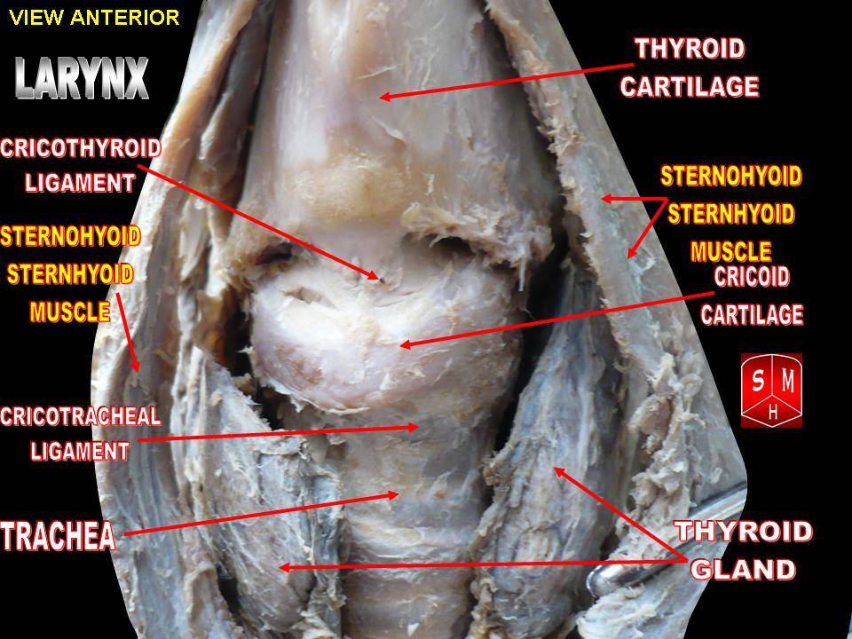
The thyroid cartilage is the largest of the nine cartilages that make up the ''laryngeal skeleton'', the cartilage structure in and around the trachea that contains the larynx. It does not completely encircle the larynx (only the cricoid cartilage encircles it). Structure The thyroid cartilage is a hyaline cartilage structure that sits in front of the larynx and above the thyroid gland. The cartilage is composed of two halves, which meet in the middle at a peak called the laryngeal prominence, also called the Adam's apple. In the midline above the prominence is the superior thyroid notch. A counterpart notch at the bottom of the cartilage is called the inferior thyroid notch. The two halves of the cartilage that make out the outer surfaces extend obliquely to cover the sides of the trachea. The posterior edge of each half articulates with the cricoid cartilage inferiorly at a joint called the cricothyroid joint. The most posterior part of the cartilage also has two projec ... [...More Info...] [...Related Items...] OR: [Wikipedia] [Google] [Baidu] |
Larynx
The larynx (), commonly called the voice box, is an organ in the top of the neck involved in breathing, producing sound and protecting the trachea against food aspiration. The opening of larynx into pharynx known as the laryngeal inlet is about 4–5 centimeters in diameter. The larynx houses the vocal cords, and manipulates pitch and volume, which is essential for phonation. It is situated just below where the tract of the pharynx splits into the trachea and the esophagus. The word ʻlarynxʼ (plural ʻlaryngesʼ) comes from the Ancient Greek word ''lárunx'' ʻlarynx, gullet, throat.ʼ Structure The triangle-shaped larynx consists largely of cartilages that are attached to one another, and to surrounding structures, by muscles or by fibrous and elastic tissue components. The larynx is lined by a ciliated columnar epithelium except for the vocal folds. The cavity of the larynx extends from its triangle-shaped inlet, to the epiglottis, and to the circular outlet at t ... [...More Info...] [...Related Items...] OR: [Wikipedia] [Google] [Baidu] |
Middle Pharyngeal Constrictor
The middle pharyngeal constrictor is a fan-shaped muscle located in the neck. It is one of three pharyngeal constrictors. Similarly to the superior and inferior pharyngeal constrictor muscles, the middle pharyngeal constrictor is innervated by a branch of the vagus nerve through the pharyngeal plexus. The middle pharyngeal constrictor is smaller than the inferior pharyngeal constrictor muscle. Structure The middle pharyngeal constrictor arises from the whole length of the upper border of the greater cornu of the hyoid bone, from the lesser cornu, and from the stylohyoid ligament. The fibers diverge from their origin: the lower ones descend beneath the constrictor inferior, the middle fibers pass transversely, and the upper fibers ascend and overlap the constrictor superior. It is inserted into the posterior median fibrous raphe, blending in the middle line with the muscle of the opposite side. Function As soon as the bolus Bolus may refer to: Geography * Bolus, Ir ... [...More Info...] [...Related Items...] OR: [Wikipedia] [Google] [Baidu] |
Stylohyoid Muscle
The stylohyoid muscle is a slender muscle, lying anterior and superior of the posterior belly of the digastric muscle. It is one of the suprahyoid muscles. It shares this muscle's innervation by the facial nerve, and functions to draw the hyoid bone backwards and elevate the tongue. Its origin is the styloid process of the temporal bone. It inserts on the body of the hyoid. Structure The stylohyoid muscle originates from the posterior and lateral surface of the styloid process of the temporal bone, near the base. Passing inferior and anterior, it inserts into the body of the hyoid bone, at its junction with the greater cornu, and just superior to the omohyoid muscle. It belongs to the group of suprahyoid muscles. It is perforated, near its insertion, by the intermediate tendon of the digastric muscle. The stylohyoid muscle has vascular supply from the lingual artery, a branch of the external carotid artery. Nerve supply A branch of the facial nerve (CN VII) innervates the s ... [...More Info...] [...Related Items...] OR: [Wikipedia] [Google] [Baidu] |


