|
Schwalbe's Line
Schwalbe's line is the anatomical line found on the interior surface of the eye's cornea, and delineates the outer limit of the corneal endothelium layer. Specifically, it represents the termination of Descemet's membrane. In many cases it can be seen via gonioscopy Gonioscopy a routine ophthalmological procedure that measures the angle between the iris and the cornea (the iridocorneal angle), using a goniolens (also known as a gonioscope) together with a slit lamp or operating microscope. Its use is import .... Some evidence suggests that the corneal endothelium actually possesses stem cells that can produce endothelial cells, especially after injury, albeit on a limited scale. References Human eye anatomy {{Eye-stub ... [...More Info...] [...Related Items...] OR: [Wikipedia] [Google] [Baidu] |
Anterior Chamber Angle - 3D Motion Parallax
Standard anatomical terms of location are used to unambiguously describe the anatomy of animals, including humans. The terms, typically derived from Latin or Greek roots, describe something in its standard anatomical position. This position provides a definition of what is at the front ("anterior"), behind ("posterior") and so on. As part of defining and describing terms, the body is described through the use of anatomical planes and anatomical axes. The meaning of terms that are used can change depending on whether an organism is bipedal or quadrupedal. Additionally, for some animals such as invertebrates, some terms may not have any meaning at all; for example, an animal that is radially symmetrical will have no anterior surface, but can still have a description that a part is close to the middle ("proximal") or further from the middle ("distal"). International organisations have determined vocabularies that are often used as standard vocabularies for subdisciplines of anatomy ... [...More Info...] [...Related Items...] OR: [Wikipedia] [Google] [Baidu] |
Cornea
The cornea is the transparent front part of the eye that covers the iris, pupil, and anterior chamber. Along with the anterior chamber and lens, the cornea refracts light, accounting for approximately two-thirds of the eye's total optical power. In humans, the refractive power of the cornea is approximately 43 dioptres. The cornea can be reshaped by surgical procedures such as LASIK. While the cornea contributes most of the eye's focusing power, its focus is fixed. Accommodation (the refocusing of light to better view near objects) is accomplished by changing the geometry of the lens. Medical terms related to the cornea often start with the prefix "'' kerat-''" from the Greek word κέρας, ''horn''. Structure The cornea has unmyelinated nerve endings sensitive to touch, temperature and chemicals; a touch of the cornea causes an involuntary reflex to close the eyelid. Because transparency is of prime importance, the healthy cornea does not have or need blood vessels with ... [...More Info...] [...Related Items...] OR: [Wikipedia] [Google] [Baidu] |
Corneal Endothelium
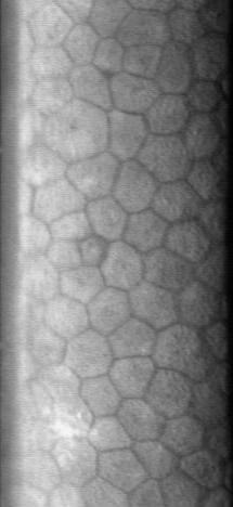
The corneal endothelium is a single layer of endothelial cells on the inner surface of the cornea. It faces the chamber formed between the cornea and the iris. The corneal endothelium are specialized, flattened, mitochondria-rich cells that line the posterior surface of the cornea and face the anterior chamber of the eye. The corneal endothelium governs fluid and solute transport across the posterior surface of the cornea and maintains the cornea in the slightly dehydrated state that is required for optical transparency. Embryology and anatomy The corneal endothelium is embryologically derived from the neural crest. The postnatal total endothelial cellularity of the cornea (approximately 300,000 cells per cornea) is achieved as early as the second trimester of gestation. Thereafter the endothelial cell density (but not the absolute number of cells) rapidly declines, as the fetal cornea grows in surface area, achieving a final adult density of approximately 2400 - 3200 cells ... [...More Info...] [...Related Items...] OR: [Wikipedia] [Google] [Baidu] |
Descemet's Membrane
Descemet's membrane ( or the Descemet membrane) is the basement membrane that lies between the corneal proper substance, also called stroma, and the endothelial layer of the cornea. It is composed of different kinds of collagen (Type IV and VIII) than the stroma. The endothelial layer is located at the posterior of the cornea. Descemet's membrane, as the basement membrane for the endothelial layer, is secreted by the single layer of squamous epithelial cells that compose the endothelial layer of the cornea. Structure Its thickness ranges from 3 μm at birth to 8–10 μm in adults.Johnson DH, Bourne WM, Campbell RJ: The ultrastructure of Descemet's membrane. I. Changes with age in normal cornea. Arch Ophthalmol 100:1942, 1982 The corneal endothelium is a single layer of squamous cells covering the surface of the cornea that faces the anterior chamber. Clinical significance Significant damage to the membrane may require a corneal transplant. Damage caused by the hereditary co ... [...More Info...] [...Related Items...] OR: [Wikipedia] [Google] [Baidu] |
Gonioscopy
Gonioscopy a routine ophthalmological procedure that measures the angle between the iris and the cornea (the iridocorneal angle), using a goniolens (also known as a gonioscope) together with a slit lamp or operating microscope. Its use is important in diagnosing and monitoring various eye conditions associated with glaucoma. The goniolens or gonioscope The goniolens allows the clinician - usually an ophthalmologist or optometrist - to view the irideocorneal angle through a mirror or prism, without which the angle is masked by total internal reflection from the ocular tissue. The mechanism for this process varies with each type of goniolens. Three examples of goniolenses are the: * Koeppe direct goniolens: this transparent device is placed directly on the cornea along with lubricating fluid, to avoid damaging its surface. The steeper curvature of this goniolens' exterior surface optically eliminates the total internal reflection problem and allows a view of the iridocorneal angl ... [...More Info...] [...Related Items...] OR: [Wikipedia] [Google] [Baidu] |


