|
Scaphoid Fossa
In the pterygoid processes of the sphenoid, above the pterygoid fossa is a small, oval, shallow depression, the scaphoid fossa, which gives origin to the Tensor veli palatini. It is not the same as and has to be distinguished from the scaphoid fossa of the external ear or pinna. References External links Diagram - look for #28(sourchere Bones of the head and neck {{musculoskeletal-stub ... [...More Info...] [...Related Items...] OR: [Wikipedia] [Google] [Baidu] |
Sphenoid Bone
The sphenoid bone is an unpaired bone of the neurocranium. It is situated in the middle of the skull towards the front, in front of the basilar part of the occipital bone. The sphenoid bone is one of the seven bones that articulate to form the orbit. Its shape somewhat resembles that of a butterfly or bat with its wings extended. Structure It is divided into the following parts: * a median portion, known as the body of sphenoid bone, containing the sella turcica, which houses the pituitary gland as well as the paired paranasal sinuses, the sphenoidal sinuses * two greater wings on the lateral side of the body and two lesser wings from the anterior side. * Pterygoid processes of the sphenoides, directed downwards from the junction of the body and the greater wings. Two sphenoidal conchae are situated at the anterior and inferior part of the body. Intrinsic ligaments of the sphenoid The more important of these are: * the pterygospinous, stretching between the spin ... [...More Info...] [...Related Items...] OR: [Wikipedia] [Google] [Baidu] |
Pterygoid Processes Of The Sphenoid
The pterygoid processes of the sphenoid (from Greek ''pteryx'', ''pterygos'', "wing"), one on either side, descend perpendicularly from the regions where the body and the greater wings of the sphenoid bone unite. Each process consists of a medial pterygoid plate and a lateral pterygoid plate, the latter of which serve as the origins of the medial and lateral pterygoid muscles. The medial pterygoid, along with the masseter allows the jaw to move in a vertical direction as it contracts and relaxes. The lateral pterygoid allows the jaw to move in a horizontal direction during mastication (chewing). Fracture of either plate are used in clinical medicine to distinguish the Le Fort fracture classification for high impact injuries to the sphenoid and maxillary bones. The superior portion of the pterygoid processes are fused anteriorly; a vertical groove, the pterygopalatine fossa, descends on the front of the line of fusion. The plates are separated below by an angular cleft, t ... [...More Info...] [...Related Items...] OR: [Wikipedia] [Google] [Baidu] |
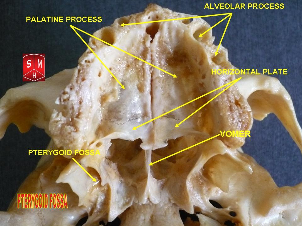
Pterygoid Fossa
The pterygoid fossa is an anatomical term for the fossa formed by the divergence of the lateral pterygoid plate and the medial pterygoid plate of the sphenoid bone. Structure The lateral and medial pterygoid plates (of the pterygoid process of the sphenoid bone) diverge behind and enclose between them a V-shaped fossa, the pterygoid fossa. This fossa faces posteriorly, and contains the medial pterygoid muscle and the tensor veli palatini muscle. See also * Pterygoid fovea * Scaphoid fossa In the pterygoid processes of the sphenoid, above the pterygoid fossa is a small, oval, shallow depression, the scaphoid fossa, which gives origin to the Tensor veli palatini. It is not the same as and has to be distinguished from the scaphoid ... * Pterygoid process References Bones of the head and neck {{musculoskeletal-stub ... [...More Info...] [...Related Items...] OR: [Wikipedia] [Google] [Baidu] |
Tensor Veli Palatini
The tensor veli palatini muscle (tensor palati or tensor muscle of the velum palatinum) is a broad, thin, ribbon-like muscle in the head that tenses the soft palate. Structure The tensor veli palatini is found anterior-lateral to the levator veli palatini muscle. It arises by a flat lamella from the scaphoid fossa at the base of the medial pterygoid plate, from the spina angularis of the sphenoid and from the lateral wall of the cartilage of the auditory tube. Descending vertically between the medial pterygoid plate and the medial pterygoid muscle, it ends in a tendon which winds around the pterygoid hamulus, being retained in this situation by some of the fibers of origin of the medial pterygoid muscle. Between the tendon and the hamulus is a small bursa. The tendon then passes medially and is inserted into the palatine aponeurosis and into the surface behind the transverse ridge on the horizontal part of the palatine bone. Nerve supply The tensor veli palatini ... [...More Info...] [...Related Items...] OR: [Wikipedia] [Google] [Baidu] |
Pinna (anatomy)
The auricle or auricula is the visible part of the ear that is outside the head. It is also called the pinna (Latin for " wing" or " fin", plural pinnae), a term that is used more in zoology. Structure The diagram shows the shape and location of most of these components: * '' antihelix'' forms a 'Y' shape where the upper parts are: ** ''Superior crus'' (to the left of the ''fossa triangularis'' in the diagram) ** ''Inferior crus'' (to the right of the ''fossa triangularis'' in the diagram) * '' Antitragus'' is below the ''tragus'' * ''Aperture'' is the entrance to the ear canal * ''Auricular sulcus'' is the depression behind the ear next to the head * ''Concha'' is the hollow next to the ear canal * Conchal angle is the angle that the back of the ''concha'' makes with the side of the head * ''Crus'' of the helix is just above the ''tragus'' * ''Cymba conchae'' is the narrowest end of the ''concha'' * External auditory meatus is the ear canal * ''Fossa triangularis'' is the ... [...More Info...] [...Related Items...] OR: [Wikipedia] [Google] [Baidu] |


