|
Rostral Migratory Stream
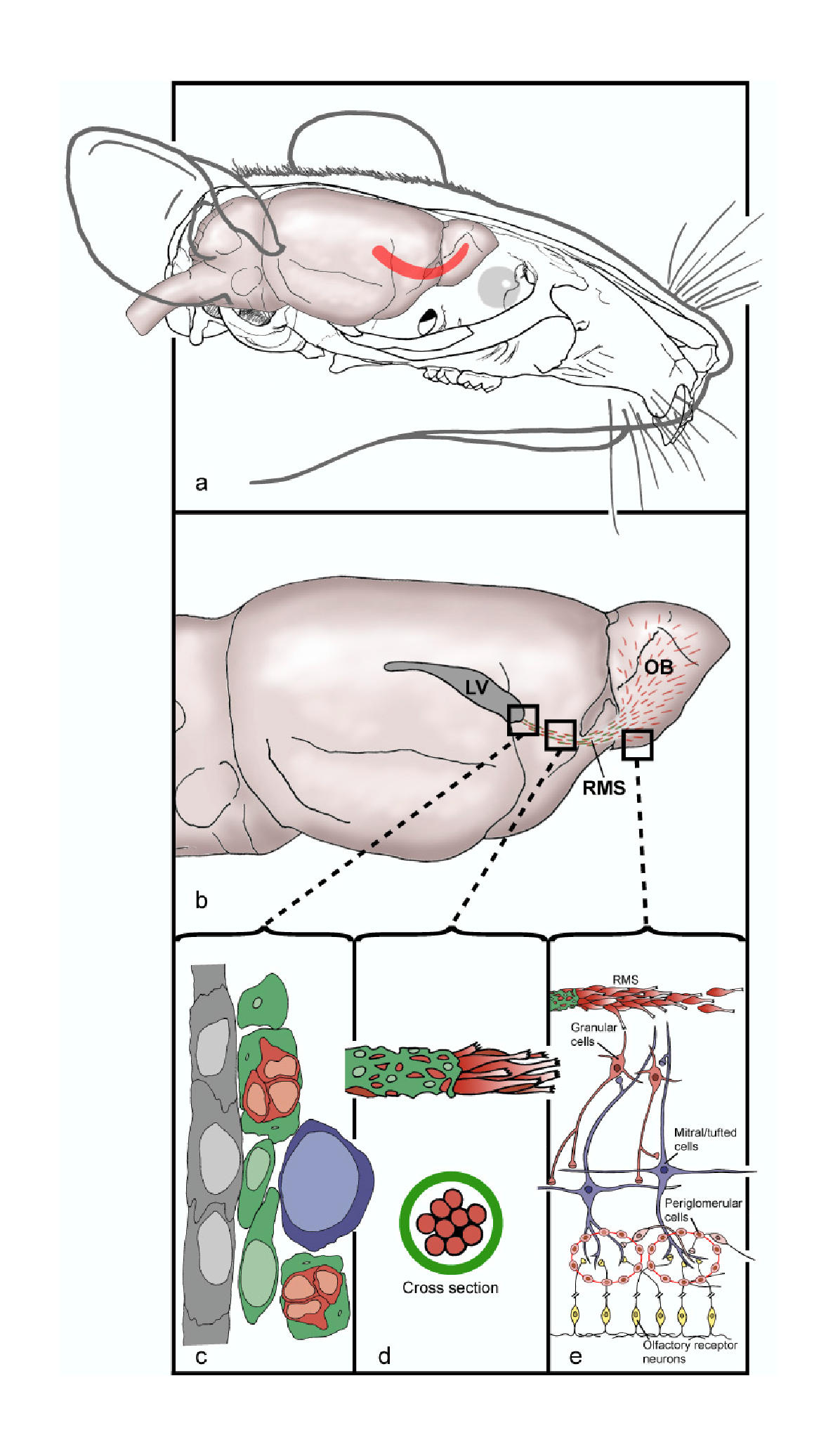
The rostral migratory stream (RMS) is a specialized migratory route found in the brain of some animals along which neuronal precursors that originated in the subventricular zone (SVZ) of the brain migrate to reach the main olfactory bulb (OB). The importance of the RMS lies in its ability to refine and even change an animal's sensitivity to smells, which explains its importance and larger size in the rodent brain as compared to the human brain, as our olfactory sense is not as developed. This pathway has been studied in the rodent, rabbit, and both the squirrel monkey and rhesus monkey. When the neurons reach the OB they differentiate into GABAergic interneurons as they are integrated into either the granule cell layer or periglomerular layer. Although it was originally believed that neurons could not regenerate in the adult brain, neurogenesis has been shown to occur in mammalian brains, including those of primates. However, neurogenesis is limited to the hippocampus and SV ... [...More Info...] [...Related Items...] OR: [Wikipedia] [Google] [Baidu] |
Rostral Migratory Stream Mouse
{{disambiguation ...
Rostral may refer to: Anatomy * Rostral (anatomical term), situated toward the oral or nasal region * Rostral bone, in ceratopsian dinosaurs * Rostral organ, of certain fish * Rostral scale, in snakes and scaled reptiles Other uses * Rostral column, a victory column commemorating a naval military victory See also * * Rostrum (other) Rostrum may refer to: * Any kind of a platform for a speaker: **dais **pulpit * Rostrum (anatomy), a beak, or anatomical structure resembling a beak, as in the mouthparts of many sucking insects * Rostrum (ship), a form of bow on naval ships * Ro ... [...More Info...] [...Related Items...] OR: [Wikipedia] [Google] [Baidu] |
Autoradiography
An autoradiograph is an image on an X-ray film or nuclear emulsion produced by the pattern of decay emissions (e.g., beta particles or gamma rays) from a distribution of a radioactive substance. Alternatively, the autoradiograph is also available as a digital image (digital autoradiography), due to the recent development of scintillation gas detectors or rare earth phosphorimaging systems. The film or emulsion is apposed to the labeled tissue section to obtain the autoradiograph (also called an autoradiogram). The ''auto-'' prefix indicates that the radioactive substance is within the sample, as distinguished from the case of historadiography or microradiography, in which the sample is marked using an external source. Some autoradiographs can be examined microscopically for localization of silver grains (such as on the interiors or exteriors of cells or organelles) in which the process is termed micro-autoradiography. For example, micro-autoradiography was used to examine whether ... [...More Info...] [...Related Items...] OR: [Wikipedia] [Google] [Baidu] |
Spinal Cord
The spinal cord is a long, thin, tubular structure made up of nervous tissue, which extends from the medulla oblongata in the brainstem to the lumbar region of the vertebral column (backbone). The backbone encloses the central canal of the spinal cord, which contains cerebrospinal fluid. The brain and spinal cord together make up the central nervous system (CNS). In humans, the spinal cord begins at the occipital bone, passing through the foramen magnum and then enters the spinal canal at the beginning of the cervical vertebrae. The spinal cord extends down to between the first and second lumbar vertebrae, where it ends. The enclosing bony vertebral column protects the relatively shorter spinal cord. It is around long in adult men and around long in adult women. The diameter of the spinal cord ranges from in the cervical and lumbar regions to in the thoracic area. The spinal cord functions primarily in the transmission of nerve signals from the motor cortex to the body, ... [...More Info...] [...Related Items...] OR: [Wikipedia] [Google] [Baidu] |
Growth Factors
A growth factor is a naturally occurring substance capable of stimulating cell proliferation, wound healing, and occasionally cellular differentiation. Usually it is a secreted protein or a steroid hormone. Growth factors are important for regulating a variety of cellular processes. Growth factors typically act as signaling molecules between cells. Examples are cytokines and hormones that bind to specific receptors on the surface of their target cells. They often promote cell differentiation and maturation, which varies between growth factors. For example, epidermal growth factor (EGF) enhances osteogenic differentiation, while fibroblast growth factors and vascular endothelial growth factors stimulate blood vessel differentiation (angiogenesis). Comparison to cytokines ''Growth factor'' is sometimes used interchangeably among scientists with the term ''cytokine.'' Historically, cytokines were associated with hematopoietic (blood and lymph forming) cells and immune syst ... [...More Info...] [...Related Items...] OR: [Wikipedia] [Google] [Baidu] |
Cytokines
Cytokines are a broad and loose category of small proteins (~5–25 kDa) important in cell signaling. Cytokines are peptides and cannot cross the lipid bilayer of cells to enter the cytoplasm. Cytokines have been shown to be involved in autocrine, paracrine and endocrine signaling as immunomodulating agents. Cytokines include chemokines, interferons, interleukins, lymphokines, and tumour necrosis factors, but generally not hormones or growth factors (despite some overlap in the terminology). Cytokines are produced by a broad range of cells, including immune cells like macrophages, B lymphocytes, T lymphocytes and mast cells, as well as endothelial cells, fibroblasts, and various stromal cells; a given cytokine may be produced by more than one type of cell. They act through cell surface receptors and are especially important in the immune system; cytokines modulate the balance between humoral and cell-based immune responses, and they regulate the maturation, growth, and resp ... [...More Info...] [...Related Items...] OR: [Wikipedia] [Google] [Baidu] |
Endothelial
The endothelium is a single layer of squamous endothelial cells that line the interior surface of blood vessels and lymphatic vessels. The endothelium forms an interface between circulating blood or lymph in the lumen and the rest of the vessel wall. Endothelial cells form the barrier between vessels and tissue and control the flow of substances and fluid into and out of a tissue. Endothelial cells in direct contact with blood are called vascular endothelial cells whereas those in direct contact with lymph are known as lymphatic endothelial cells. Vascular endothelial cells line the entire circulatory system, from the heart to the smallest capillaries. These cells have unique functions that include fluid filtration, such as in the glomerulus of the kidney, blood vessel tone, hemostasis, neutrophil recruitment, and hormone trafficking. Endothelium of the interior surfaces of the heart chambers is called endocardium. An impaired function can lead to serious health issues throug ... [...More Info...] [...Related Items...] OR: [Wikipedia] [Google] [Baidu] |
Basal Lamina
The basal lamina is a layer of extracellular matrix secreted by the epithelial cells, on which the epithelium sits. It is often incorrectly referred to as the basement membrane, though it does constitute a portion of the basement membrane. The basal lamina is visible only with the electron microscope, where it appears as an electron-dense layer that is 20–100 nm thick (with some exceptions that are thicker, such as basal lamina in lung alveoli and renal glomeruli). Structure The layers of the basal lamina ("BL") and those of the basement membrane ("BM") are described below: Anchoring fibrils composed of type VII collagen extend from the basal lamina into the underlying reticular lamina and loop around collagen bundles. Although found beneath all basal laminae, they are especially numerous in stratified squamous cells of the skin. These layers should not be confused with the lamina propria, which is found outside the basal lamina. Basement membrane The basement membra ... [...More Info...] [...Related Items...] OR: [Wikipedia] [Google] [Baidu] |
Gap Junctions
Gap junctions are specialized intercellular connections between a multitude of animal cell-types. They directly connect the cytoplasm of two cells, which allows various molecules, ions and electrical impulses to directly pass through a regulated gate between cells. One gap junction channel is composed of two protein hexamers (or hemichannels) called connexons in vertebrates and innexons in invertebrates. The hemichannel pair connect across the intercellular space bridging the gap between two cells. Gap junctions are analogous to the plasmodesmata that join plant cells. Gap junctions occur in virtually all tissues of the body, with the exception of adult fully developed skeletal muscle and mobile cell types such as sperm or erythrocytes. Gap junctions are not found in simpler organisms such as sponges and slime molds. A gap junction may also be called a ''nexus'' or ''macula communicans''. While an ephapse has some similarities to a gap junction, by modern definition the tw ... [...More Info...] [...Related Items...] OR: [Wikipedia] [Google] [Baidu] |
Astrocytes
Astrocytes (from Ancient Greek , , "star" + , , "cavity", "cell"), also known collectively as astroglia, are characteristic star-shaped glial cells in the brain and spinal cord. They perform many functions, including biochemical control of endothelial cells that form the blood–brain barrier, provision of nutrients to the nervous tissue, maintenance of extracellular ion balance, regulation of cerebral blood flow, and a role in the repair and scarring process of the brain and spinal cord following infection and traumatic injuries. The proportion of astrocytes in the brain is not well defined; depending on the counting technique used, studies have found that the astrocyte proportion varies by region and ranges from 20% to 40% of all glia. Another study reports that astrocytes are the most numerous cell type in the brain. Astrocytes are the major source of cholesterol in the central nervous system. Apolipoprotein E transports cholesterol from astrocytes to neurons and other glial ... [...More Info...] [...Related Items...] OR: [Wikipedia] [Google] [Baidu] |
BDNF
Brain-derived neurotrophic factor (BDNF), or abrineurin, is a protein found in the and the periphery. that, in humans, is encoded by the ''BDNF'' gene. BDNF is a member of the neurotrophin family of growth factors, which are related to the canonical nerve growth factor (NGF), a family which also includes NT-3 and NT-4/NT-5. Neurotrophic factors are found in the brain and the periphery. BDNF was first isolated from a pig brain in 1982 by Yves-Alain Barde and Hans Thoenen. BDNF activates the TrkB tyrosine kinase receptor. Function BDNF acts on certain neurons of the central nervous system and the peripheral nervous system expressing TrkB, helping to support survival of existing neurons, and encouraging growth and differentiation of new neurons and synapses. In the brain it is active in the hippocampus, cortex, and basal forebrain—areas vital to learning, memory, and higher thinking. BDNF is also expressed in the retina, kidneys, prostate, motor neurons, and skeletal m ... [...More Info...] [...Related Items...] OR: [Wikipedia] [Google] [Baidu] |
Striatum
The striatum, or corpus striatum (also called the striate nucleus), is a nucleus (a cluster of neurons) in the subcortical basal ganglia of the forebrain. The striatum is a critical component of the motor and reward systems; receives glutamatergic and dopaminergic inputs from different sources; and serves as the primary input to the rest of the basal ganglia. Functionally, the striatum coordinates multiple aspects of cognition, including both motor and action planning, decision-making, motivation, reinforcement, and reward perception. The striatum is made up of the caudate nucleus and the lentiform nucleus. The lentiform nucleus is made up of the larger putamen, and the smaller globus pallidus. Strictly speaking the globus pallidus is part of the striatum. It is common practice, however, to implicitly exclude the globus pallidus when referring to striatal structures. In primates, the striatum is divided into a ventral striatum, and a dorsal striatum, subdivisions that are ... [...More Info...] [...Related Items...] OR: [Wikipedia] [Google] [Baidu] |
Pericyte
Pericytes (previously known as Rouget cells) are multi-functional mural cells of the microcirculation that wrap around the endothelial cells that line the capillaries throughout the body. Pericytes are embedded in the basement membrane of blood capillaries, where they communicate with endothelial cells by means of both direct physical contact and paracrine signaling. The morphology, distribution, density and molecular fingerprints of pericytes vary between organs and vascular beds. Pericytes help to maintain homeostatic and hemostatic functions in the brain, one of the organs with higher pericyte coverage, and also sustain the blood–brain barrier. These cells are also a key component of the neurovascular unit, which includes endothelial cells, astrocytes, and neurons. Pericytes have been postulated to regulate capillary blood flow and the clearance and phagocytosis of cellular debris ''in vitro.'' Pericytes stabilize and monitor the maturation of endothelial cells by means of ... [...More Info...] [...Related Items...] OR: [Wikipedia] [Google] [Baidu] |


