|
Ring-enhancing Lesion
A ring-enhancing lesion is an abnormal radiologic sign on MRI or CT scans obtained using radiocontrast. On the image, there is an area of decreased density (see radiodensity) surrounded by a bright rim from concentration of the enhancing contrast dye. This enhancement may represent breakdown of the blood-brain barrier and the development of an inflammatory capsule. This can be a finding in numerous disease states. In the brain, it can occur with an early brain abscess as well as in ''Nocardia'' infections associated with lung cavitary lesions. In patients with HIV, the major differential is between CNS lymphoma and CNS toxoplasmosis Toxoplasmosis is a parasitic disease caused by ''Toxoplasma gondii'', an apicomplexan. Infections with toxoplasmosis are associated with a variety of neuropsychiatric and behavioral conditions. Occasionally, people may have a few weeks or months ..., with CT imaging being the appropriate next step to differentiate between the two conditions.Fauci A. ... [...More Info...] [...Related Items...] OR: [Wikipedia] [Google] [Baidu] |
Glioblastoma Multiforme
Glioblastoma, previously known as glioblastoma multiforme (GBM), is one of the most aggressive types of cancer that begin within the brain. Initially, signs and symptoms of glioblastoma are nonspecific. They may include headaches, personality changes, nausea, and symptoms similar to those of a stroke. Symptoms often worsen rapidly and may progress to unconsciousness. The cause of most cases of glioblastoma is not known. Uncommon risk factors include genetic disorders, such as neurofibromatosis and Li–Fraumeni syndrome, and previous radiation therapy. Glioblastomas represent 15% of all brain tumors. They can either start from normal brain cells or develop from an existing low-grade astrocytoma. The diagnosis typically is made by a combination of a CT scan, MRI scan, and tissue biopsy. There is no known method of preventing the cancer. Treatment usually involves surgery, after which chemotherapy and radiation therapy are used. The medication temozolomide is frequently used ... [...More Info...] [...Related Items...] OR: [Wikipedia] [Google] [Baidu] |
Radiologic Sign
A radiologic sign is an objective indication of some medical fact (that is, a medical sign) that is detected by a physician during radiologic examination with medical imaging (for example, via an X-ray, CT scan, MRI scan, or sonographic scan). Examples * Double decidual sac sign * Face of the giant panda sign * Football sign * Golden S sign * Hampton's hump * Hilum overlay sign * Kerley lines * Mickey Mouse sign * Omental cake * Peribronchial cuffing * Pneumatosis intestinalis * Rigler's sign * Westermark sign In chest radiography, the Westermark sign is a sign that represents a focus of oligemia (hypovolemia) (leading to collapse of vessel) seen distal to a pulmonary embolism (PE). While the chest x-ray is normal in the majority of PE cases, the Westerm ... References See also List of radiologic signs {{Radiologic signs * ... [...More Info...] [...Related Items...] OR: [Wikipedia] [Google] [Baidu] |
CT Scan
A computed tomography scan (CT scan; formerly called computed axial tomography scan or CAT scan) is a medical imaging technique used to obtain detailed internal images of the body. The personnel that perform CT scans are called radiographers or radiology technologists. CT scanners use a rotating X-ray tube and a row of detectors placed in a gantry (medical), gantry to measure X-ray Attenuation#Radiography, attenuations by different tissues inside the body. The multiple X-ray measurements taken from different angles are then processed on a computer using tomographic reconstruction algorithms to produce Tomography, tomographic (cross-sectional) images (virtual "slices") of a body. CT scans can be used in patients with metallic implants or pacemakers, for whom magnetic resonance imaging (MRI) is Contraindication, contraindicated. Since its development in the 1970s, CT scanning has proven to be a versatile imaging technique. While CT is most prominently used in medical diagnosis, ... [...More Info...] [...Related Items...] OR: [Wikipedia] [Google] [Baidu] |
Radiocontrast
Radiocontrast agents are substances used to enhance the visibility of internal structures in X-ray-based imaging techniques such as computed tomography (contrast CT), projectional radiography, and fluoroscopy. Radiocontrast agents are typically iodine, or more rarely barium sulfate. The contrast agents absorb external X-rays, resulting in decreased exposure on the X-ray detector. This is different from radiopharmaceuticals used in nuclear medicine which emit radiation. Magnetic resonance imaging (MRI) functions through different principles and thus MRI contrast agents have a different mode of action. These compounds work by altering the magnetic properties of nearby hydrogen nuclei. Types and uses Radiocontrast agents used in X-ray examinations can be grouped in positive (iodinated agents, barium sulfate), and negative agents (air, carbon dioxide, methylcellulose). Iodine (circulatory system) Iodinated contrast contains iodine. It is the main type of radiocontrast used for intr ... [...More Info...] [...Related Items...] OR: [Wikipedia] [Google] [Baidu] |
Radiodensity
Radiodensity (or radiopacity) is opacity to the radio wave and X-ray portion of the electromagnetic spectrum: that is, the relative inability of those kinds of electromagnetic radiation to pass through a particular material. Radiolucency or hypodensity indicates greater passage (greater transradiancy) to X-ray photonsNovelline, Robert. ''Squire's Fundamentals of Radiology''. Harvard University Press. 5th edition. 1997. . and is the analogue of transparency and translucency with visible light. Materials that inhibit the passage of electromagnetic radiation are called radiodense or radiopaque, while those that allow radiation to pass more freely are referred to as radiolucent. Radiopaque volumes of material have white appearance on radiographs, compared with the relatively darker appearance of radiolucent volumes. For example, on typical radiographs, bones look white or light gray (radiopaque), whereas muscle and skin look black or dark gray, being mostly invisible (radiolucent). Th ... [...More Info...] [...Related Items...] OR: [Wikipedia] [Google] [Baidu] |
Brain Abscess
Brain abscess (or cerebral abscess) is an abscess caused by inflammation and collection of infected material, coming from local (ear infection, dental abscess, infection of paranasal sinuses, infection of the mastoid air cells of the temporal bone, epidural abscess) or remote ( lung, heart, kidney etc.) infectious sources, within the brain tissue. The infection may also be introduced through a skull fracture following a head trauma or surgical procedures. Brain abscess is usually associated with congenital heart disease in young children. It may occur at any age but is most frequent in the third decade of life. Signs and symptoms Fever, headache, and neurological problems, while classic, only occur in 20% of people with brain abscess. The famous triad of fever, headache and focal neurologic findings are highly suggestive of brain abscess. These symptoms are caused by a combination of increased intracranial pressure due to a space-occupying lesion (headache, vomiting, confusion, ... [...More Info...] [...Related Items...] OR: [Wikipedia] [Google] [Baidu] |
Nocardia
''Nocardia'' is a genus of weakly staining Gram-positive, catalase-positive, rod-shaped bacteria. It forms partially acid-fast beaded branching filaments (acting as fungi, but being truly bacteria). It contains a total of 85 species. Some species are nonpathogenic, while others are responsible for nocardiosis. ''Nocardia'' species are found worldwide in soil rich in organic matter. In addition, they are oral microflora found in healthy gingiva, as well as periodontal pockets. Most ''Nocardia'' infections are acquired by inhalation of the bacteria or through traumatic introduction. Culture and staining ''Nocardia'' colonies have a variable appearance, but most species appear to have aerial hyphae when viewed with a dissecting microscope, particularly when they have been grown on nutritionally limiting media. ''Nocardia'' grow slowly on nonselective culture media, and are strict aerobes with the ability to grow in a wide temperature range. Some species are partially acid-fast ... [...More Info...] [...Related Items...] OR: [Wikipedia] [Google] [Baidu] |
CNS Lymphoma
Primary central nervous system lymphoma (PCNSL), also termed primary diffuse large B-cell lymphoma of the central nervous system (DLBCL-CNS), is a primary intracranial tumor appearing mostly in patients with severe immunodeficiency (typically patients with AIDS). It is a subtype and one of the most aggressive of the diffuse large B-cell lymphomas. PCNSLs represent around 20% of all cases of lymphomas in HIV infections. (Other types are Burkitt's lymphomas and immunoblastic lymphomas). Primary CNS lymphoma is highly associated with Epstein-Barr virus (EBV) infection (> 90%) in immunodeficient patients (such as those with AIDS and those immunosuppressed), and does not have a predilection for any particular age group. Mean CD4+ count at time of diagnosis is ~50/µL. In immunocompromised patients, prognosis is usually poor. In immunocompetent patients (that is, patients who do not have AIDS or some other acquired or secondary immunodeficiency), there is rarely an association with ... [...More Info...] [...Related Items...] OR: [Wikipedia] [Google] [Baidu] |
Toxoplasmosis
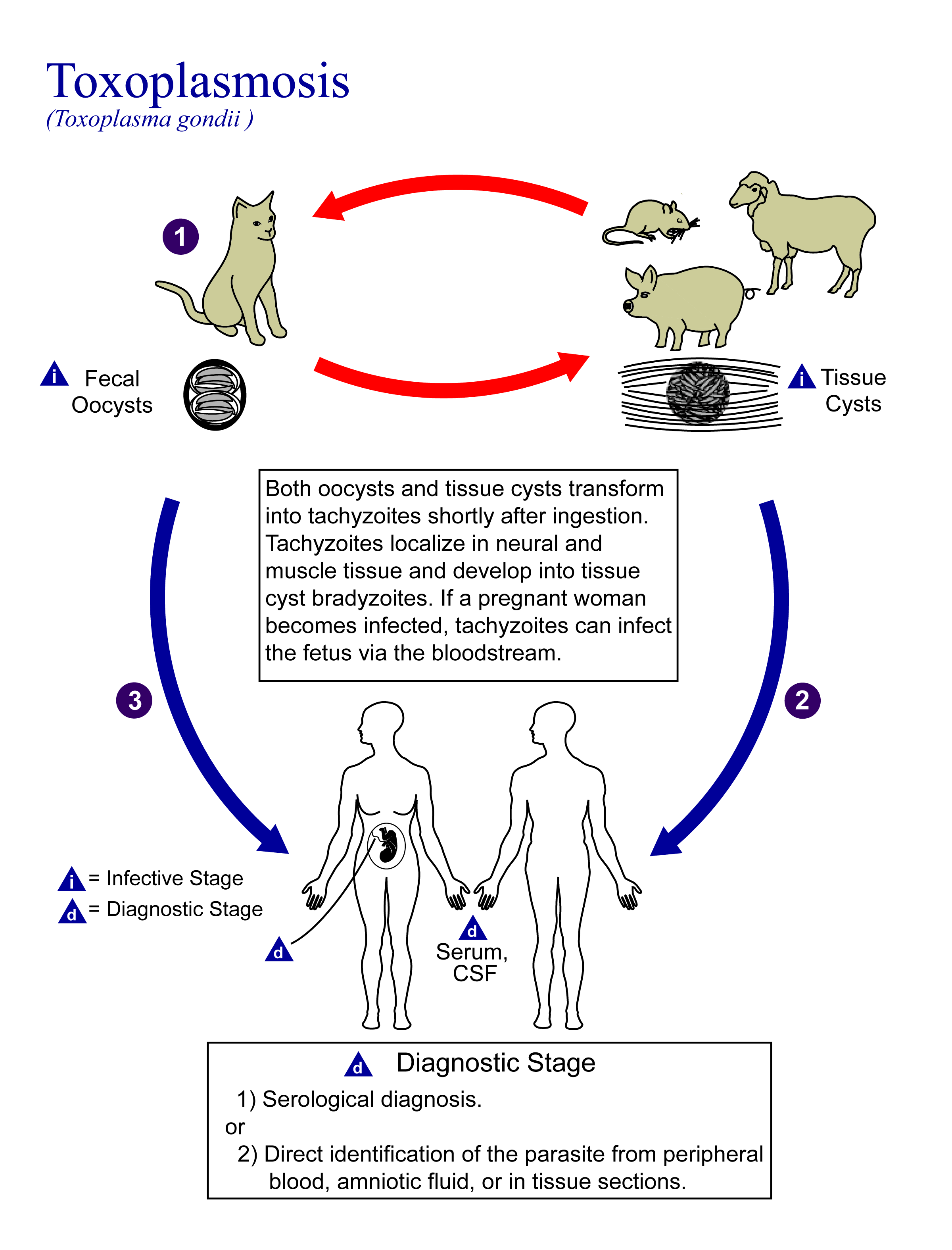
Toxoplasmosis is a parasitic disease caused by ''Toxoplasma gondii'', an apicomplexan. Infections with toxoplasmosis are associated with a variety of neuropsychiatric and behavioral conditions. Occasionally, people may have a few weeks or months of mild, flu-like illness such as muscle aches and tender lymph nodes. In a small number of people, eye problems may develop. In those with a weak immune system, severe symptoms such as seizures and poor coordination may occur. If a person becomes infected during pregnancy, a condition known as congenital toxoplasmosis may affect the child. Toxoplasmosis is usually spread by eating poorly cooked food that contains cysts, exposure to infected cat feces, and from an infected woman to their baby during pregnancy. Rarely, the disease may be spread by blood transfusion. It is not otherwise spread between people. The parasite is known to reproduce sexually only in the cat family. However, it can infect most types of warm-blooded animals, in ... [...More Info...] [...Related Items...] OR: [Wikipedia] [Google] [Baidu] |
Neuroimaging
Neuroimaging is the use of quantitative (computational) techniques to study the structure and function of the central nervous system, developed as an objective way of scientifically studying the healthy human brain in a non-invasive manner. Increasingly it is also being used for quantitative studies of brain disease and psychiatric illness. Neuroimaging is a highly multidisciplinary research field and is not a medical specialty. Neuroimaging differs from neuroradiology which is a medical specialty and uses brain imaging in a clinical setting. Neuroradiology is practiced by radiologists who are medical practitioners. Neuroradiology primarily focuses on identifying brain lesions, such as vascular disease, strokes, tumors and inflammatory disease. In contrast to neuroimaging, neuroradiology is qualitative (based on subjective impressions and extensive clinical training) but sometimes uses basic quantitative methods. Functional brain imaging techniques, such as functional magnet ... [...More Info...] [...Related Items...] OR: [Wikipedia] [Google] [Baidu] |


