|
Retrolental Fibroplasia
Retinopathy of prematurity (ROP), also called retrolental fibroplasia (RLF) and Terry syndrome, is a disease of the eye Eyes are organs of the visual system. They provide living organisms with vision, the ability to receive and process visual detail, as well as enabling several photo response functions that are independent of vision. Eyes detect light and conv ... affecting Preterm birth, prematurely born babies generally having received Neonatal intensive care unit, neonatal intensive care, in which oxygen therapy is used due to the premature development of their lungs. It is thought to be caused by disorganized growth of retinal blood vessels which may result in scarring and retinal detachment. ROP can be mild and may resolve spontaneously, but it may lead to blindness in serious cases. Thus, all preterm babies are at risk for ROP, and very low birth-weight is an additional risk factor. Both oxygen toxicity and relative Hypoxia (medical), hypoxia can contribute to the dev ... [...More Info...] [...Related Items...] OR: [Wikipedia] [Google] [Baidu] |
Human Eye
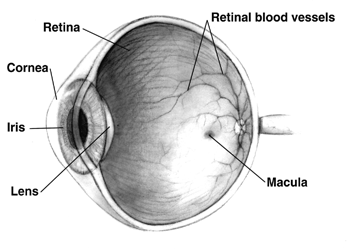
The human eye is a sensory organ, part of the sensory nervous system, that reacts to visible light and allows humans to use visual information for various purposes including seeing things, keeping balance, and maintaining circadian rhythm. The eye can be considered as a living optical device. It is approximately spherical in shape, with its outer layers, such as the outermost, white part of the eye (the sclera) and one of its inner layers (the pigmented choroid) keeping the eye essentially light tight except on the eye's optic axis. In order, along the optic axis, the optical components consist of a first lens (the cornea—the clear part of the eye) that accomplishes most of the focussing of light from the outside world; then an aperture (the pupil) in a diaphragm (the iris—the coloured part of the eye) that controls the amount of light entering the interior of the eye; then another lens (the crystalline lens) that accomplishes the remaining focussing of light into ... [...More Info...] [...Related Items...] OR: [Wikipedia] [Google] [Baidu] |
Risk Factor
In epidemiology, a risk factor or determinant is a variable associated with an increased risk of disease or infection. Due to a lack of harmonization across disciplines, determinant, in its more widely accepted scientific meaning, is often used as a synonym. The main difference lies in the realm of practice: medicine (clinical practice) versus public health. As an example from clinical practice, low ingestion of dietary sources of vitamin C is a known risk factor for developing scurvy. Specific to public health policy, a determinant is a health risk that is general, abstract, related to inequalities, and difficult for an individual to control. For example, poverty is known to be a determinant of an individual's standard of health. Correlation vs causation Risk factors or determinants are correlational and not necessarily causal, because correlation does not prove causation. For example, being young cannot be said to cause measles, but young people have a higher rate of meas ... [...More Info...] [...Related Items...] OR: [Wikipedia] [Google] [Baidu] |
Ophthalmoscope
Ophthalmoscopy, also called funduscopy, is a test that allows a health professional to see inside the fundus of the eye and other structures using an ophthalmoscope (or funduscope). It is done as part of an eye examination and may be done as part of a routine physical examination. It is crucial in determining the health of the retina, optic disc, and vitreous humor. The pupil is a hole through which the eye's interior will be viewed. Opening the pupil wider (dilating it) is a simple and effective way to better see the structures behind it. Therefore, dilation of the pupil ( mydriasis) is often accomplished with medicated eye drops before funduscopy. However, although dilated fundus examination is ideal, undilated examination is more convenient and is also helpful (albeit not as comprehensive), and it is the most common type in primary care. An alternative or complement to ophthalmoscopy is to perform a fundus photography, where the image can be analysed later by a professional. ... [...More Info...] [...Related Items...] OR: [Wikipedia] [Google] [Baidu] |
Pupillary Dilation
Pupillary response is a physiological response that varies the size of the pupil, via the optic and oculomotor cranial nerve. A constriction response (miosis), is the narrowing of the pupil, which may be caused by scleral buckles or drugs such as opiates/opioids or anti-hypertension medications. Constriction of the pupil occurs when the circular muscle, controlled by the parasympathetic nervous system (PSNS), contracts, and also to an extent when the radial muscle relaxes. A dilation response (mydriasis), is the widening of the pupil and may be caused by adrenaline; anticholinergic agents; stimulant drugs such as MDMA, cocaine, and amphetamines; and some hallucinogenics (e.g. LSD). Dilation of the pupil occurs when the smooth cells of the radial muscle, controlled by the sympathetic nervous system (SNS), contract, and also when the cells of the iris sphincter muscle relax. The responses can have a variety of causes, from an involuntary reflex reaction to exposure or inexposure ... [...More Info...] [...Related Items...] OR: [Wikipedia] [Google] [Baidu] |
Gestation
Gestation is the period of development during the carrying of an embryo, and later fetus, inside viviparous animals (the embryo develops within the parent). It is typical for mammals, but also occurs for some non-mammals. Mammals during pregnancy can have one or more gestations at the same time, for example in a multiple birth. The time interval of a gestation is called the '' gestation period''. In obstetrics, ''gestational age'' refers to the time since the onset of the last menses, which on average is fertilization age plus two weeks. Mammals In mammals, pregnancy begins when a zygote (fertilized ovum) implants in the female's uterus and ends once the fetus leaves the uterus during labor or an abortion (whether induced or spontaneous). Humans In humans, pregnancy can be defined clinically or biochemically. Clinically, pregnancy starts from first day of the mother's last period. Biochemically, pregnancy starts when a woman's human chorionic gonadotropin (hCG) levels ... [...More Info...] [...Related Items...] OR: [Wikipedia] [Google] [Baidu] |
Persistent Hyperplastic Primary Vitreous
Persistent hyperplastic primary vitreous (PHPV), also known as persistent fetal vasculature (PFV), is a rare congenital developmental anomaly of the eye that results following failure of the embryological, primary vitreous and hyaloid vasculature to regress. It can be present in three forms: purely anterior (persistent tunica vasculosa lentis and persistent posterior fetal fibrovascular sheath of the lens), purely posterior (falciform retinal septum and ablatio falcicormis congenita) and a combination of both. Most examples of PHPV are unilateral and non-hereditary. When bilateral, PHPV may follow an autosomal recessive or autosomal dominant inheritance pattern. Symptoms The primary vitreous used in formation of the eye during fetal development remains in the eye upon birth and is hazy and scarred. The symptoms are leukocoria, strabismus, nystagmus and blurred vision, blindness. Causes #Trisomy 13 (Patau syndrome) #Norrie disease # Walker-Warburg syndrome #Autosomal dominant #Auto ... [...More Info...] [...Related Items...] OR: [Wikipedia] [Google] [Baidu] |
Persistent Fetal Vasculature Syndrome
Persistent hyperplastic primary vitreous (PHPV), also known as persistent fetal vasculature (PFV), is a rare congenital developmental anomaly of the eye that results following failure of the embryological, primary vitreous and hyaloid vasculature to regress. It can be present in three forms: purely anterior (persistent tunica vasculosa lentis and persistent posterior fetal fibrovascular sheath of the lens), purely posterior (falciform retinal septum and ablatio falcicormis congenita) and a combination of both. Most examples of PHPV are unilateral and non-hereditary. When bilateral, PHPV may follow an autosomal recessive or autosomal dominant inheritance pattern. Symptoms The primary vitreous used in formation of the eye during fetal development remains in the eye upon birth and is hazy and scarred. The symptoms are leukocoria, strabismus, nystagmus and blurred vision, blindness. Causes #Trisomy 13 (Patau syndrome) #Norrie disease # Walker-Warburg syndrome #Autosomal dominant #Auto ... [...More Info...] [...Related Items...] OR: [Wikipedia] [Google] [Baidu] |
Familial Exudative Vitreoretinopathy
Familial exudative vitreoretinopathy (FEVR, pronounced as fever) is a genetic disorder affecting the growth and development of blood vessels in the retina of the eye. This disease can lead to visual impairment and sometimes complete blindness in one or both eyes. FEVR is characterized by incomplete vascularization of the peripheral retina. This can lead to the growth of new blood vessels which are prone to leakage and hemorrhage and can cause retinal folds, tears, and detachments. Treatment involves laser photocoagulation of the avascular portions of the retina to reduce new blood vessel growth and risk of complications including leakage of retinal blood vessels and retinal detachments. Information Pathophysiology FEVR is caused by genetic defects involving the regulation of blood vessel growth in developing eyes. As a result, there is poor blood vessel growth to the periphery of the retina. The lack of blood supply to the peripheral retina triggers the release of molecules tha ... [...More Info...] [...Related Items...] OR: [Wikipedia] [Google] [Baidu] |
Persistent Tunica Vasculosa Lentis
Persistent tunica vasculosa lentis is a congenital ocular anomaly. It is a form of persistent hyperplastic primary vitreous (PHPV). It is a developmental disorder of the vitreous. It is usually unilateral and first noticed in the neonatal period. It may be associated with microphthalmos, cataracts, and increased intraocular pressure. Elongated ciliary processes are visible through the dilated pupil. A USG B-scan confirms diagnosis in the presence of a cataract. See also * Persistent fetal vasculature *Tunica vasculosa lentis The tunica vasculosa lentis is an extensive capillary network, spreading over the posterior and lateral surfaces of the lens of the eye. It disappears shortly after birth. See also * Persistent tunica vasculosa lentis Persistent tunica vasc ... References * http://www.djo.harvard.edu/files/1568.pdf * Wright KW, Speigel PH, Buckley EG. Pediatric Ophthalmology and Strabismus. Springer; US, New York: 2003. p. 17. Eye diseases {{eye-d ... [...More Info...] [...Related Items...] OR: [Wikipedia] [Google] [Baidu] |
Tortuosity
Tortuosity is widely used as a critical parameter to predict transport properties of porous media, such as rocks and soils. But unlike other standard microstructural properties, the concept of tortuosity is vague with multiple definitions and various evaluation methods introduced in different contexts. Hydraulic, electrical, diffusional, and thermal tortuosities are defined to describe different transport processes in porous media, while geometrical tortuosity is introduced to characterize the morphological property of porous microstructures. Tortuosity in 2-D Subjective estimation (sometimes aided by optometric grading scales) is often used. The simplest mathematical method to estimate tortuosity is the arc-chord ratio: the ratio of the length of the curve (''C'') to the distance between its ends (''L''): :\tau = \frac Arc-chord ratio equals 1 for a straight line and is infinite for a circle. Another method, proposed in 1999, is to estimate the tortuosity as the integral of ... [...More Info...] [...Related Items...] OR: [Wikipedia] [Google] [Baidu] |
Myopia
Near-sightedness, also known as myopia and short-sightedness, is an eye disease where light focuses in front of, instead of on, the retina. As a result, distant objects appear blurry while close objects appear normal. Other symptoms may include headaches and eye strain. Severe near-sightedness is associated with an increased risk of retinal detachment, cataracts, and glaucoma. The underlying mechanism involves the length of the eyeball growing too long or less commonly the lens being too strong. It is a type of refractive error. Diagnosis is by eye examination. Tentative evidence indicates that the risk of near-sightedness can be decreased by having young children spend more time outside. This decrease in risk may be related to natural light exposure. Near-sightedness can be corrected with eyeglasses, contact lenses, or a refractive surgery. Eyeglasses are the easiest and safest method of correction. Contact lenses can provide a wider field of vision, but are associated with ... [...More Info...] [...Related Items...] OR: [Wikipedia] [Google] [Baidu] |


