|
Retinal Ganglion Cells
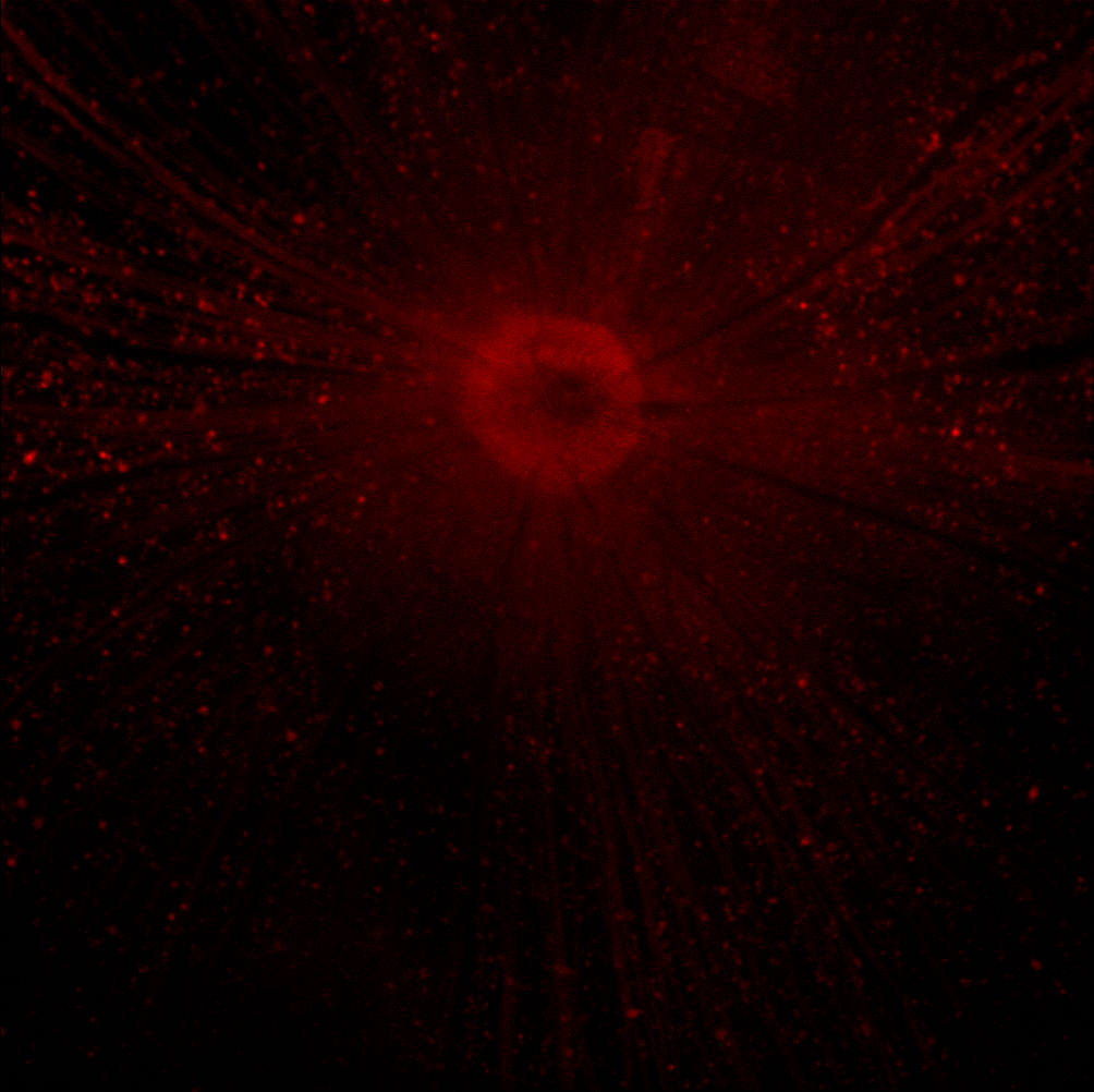
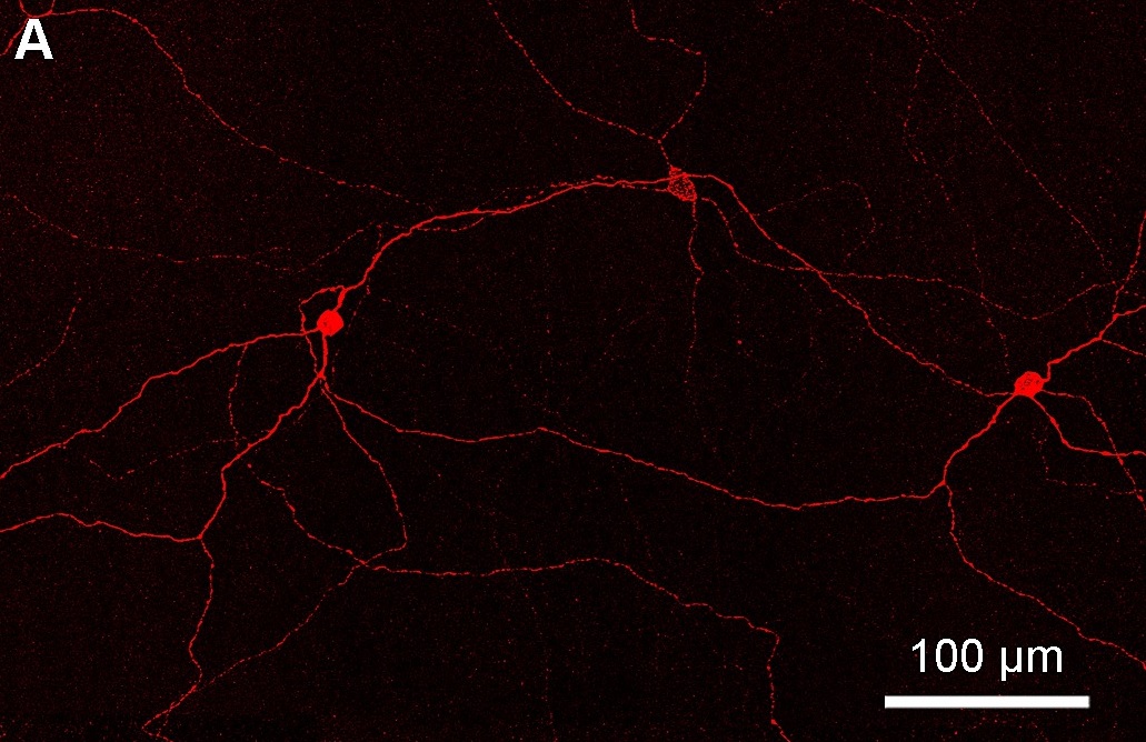
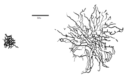
A retinal ganglion cell (RGC) is a type of neuron located near the inner surface (the ganglion cell layer) of the retina of the eye. It receives visual information from photoreceptors via two intermediate neuron types: bipolar cells and retina amacrine cells. Retina amacrine cells, particularly narrow field cells, are important for creating functional subunits within the ganglion cell layer and making it so that ganglion cells can observe a small dot moving a small distance. Retinal ganglion cells collectively transmit image-forming and non-image forming visual information from the retina in the form of action potential to several regions in the thalamus, hypothalamus, and mesencephalon, or midbrain. Retinal ganglion cells vary significantly in terms of their size, connections, and responses to visual stimulation but they all share the defining property of having a long axon that extends into the brain. These axons form the optic nerve, optic chiasm, and optic tract. A small pe ... [...More Info...] [...Related Items...] OR: [Wikipedia] [Google] [Baidu] |
Neuron
A neuron, neurone, or nerve cell is an electrically excitable cell that communicates with other cells via specialized connections called synapses. The neuron is the main component of nervous tissue in all animals except sponges and placozoa. Non-animals like plants and fungi do not have nerve cells. Neurons are typically classified into three types based on their function. Sensory neurons respond to stimuli such as touch, sound, or light that affect the cells of the sensory organs, and they send signals to the spinal cord or brain. Motor neurons receive signals from the brain and spinal cord to control everything from muscle contractions to glandular output. Interneurons connect neurons to other neurons within the same region of the brain or spinal cord. When multiple neurons are connected together, they form what is called a neural circuit. A typical neuron consists of a cell body (soma), dendrites, and a single axon. The soma is a compact structure, and the axon and dend ... [...More Info...] [...Related Items...] OR: [Wikipedia] [Google] [Baidu] |
Circadian Rhythm
A circadian rhythm (), or circadian cycle, is a natural, internal process that regulates the sleep–wake cycle and repeats roughly every 24 hours. It can refer to any process that originates within an organism (i.e., Endogeny (biology), endogenous) and responds to the environment (Entrainment (chronobiology), entrained by the environment). These 24-hour rhythms are driven by a circadian clock, and they have been widely observed in animals, plants, fungi and cyanobacteria. The term ''circadian'' comes from the Latin ''wikt:circa#Latin, circa'', meaning "approximately", and ''dies'', meaning "day". Processes with 24-hour cycles are more generally called diurnal rhythms; diurnal rhythms should not be called circadian rhythms unless they can be confirmed as endogenous, and not environmental. Although circadian rhythms are endogenous, they are adjusted to the local environment by external cues called zeitgebers (German for "time givers"), which include light, temperature and redox cy ... [...More Info...] [...Related Items...] OR: [Wikipedia] [Google] [Baidu] |
Superior Colliculus
In neuroanatomy, the superior colliculus () is a structure lying on the roof of the mammalian midbrain. In non-mammalian vertebrates, the homologous structure is known as the optic tectum, or optic lobe. The adjective form ''tectal'' is commonly used for both structures. In mammals, the superior colliculus forms a major component of the midbrain. It is a paired structure and together with the paired inferior colliculi forms the corpora quadrigemina. The superior colliculus is a layered structure, with a pattern that is similar to all mammals. The layers can be grouped into the superficial layers ( stratum opticum and above) and the deeper remaining layers. Neurons in the superficial layers receive direct input from the retina and respond almost exclusively to visual stimuli. Many neurons in the deeper layers also respond to other modalities, and some respond to stimuli in multiple modalities. The deeper layers also contain a population of motor-related neurons, capable of activat ... [...More Info...] [...Related Items...] OR: [Wikipedia] [Google] [Baidu] |
Photosensitive Ganglion Cell
Intrinsically photosensitive retinal ganglion cells (ipRGCs), also called photosensitive retinal ganglion cells (pRGC), or melanopsin-containing retinal ganglion cells (mRGCs), are a type of neuron in the retina of the mammalian eye. The presence of (something like) ipRGCs was first suspected in 1927 when rodless, coneless mice still responded to a light stimulus through pupil constriction, This implied that rods and cones are not the only light-sensitive neurons in the retina. Yet research on these cells did not advance until the 1980s. Recent research has shown that these retinal ganglion cells, unlike other retinal ganglion cells, are intrinsically photosensitive due to the presence of melanopsin, a light-sensitive protein. Therefore they constitute a third class of photoreceptors, in addition to rod and cone cells. Overview Compared to the rods and cones, the ipRGCs respond more sluggishly and signal the presence of light over the long term. They represent a very small su ... [...More Info...] [...Related Items...] OR: [Wikipedia] [Google] [Baidu] |
Bistratified Cell
Bistratified ganglion cell can refer to either of two kinds of retinal ganglion cells whose cell body is located in the ganglion cell layer of the retina, the small-field bistratified ganglion cell, also known as small bistratified cell (SBC), and the large-field bistratified ganglion cell or large bistratified cell (LBC). Bistratified cells receive their input from bipolar cells and amacrine cells. The bistratified cells project their axons through the optic nerve and optic tract to the koniocellular layers in the lateral geniculate nucleus (LGN), synapsing with koniocellular cells. Koniocellular means "cells as small as dust"; their small size made them hard to find. About 8 to 10% of retinal ganglion cells are bistratified cells. They receive inputs from intermediate numbers of rods and cones. They have moderate spatial resolution, moderate conduction velocity, and can respond to moderate-contrast stimuli. They may be involved in color vision. See also *Midget cell *Parasol c ... [...More Info...] [...Related Items...] OR: [Wikipedia] [Google] [Baidu] |
Magnocellular Cell
Magnocellular cells, also called M-cells, are neurons located within the Adina magnocellular layer of the lateral geniculate nucleus of the thalamus. The cells are part of the visual system. They are termed "magnocellular" since they are characterized by their relatively large size compared to parvocellular cells. Structure The full details of the flow of signaling from the eye to the visual cortex of the brain that result in the experience of vision are incompletely understood. Many aspects are subject to active controversy and the disruption of new evidence. In the visual system, signals mostly travel from the retina to the lateral geniculate nucleus (LGN) and then to the visual cortex. In humans the LGN is normally described as having six distinctive layers. The inner two layers, (1 and 2) are magnocellular cell (M cell) layers, while the outer four layers, (3,4,5 and 6), are parvocellular cell (P cell) layers. An additional set of neurons, known as the koniocellular cell ... [...More Info...] [...Related Items...] OR: [Wikipedia] [Google] [Baidu] |
Parasol Cell
A parasol cell, sometimes called an M cell or M ganglion cell, is one type of retinal ganglion cell (RGC) located in the ganglion cell layer of the retina. These cells project to magnocellular cells in the lateral geniculate nucleus (LGN) as part of the magnocellular pathway in the visual system. They have large cell bodies as well as extensive branching dendrite networks and as such have large Receptive field, receptive fields. Relative to other RGCs, they have fast conduction velocities. While they do show clear center-surround antagonism (known as spatial opponency), they receive no information about color (absence of chromatic opponency). Parasol ganglion cells contribute information about the motion and depth of objects to the visual system. Parasol ganglion cells in the Magnocellular pathway Parasol ganglion cells are the first step in the magnocellular pathway of the visual system. They project from the retina via the optic nerve to the two most ventral layers of the LGN, ... [...More Info...] [...Related Items...] OR: [Wikipedia] [Google] [Baidu] |
Parvocellular Cell
Parvocellular cells, also called P-cells, are neurons located within the parvocellular layers of the lateral geniculate nucleus (LGN) of the thalamus. "''Parvus''" is Latin for "small", and the name "parvocellular" refers to the small size of the cell compared to the larger magnocellular cells. Phylogenetically, parvocellular neurons are more modern than magnocellular ones. Function The parvocellular neurons of the visual system receive their input from midget cells, a type of retinal ganglion cell, whose axons are exiting the optic tract. These synapses occur in one of the four dorsal parvocellular layers of the lateral geniculate nucleus. The information from each eye is kept separate at this point, and continues to be segregated until processing in the visual cortex. The electrically-encoded visual information leaves the parvocellular cells via relay cells in the optic radiations, traveling to the primary visual cortex layer 4C-β. The parvocellular neurons are sensiti ... [...More Info...] [...Related Items...] OR: [Wikipedia] [Google] [Baidu] |
Midget Cell
A midget cell is one type of retinal ganglion cell (RGC). Midget cells originate in the ganglion cell layer of the retina, and project to the parvocellular layers of the lateral geniculate nucleus (LGN). The axons of midget cells travel through the optic nerve and optic tract, ultimately synapsing with parvocellular cells in the LGN. These cells are known as midget retinal ganglion cells due to the small sizes of their dendrite, dendritic trees and cell bodies. About 80% of RGCs are midget cells. They receive inputs from relatively few rods and cones. In many cases, they are connected to midget Bipolar cell of the retina, bipolar cells, which are linked to one cone each. These neurons show roughly circular Receptive field, receptive fields with antagonistic center and surround; this property is known as spatial opponency and these neurons are typically divided into ON- or OFF-center, depending on whether they are excited or inhibited by photons falling on the center of their r ... [...More Info...] [...Related Items...] OR: [Wikipedia] [Google] [Baidu] |
50x RGC Axotomy 1 Day
5 (five) is a number, numeral and digit. It is the natural number, and cardinal number, following 4 and preceding 6, and is a prime number. It has attained significance throughout history in part because typical humans have five digits on each hand. In mathematics 5 is the third smallest prime number, and the second super-prime. It is the first safe prime, the first good prime, the first balanced prime, and the first of three known Wilson primes. Five is the second Fermat prime and the third Mersenne prime exponent, as well as the third Catalan number, and the third Sophie Germain prime. Notably, 5 is equal to the sum of the ''only'' consecutive primes, 2 + 3, and is the only number that is part of more than one pair of twin primes, ( 3, 5) and (5, 7). It is also a sexy prime with the fifth prime number and first prime repunit, 11. Five is the third factorial prime, an alternating factorial, and an Eisenstein prime with no imaginary part and real part of the form 3p ... [...More Info...] [...Related Items...] OR: [Wikipedia] [Google] [Baidu] |
Fovea Centralis
The fovea centralis is a small, central pit composed of closely packed cones in the eye. It is located in the center of the macula lutea of the retina. The fovea is responsible for sharp central vision (also called foveal vision), which is necessary in humans for activities for which visual detail is of primary importance, such as reading and driving. The fovea is surrounded by the ''parafovea'' belt and the ''perifovea'' outer region. The parafovea is the intermediate belt, where the ganglion cell layer is composed of more than five layers of cells, as well as the highest density of cones; the perifovea is the outermost region where the ganglion cell layer contains two to four layers of cells, and is where visual acuity is below the optimum. The perifovea contains an even more diminished density of cones, having 12 per 100 micrometres versus 50 per 100 micrometres in the most central fovea. That, in turn, is surrounded by a larger peripheral area, which delivers highly compres ... [...More Info...] [...Related Items...] OR: [Wikipedia] [Google] [Baidu] |
Photoreceptor Cell
A photoreceptor cell is a specialized type of neuroepithelial cell found in the retina that is capable of visual phototransduction. The great biological importance of photoreceptors is that they convert light (visible electromagnetic radiation) into signals that can stimulate biological processes. To be more specific, photoreceptor proteins in the cell absorb photons, triggering a change in the cell's membrane potential. There are currently three known types of photoreceptor cells in mammalian eyes: rods, cones, and intrinsically photosensitive retinal ganglion cells. The two classic photoreceptor cells are rods and cones, each contributing information used by the visual system to form an image of the environment, sight. Rods primarily mediate scotopic vision (dim conditions) whereas cones primarily mediate to photopic vision (bright conditions), but the processes in each that supports phototransduction is similar. A third class of mammalian photoreceptor cell was discovered ... [...More Info...] [...Related Items...] OR: [Wikipedia] [Google] [Baidu] |


