|
Bistratified Cell
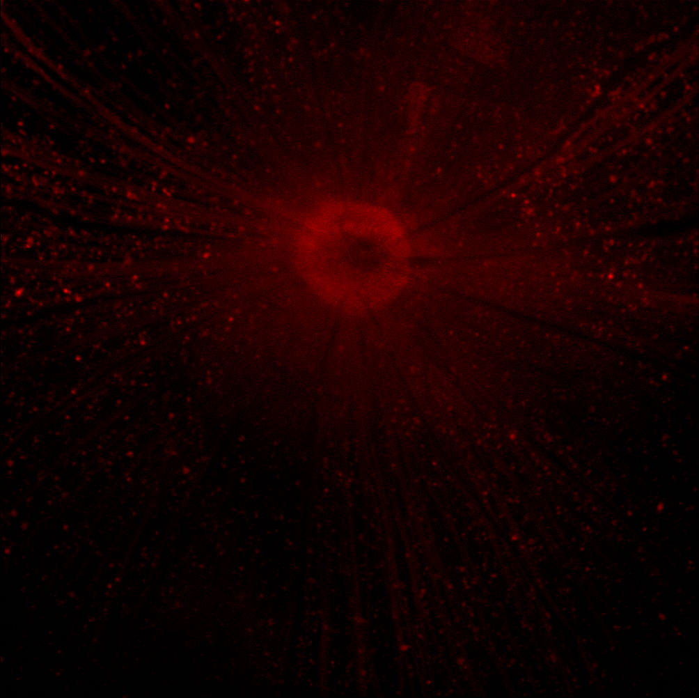
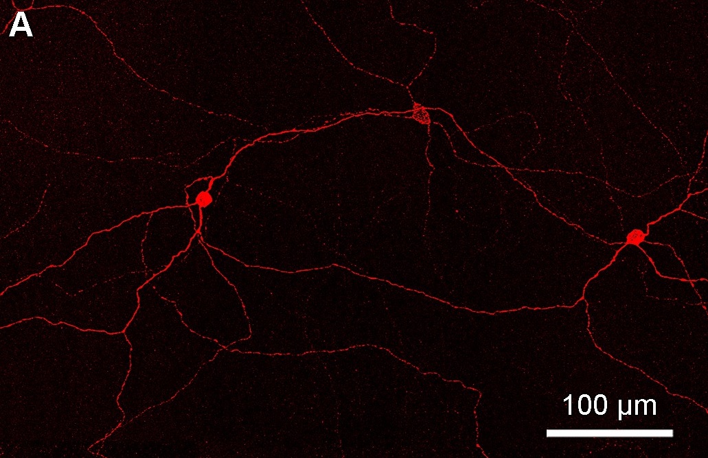
Bistratified ganglion cell can refer to either of two kinds of retinal ganglion cells whose cell body is located in the ganglion cell layer of the retina, the small-field bistratified ganglion cell, also known as small bistratified cell (SBC), and the large-field bistratified ganglion cell or large bistratified cell (LBC). Bistratified cells receive their input from bipolar cells and amacrine cells. The bistratified cells project their axons through the optic nerve and optic tract to the koniocellular layers in the lateral geniculate nucleus (LGN), synapsing with koniocellular cells. Koniocellular means "cells as small as dust"; their small size made them hard to find. About 8 to 10% of retinal ganglion cells are bistratified cells. They receive inputs from intermediate numbers of rods and cones. They have moderate spatial resolution, moderate conduction velocity, and can respond to moderate-contrast stimuli. They may be involved in color vision. See also *Midget cell *Parasol c ... [...More Info...] [...Related Items...] OR: [Wikipedia] [Google] [Baidu] |
Retinal Ganglion Cell
A retinal ganglion cell (RGC) is a type of neuron located near the inner surface (the ganglion cell layer) of the retina of the human eye, eye. It receives visual information from photoreceptor cell, photoreceptors via two intermediate neuron types: Bipolar cell of the retina, bipolar cells and retina amacrine cells. Retina amacrine cells, particularly narrow field cells, are important for creating functional subunits within the ganglion cell layer and making it so that ganglion cells can observe a small dot moving a small distance. Retinal ganglion cells collectively transmit image-forming and non-image forming visual information from the retina in the form of action potential to several regions in the thalamus, hypothalamus, and mesencephalon, or midbrain. Retinal ganglion cells vary significantly in terms of their size, connections, and responses to visual stimulation but they all share the defining property of having a long axon that extends into the brain. These axons form th ... [...More Info...] [...Related Items...] OR: [Wikipedia] [Google] [Baidu] |
Ganglion Cell Layer
The ganglion cell layer (ganglionic layer) is a layer of the retina that consists of retinal ganglion cells and displaced amacrine cells. The cells are somewhat flask-shaped; the rounded internal surface of each resting on the stratum opticum, and sending off an axon which is prolonged into it. From the opposite end numerous dendrites extend into the inner plexiform layer, where they branch and form flattened arborizations at different levels. The ganglion cells vary much in size, and the dendrites of the smaller ones as a rule arborize in the inner plexiform layer as soon as they enter it; while those of the larger cells ramify close to the inner nuclear layer The inner nuclear layer or layer of inner granules, of the retina, is made up of a number of closely packed cells, of which there are three varieties, viz.: bipolar cells, horizontal cells, and amacrine cells. Bipolar cells The bipolar cells, by .... References External links * Human eye anatomy {{eye-s ... [...More Info...] [...Related Items...] OR: [Wikipedia] [Google] [Baidu] |
Retina
The retina (from la, rete "net") is the innermost, light-sensitive layer of tissue of the eye of most vertebrates and some molluscs. The optics of the eye create a focused two-dimensional image of the visual world on the retina, which then processes that image within the retina and sends nerve impulses along the optic nerve to the visual cortex to create visual perception. The retina serves a function which is in many ways analogous to that of the film or image sensor in a camera. The neural retina consists of several layers of neurons interconnected by synapses and is supported by an outer layer of pigmented epithelial cells. The primary light-sensing cells in the retina are the photoreceptor cells, which are of two types: rods and cones. Rods function mainly in dim light and provide monochromatic vision. Cones function in well-lit conditions and are responsible for the perception of colour through the use of a range of opsins, as well as high-acuity vision used for task ... [...More Info...] [...Related Items...] OR: [Wikipedia] [Google] [Baidu] |
Bipolar Cell Of The Retina
As a part of the retina, bipolar cells exist between photoreceptors (rod cells and cone cells) and ganglion cells. They act, directly or indirectly, to transmit signals from the photoreceptors to the ganglion cells. Structure Bipolar cells are so-named as they have a central body from which two sets of processes arise. They can synapse with either rods or cones (rod/cone mixed input BCs have been found in teleost fish but not mammals), and they also accept synapses from horizontal cells. The bipolar cells then transmit the signals from the photoreceptors or the horizontal cells, and pass it on to the ganglion cells directly or indirectly (via amacrine cells). Unlike most neurons, bipolar cells communicate via graded potentials, rather than action potentials. Function Bipolar cells receive synaptic input from either rods or cones, or both rods and cones, though they are generally designated rod bipolar or cone bipolar cells. There are roughly 10 distinct forms of cone bipolar ce ... [...More Info...] [...Related Items...] OR: [Wikipedia] [Google] [Baidu] |
Amacrine Cell
Amacrine cells are interneurons in the retina. They are named from the Greek roots ''a–'' ("non"), ''makr–'' ("long") and ''in–'' ("fiber"), because of their short neuronal processes. Amacrine cells are inhibitory neurons, and they project their dendritic arbors onto the inner plexiform layer (IPL), they interact with retinal ganglion cells and/or bipolar cells. Structure Amacrine cells operate at inner plexiform layer (IPL), the second synaptic retinal layer where bipolar cells and retinal ganglion cells form synapses. There are at least 33 different subtypes of amacrine cells based just on their dendrite morphology and stratification. Like horizontal cells, amacrine cells work laterally, but whereas horizontal cells are connected to the output of rod and cone cells, amacrine cells affect the output from bipolar cells, and are often more specialized. Each type of amacrine cell releases one or several neurotransmitters where it connects with other cells. They are often ... [...More Info...] [...Related Items...] OR: [Wikipedia] [Google] [Baidu] |
Optic Nerve
In neuroanatomy, the optic nerve, also known as the second cranial nerve, cranial nerve II, or simply CN II, is a paired cranial nerve that transmits visual system, visual information from the retina to the brain. In humans, the optic nerve is derived from optic stalks during the seventh week of development and is composed of retinal ganglion cell axons and glial cells; it extends from the optic disc to the optic chiasma and continues as the optic tract to the lateral geniculate nucleus, Pretectal area, pretectal nuclei, and superior colliculus. Structure The optic nerve has been classified as the second of twelve paired cranial nerves, but it is technically part of the central nervous system, rather than the peripheral nervous system because it is derived from an out-pouching of the diencephalon (optic stalks) during embryonic development. As a consequence, the fibers of the optic nerve are covered with myelin produced by oligodendrocytes, rather than Schwann cells of the per ... [...More Info...] [...Related Items...] OR: [Wikipedia] [Google] [Baidu] |
Optic Tract
In neuroanatomy, the optic tract () is a part of the visual system in the brain. It is a continuation of the optic nerve that relays information from the optic chiasm to the ipsilateral lateral geniculate nucleus (LGN), pretectal nuclei, and superior colliculus. It is composed of two individual tracts, the left optic tract and the right optic tract, each of which conveys visual information exclusive to its respective contralateral half of the visual field. Each of these tracts is derived from a combination of temporal and nasal retinal fibers from each eye that corresponds to one half of the visual field. In more specific terms, the optic tract contains fibers from the ipsilateral temporal hemiretina and contralateral nasal hemiretina. Visual system The optic tract carries retinal information relating to the whole visual field. Specifically, the left optic tract corresponds to the right visual field, while the right optic tract corresponds to the left visual field. To form the ri ... [...More Info...] [...Related Items...] OR: [Wikipedia] [Google] [Baidu] |
Lateral Geniculate Nucleus
In neuroanatomy, the lateral geniculate nucleus (LGN; also called the lateral geniculate body or lateral geniculate complex) is a structure in the thalamus and a key component of the mammalian visual pathway. It is a small, ovoid, ventral projection of the thalamus where the thalamus connects with the optic nerve. There are two LGNs, one on the left and another on the right side of the thalamus. In humans, both LGNs have six layers of neurons (grey matter) alternating with optic fibers (white matter). The LGN receives information directly from the ascending retinal ganglion cells via the optic tract and from the reticular activating system. Neurons of the LGN send their axons through the optic radiation, a direct pathway to the primary visual cortex. In addition, the LGN receives many strong feedback connections from the primary visual cortex. In humans as well as other mammals, the two strongest pathways linking the eye to the brain are those projecting to the dorsal part of th ... [...More Info...] [...Related Items...] OR: [Wikipedia] [Google] [Baidu] |
Koniocellular Cell
A koniocellular cell (konio: Greek, ''dust'' or ''poison'', also known as K cell) is a neuron with a small cell body that is located in the koniocellular layer of the lateral geniculate nucleus (LGN) in primates, including humans. Koniocellular layers are located ventral to each parvocellular and magnocellular layer of the LGN. Even if the quantity of neurons is approximately equal to the number of magnocellular cells the koniocellular layers are much thinner due to their size. In comparison to the parvocellular and magnocellular system, fewer studies have been conducted to investigate the koniocellular system. Koniocellular cells are a heterogeneous population differing in many aspects, such as response properties and connectivity. Structure K cells are neurochemically and anatomically distinct from M and P cells. There are three proteins by which K cells can be clearly distinguished: * Calbindin (28kDa calcium binding protein, CALB) * The alpha subunit of type II calmodulin- ... [...More Info...] [...Related Items...] OR: [Wikipedia] [Google] [Baidu] |
Midget Cell
A midget cell is one type of retinal ganglion cell (RGC). Midget cells originate in the ganglion cell layer of the retina, and project to the parvocellular layers of the lateral geniculate nucleus (LGN). The axons of midget cells travel through the optic nerve and optic tract, ultimately synapsing with parvocellular cells in the LGN. These cells are known as midget retinal ganglion cells due to the small sizes of their dendrite, dendritic trees and cell bodies. About 80% of RGCs are midget cells. They receive inputs from relatively few rods and cones. In many cases, they are connected to midget Bipolar cell of the retina, bipolar cells, which are linked to one cone each. These neurons show roughly circular Receptive field, receptive fields with antagonistic center and surround; this property is known as spatial opponency and these neurons are typically divided into ON- or OFF-center, depending on whether they are excited or inhibited by photons falling on the center of their r ... [...More Info...] [...Related Items...] OR: [Wikipedia] [Google] [Baidu] |
Parasol Cell
A parasol cell, sometimes called an M cell or M ganglion cell, is one type of retinal ganglion cell (RGC) located in the ganglion cell layer of the retina. These cells project to magnocellular cells in the lateral geniculate nucleus (LGN) as part of the magnocellular pathway in the visual system. They have large cell bodies as well as extensive branching dendrite networks and as such have large Receptive field, receptive fields. Relative to other RGCs, they have fast conduction velocities. While they do show clear center-surround antagonism (known as spatial opponency), they receive no information about color (absence of chromatic opponency). Parasol ganglion cells contribute information about the motion and depth of objects to the visual system. Parasol ganglion cells in the Magnocellular pathway Parasol ganglion cells are the first step in the magnocellular pathway of the visual system. They project from the retina via the optic nerve to the two most ventral layers of the LGN, ... [...More Info...] [...Related Items...] OR: [Wikipedia] [Google] [Baidu] |
Photosensitive Ganglion Cell
Intrinsically photosensitive retinal ganglion cells (ipRGCs), also called photosensitive retinal ganglion cells (pRGC), or melanopsin-containing retinal ganglion cells (mRGCs), are a type of neuron in the retina of the mammalian eye. The presence of (something like) ipRGCs was first suspected in 1927 when rodless, coneless mice still responded to a light stimulus through pupil constriction, This implied that rods and cones are not the only light-sensitive neurons in the retina. Yet research on these cells did not advance until the 1980s. Recent research has shown that these retinal ganglion cells, unlike other retinal ganglion cells, are intrinsically photosensitive due to the presence of melanopsin, a light-sensitive protein. Therefore they constitute a third class of photoreceptors, in addition to rod and cone cells. Overview Compared to the rods and cones, the ipRGCs respond more sluggishly and signal the presence of light over the long term. They represent a very small su ... [...More Info...] [...Related Items...] OR: [Wikipedia] [Google] [Baidu] |


