|
Renal Portal System
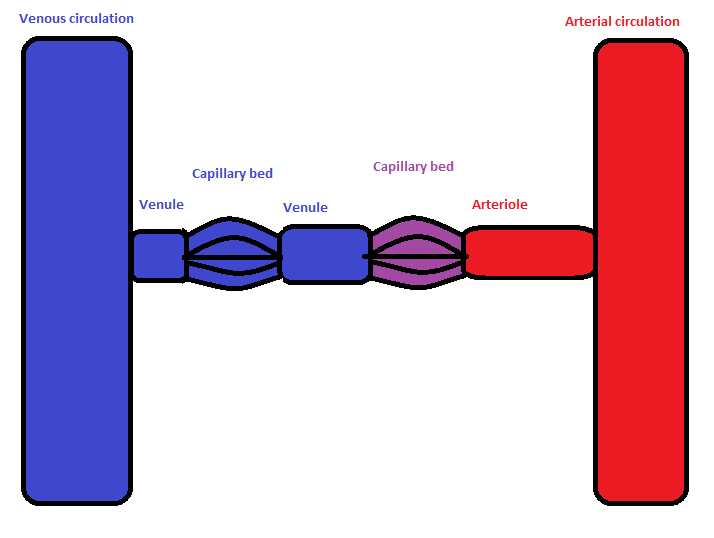
A renal portal system is a portal venous system found in most vertebrates excluding hagfish and lampreys. Its function is to supply blood to renal tubules when glomerular filtration is absent or downregulated. Description The main channel is the renal portal vein, developed from the posterior cardinal vein, which brings venous blood circulation from the tail and groin to the kidney, where it is shunted into a capillary network around the convoluted tubules. The blood then enters the renal vein, passing either through the subcardinal veins and into the posterior cardinal veins or through the posterior vena cava. Variations In lungfish and tetrapods, the renal portal vein is joined by a vein traveling upwards from the abdominal vein, which can bring venous blood from the hind limbs and ventral body wall into the renal portal system, or alternatively, enable blood from the tail and groin to pass into the hepatic portal system, already served by blood from the gut, via the hepatic p ... [...More Info...] [...Related Items...] OR: [Wikipedia] [Google] [Baidu] |
Portal Venous System
In the circulatory system of animals, a portal venous system occurs when a capillary bed pools into another capillary bed through veins, without first going through the heart. Both capillary beds and the blood vessels that connect them are considered part of the portal venous system. They are relatively uncommon as the majority of capillary beds drain into veins which then drain into the heart, not into another capillary bed. Portal venous systems are considered venous because the blood vessels that join the two capillary beds are either veins or venules. Examples of such systems include the hepatic portal system, the hypophyseal portal system, and (in non-mammals) the renal portal system. Unqualified, ''portal venous system'' often refers to the hepatic portal system. For this reason, ''portal vein'' most commonly refers to the hepatic portal vein. The functional significance of such a system is that it transports products of one region directly to another region in relati ... [...More Info...] [...Related Items...] OR: [Wikipedia] [Google] [Baidu] |
Frogs
A frog is any member of a diverse and largely carnivorous group of short-bodied, tailless amphibians composing the order Anura (ανοὐρά, literally ''without tail'' in Ancient Greek). The oldest fossil "proto-frog" ''Triadobatrachus'' is known from the Early Triassic of Madagascar, but molecular clock dating suggests their split from other amphibians may extend further back to the Permian, 265 million years ago. Frogs are widely distributed, ranging from the tropics to subarctic regions, but the greatest concentration of species diversity is in tropical rainforest. Frogs account for around 88% of extant amphibian species. They are also one of the five most diverse vertebrate orders. Warty frog species tend to be called toads, but the distinction between frogs and toads is informal, not from taxonomy or evolutionary history. An adult frog has a stout body, protruding eyes, anteriorly-attached tongue, limbs folded underneath, and no tail (the tail of tailed frogs is an ex ... [...More Info...] [...Related Items...] OR: [Wikipedia] [Google] [Baidu] |
Azygos Vein
The azygos vein is a vein running up the right side of the thoracic vertebral column draining itself towards the superior vena cava. It connects the systems of superior vena cava and inferior vena cava and can provide an alternative path for blood to the right atrium when either of the venae cavae is blocked. Structure The azygos vein transports deoxygenated blood from the posterior walls of the thorax and abdomen into the superior vena cava. It is formed by the union of the ascending lumbar veins with the right subcostal veins at the level of the 12th thoracic vertebra, ascending to the right of the descending aorta and thoracic duct, passing behind the right crus of diaphragm, anterior to the vertebral bodies of T12 to T5 and right posterior intercostal arteries. At the level of T4 vertebrae, it arches over the root of the right lung from behind to the front to join the superior vena cava. The trachea and oesophagus is located medially to the arch of the azygous vein. The ... [...More Info...] [...Related Items...] OR: [Wikipedia] [Google] [Baidu] |
Suprarenal Veins
The suprarenal veins are two in number: * the ''right'' ends in the inferior vena cava. * the ''left'' ends in the left renal or left inferior phrenic vein. They receive blood from the adrenal glands and will sometimes form anastomoses with the inferior phrenic veins The inferior phrenic veins drain the diaphragm and follow the course of the inferior phrenic arteries; * the right ends in the inferior vena cava; * the left is often represented by two branches, ** one of which ends in the left renal or suprar .... Additional images File:Gray480.png, Diagram showing completion of development of the parietal veins File:Gray1183.png, Suprarenal glands viewed from the front File:Gray1184.png, Suprarenal glands viewed from behind References External links Veins of the torso Adrenal gland {{circulatory-stub ... [...More Info...] [...Related Items...] OR: [Wikipedia] [Google] [Baidu] |
Gonadal Vein
In medicine, gonadal vein refers to the blood vessel that carries blood away from the gonad (testis, ovary) toward the heart. These are different arteries in women (ovarian vein) and men (testicular vein), but share the same embryological origin. The termination of the two gonadal veins in an individual is usually asymmetrical, with the left one draining into the left renal vein, and the right one draining into the inferior vena cava. Anatomy Fate The left gonadal vein usually empties into (inferior aspect of) the ipsilateral renal vein proximally to where the renal vein crossing over the aorta. The right gonadal vein typically empties directly into the (right anterolateral aspect of) inferior vena cava, joining it at an acute angle, some 2 cm inferior to the ipsilateral renal vein The renal veins are large-calibre veins that drain blood filtered by the kidneys into the inferior vena cava. There is one renal vein draining each kidney. Because the inferior vena cava is ... [...More Info...] [...Related Items...] OR: [Wikipedia] [Google] [Baidu] |
Renal Vein
The renal veins are large-calibre veins that drain blood filtered by the kidneys into the inferior vena cava. There is one renal vein draining each kidney. Because the inferior vena cava is on the right half of the body, the left renal vein is longer than the right one. Structure One renal vein drains each kidney. A renal vein is situated anterior to its corresponding accompanying renal artery. The renal veins empty into the inferior vena cava, entering it at nearly a 90° angle. Due to the right-ward displacement of the inferior vena cava from the midline, the left renal vein is some 3 times longer than the right one (~7.5 cm and ~2.5 cm, respectively). The renal vein divides into 4 divisions upon entering the kidney: * the anterior branch which receives blood from the anterior portion of the kidney and, * the posterior branch which receives blood from the posterior portion. Tributaries Because the tributaries of the inferior vena cava are not bilaterally symmetrical, the l ... [...More Info...] [...Related Items...] OR: [Wikipedia] [Google] [Baidu] |
Hepatic Vein
In human anatomy, the hepatic veins are the veins that drain venous blood from the liver into the inferior vena cava (as opposed to the hepatic portal vein which conveys blood from the gastrointestinal organs to the liver). There are usually three large upper hepatic veins draining from the left, middle, and right parts of the liver, as well as a number (6-20) of lower hepatic veins. All hepatic veins are valveless. Structure All the hepatic veins drain into the inferior vena cava. The hepatic veins are divided into an upper and a lower group. Upper group The upper group consists of three hepatic veins - the right, middle, and left hepatic veins - draining the central veins from the right, middle, and left regions of the liver and are larger than the lower group of veins. The veins of the upper group drain into the suprahepatic part of the inferior vena cava (i.e. part superior to the liver). Right hepatic vein The right hepatic vein is the longest and largest of all the h ... [...More Info...] [...Related Items...] OR: [Wikipedia] [Google] [Baidu] |
Posterior Vena Cava
The inferior vena cava is a large vein that carries the deoxygenated blood from the lower and middle body into the right atrium of the heart. It is formed by the joining of the right and the left common iliac veins, usually at the level of the fifth lumbar vertebra. The inferior vena cava is the lower (" inferior") of the two venae cavae, the two large veins that carry deoxygenated blood from the body to the right atrium of the heart: the inferior vena cava carries blood from the lower half of the body whilst the superior vena cava carries blood from the upper half of the body. Together, the venae cavae (in addition to the coronary sinus, which carries blood from the muscle of the heart itself) form the venous counterparts of the aorta. It is a large retroperitoneal vein that lies posterior to the abdominal cavity and runs along the right side of the vertebral column. It enters the right auricle at the lower right, back side of the heart. The name derives from la, vena, ... [...More Info...] [...Related Items...] OR: [Wikipedia] [Google] [Baidu] |
Tail
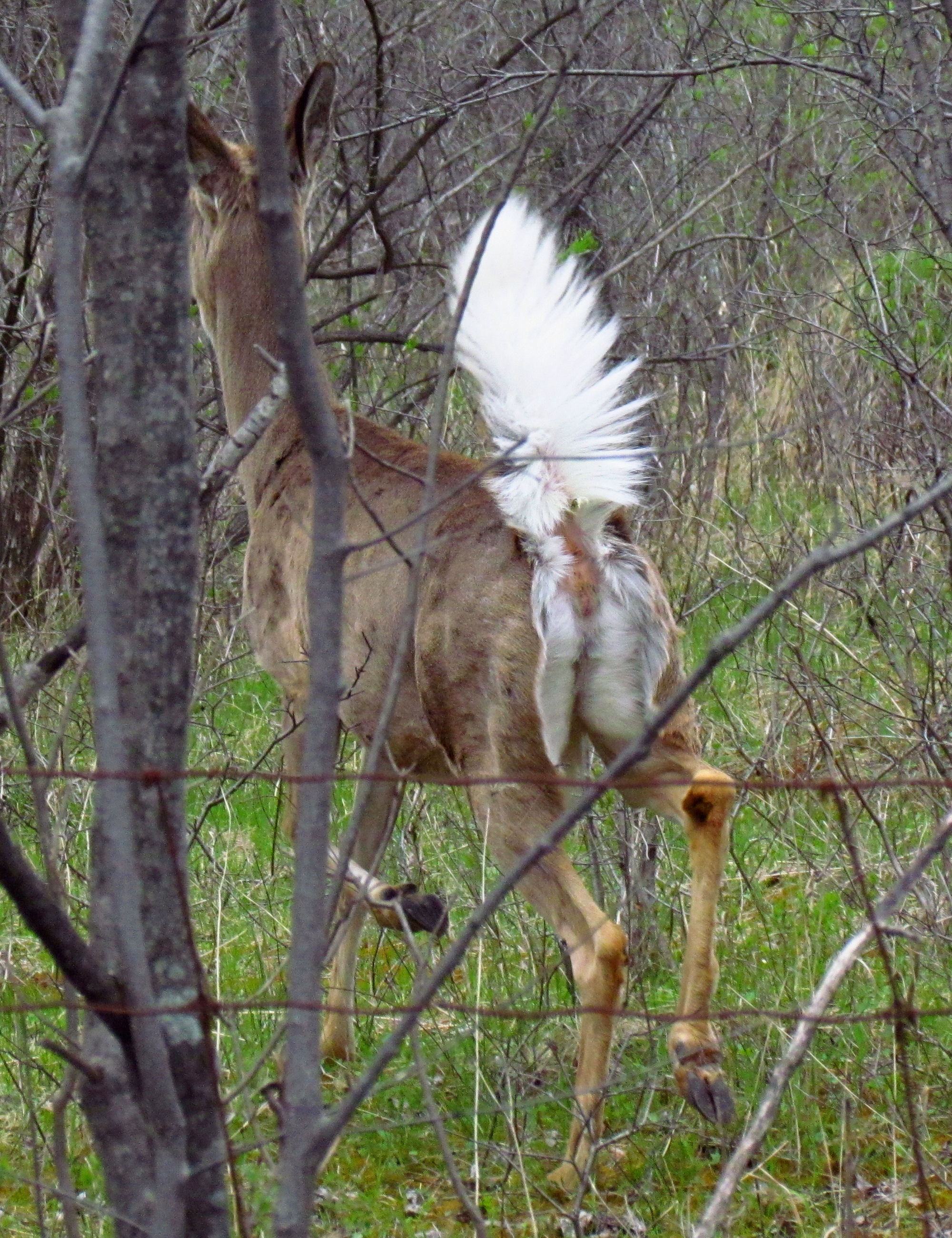
The tail is the section at the rear end of certain kinds of animals’ bodies; in general, the term refers to a distinct, flexible appendage to the torso. It is the part of the body that corresponds roughly to the sacrum and coccyx in mammals, reptiles, and birds. While tails are primarily a feature of vertebrates, some invertebrates including scorpions and springtails, as well as snails and slugs, have tail-like appendages that are sometimes referred to as tails. Tailed objects are sometimes referred to as "caudate" and the part of the body associated with or proximal to the tail are given the adjective "caudal". Function Animal tails are used in a variety of ways. They provide a source of locomotion for fish and some other forms of marine life. Many land animals use their tails to brush away flies and other biting insects. Most canines use their tails to comunicate mood and intention . Some species, including cats and kangaroos, use their tails for balance; and some, such ... [...More Info...] [...Related Items...] OR: [Wikipedia] [Google] [Baidu] |
Bird
Birds are a group of warm-blooded vertebrates constituting the class Aves (), characterised by feathers, toothless beaked jaws, the laying of hard-shelled eggs, a high metabolic rate, a four-chambered heart, and a strong yet lightweight skeleton. Birds live worldwide and range in size from the bee hummingbird to the ostrich. There are about ten thousand living species, more than half of which are passerine, or "perching" birds. Birds have whose development varies according to species; the only known groups without wings are the extinct moa and elephant birds. Wings, which are modified forelimbs, gave birds the ability to fly, although further evolution has led to the loss of flight in some birds, including ratites, penguins, and diverse endemic island species. The digestive and respiratory systems of birds are also uniquely adapted for flight. Some bird species of aquatic environments, particularly seabirds and some waterbirds, have further evolved for swimming. B ... [...More Info...] [...Related Items...] OR: [Wikipedia] [Google] [Baidu] |
Metarteriole
A metarteriole is a short microvessel in the microcirculation that links arterioles and capillaries. Instead of a continuous tunica media, they have individual smooth muscle cells placed a short distance apart, each forming a precapillary sphincter that encircles the entrance to that capillary bed. Constriction of these sphincters reduces or shuts off blood flow through their respective capillary beds. This allows the blood to be diverted to elsewhere in the body. Metarterioles exist in the ''mesenteric microcirculation'', and the name was originally conceived only to define the "''thoroughfare channels ''" between arterioles and venules A venule is a very small blood vessel in the microcirculation that allows blood to return from the capillary beds to drain into the larger blood vessels, the veins. Venules range from 7μm to 1mm in diameter. Veins contain approximately 70% of t .... In recent times the term has often been used instead to describe the smallest arterioles direct ... [...More Info...] [...Related Items...] OR: [Wikipedia] [Google] [Baidu] |
Amniotes
Amniotes are a clade of tetrapod vertebrates that comprises sauropsids (including all reptiles and birds, and extinct parareptiles and non-avian dinosaurs) and synapsids (including pelycosaurs and therapsids such as mammals). They are distinguished from the other tetrapod clade — the amphibians — by the development of three extraembryonic membranes ( amnion for embryoic protection, chorion for gas exchange, and allantois for metabolic waste disposal or storage), thicker and more keratinized skin, and costal respiration (breathing by expanding/constricting the rib cage). All three main features listed above, namely the presence of an amniotic buffer, water-impermeable cutes and a robust respiratory system, are very important for amniotes to live on land as true terrestrial animals – the ability to reproduce in locations away from water bodies, better homeostasis in drier environments, and more efficient air respiration to power terrestrial locomotions, although they migh ... [...More Info...] [...Related Items...] OR: [Wikipedia] [Google] [Baidu] |

_Ranomafana.jpg)


.jpg)
.jpg)