|
Renal Calyx
The renal calyces are chambers of the kidney through which urine passes. The minor calyces surround the apex of the renal pyramids. Urine formed in the kidney passes through a renal papilla at the apex into the minor calyx; two or three minor calyces converge to form a major calyx, through which urine passes before continuing through the renal pelvis into the ureter. Function Peristalsis of the smooth muscle originating in pace-maker cells originating in the walls of the calyces propels urine through the renal pelvis and ureters to the bladder. The initiation is caused by the increase in volume that stretches the walls of the calyces. This causes them to fire impulses which stimulate rhythmical contraction and relaxation, called peristalsis. Parasympathetic innervation enhances the peristalsis while sympathetic innervation inhibits it. Clinical significance A " staghorn calculus" is a kidney stone that may extend into the renal calyces. A renal diverticulum is diverticulum of ... [...More Info...] [...Related Items...] OR: [Wikipedia] [Google] [Baidu] |
Ureteric Bud
The ureteric bud, also known as the metanephric diverticulum, is a protrusion from the mesonephric duct during the development of the urinary and reproductive organs. It later develops into a conduit for urine drainage from the kidneys, which, in contrast, originate from the metanephric blastema The metanephrogenic blastema or metanephric blastema (or metanephric mesenchyme, or metanephric mesoderm) is one of the two embryological structures that give rise to the kidney, the other being the ureteric bud. The metanephric blastema mostly de .... References {{Authority control Embryology of urogenital system ... [...More Info...] [...Related Items...] OR: [Wikipedia] [Google] [Baidu] |
Peristalsis
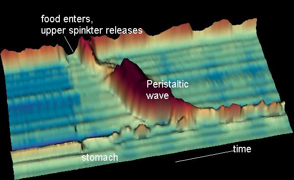
Peristalsis ( , ) is a radially symmetrical contraction and relaxation of muscles that propagate in a wave down a tube, in an anterograde direction. Peristalsis is progression of coordinated contraction of involuntary circular muscles, which is preceded by a simultaneous contraction of the longitudinal muscle and relaxation of the circular muscle in the lining of the gut. In much of a digestive tract such as the human gastrointestinal tract, smooth muscle tissue contracts in sequence to produce a peristaltic wave, which propels a ball of food (called a bolus before being transformed into chyme in the stomach) along the tract. The peristaltic movement comprises relaxation of circular smooth muscles, then their contraction behind the chewed material to keep it from moving backward, then longitudinal contraction to push it forward. Earthworms use a similar mechanism to drive their locomotion, and some modern machinery imitate this design. The word comes from New Latin and is ... [...More Info...] [...Related Items...] OR: [Wikipedia] [Google] [Baidu] |
Renal Medulla
The renal medulla is the innermost part of the kidney. The renal medulla is split up into a number of sections, known as the renal pyramids. Blood enters into the kidney via the renal artery, which then splits up to form the segmental arteries which then branch to form interlobar arteries. The interlobar arteries each in turn branch into arcuate arteries, which in turn branch to form interlobular arteries, and these finally reach the glomeruli. At the glomerulus the blood reaches a highly disfavourable pressure gradient and a large exchange surface area, which forces the serum portion of the blood out of the vessel and into the renal tubules. Flow continues through the renal tubules, including the proximal tubule, the Loop of Henle, through the distal tubule and finally leaves the kidney by means of the collecting duct, leading to the renal pelvis, the dilated portion of the ureter. The renal medulla (Latin: ''medulla renis'' 'marrow of the kidney') contains the structure ... [...More Info...] [...Related Items...] OR: [Wikipedia] [Google] [Baidu] |
Renal Calyces
The renal calyces are chambers of the kidney through which urine passes. The minor calyces surround the apex of the renal pyramids. Urine formed in the kidney passes through a renal papilla at the apex into the minor calyx; two or three minor calyces converge to form a major calyx, through which urine passes before continuing through the renal pelvis into the ureter. Function Peristalsis of the smooth muscle originating in pace-maker cells originating in the walls of the calyces propels urine through the renal pelvis and ureters to the bladder. The initiation is caused by the increase in volume that stretches the walls of the calyces. This causes them to fire impulses which stimulate rhythmical contraction and relaxation, called peristalsis. Parasympathetic innervation enhances the peristalsis while sympathetic innervation inhibits it. Clinical significance A " staghorn calculus" is a kidney stone that may extend into the renal calyces. A renal diverticulum is diverticulum of ... [...More Info...] [...Related Items...] OR: [Wikipedia] [Google] [Baidu] |
Kidney Stone
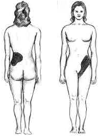
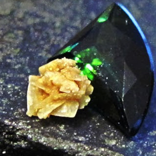
Kidney stone disease, also known as nephrolithiasis or urolithiasis, is a crystallopathy where a solid piece of material (kidney stone) develops in the urinary tract. Kidney stones typically form in the kidney and leave the body in the urine stream. A small stone may pass without causing symptoms. If a stone grows to more than , it can cause blockage of the ureter, resulting in sharp and severe pain in the lower back or abdomen. A stone may also result in blood in the urine, vomiting, or painful urination. About half of people who have had a kidney stone will have another within ten years. Most stones form by a combination of genetics and environmental factors. Risk factors include high urine calcium levels, obesity, certain foods, some medications, calcium supplements, hyperparathyroidism, gout and not drinking enough fluids. Stones form in the kidney when minerals in urine are at high concentration. The diagnosis is usually based on symptoms, urine testing, and me ... [...More Info...] [...Related Items...] OR: [Wikipedia] [Google] [Baidu] |
Staghorn Calculus
Kidney stone disease, also known as nephrolithiasis or urolithiasis, is a crystallopathy where a solid piece of material (kidney stone) develops in the urinary tract. Kidney stones typically form in the kidney and leave the body in the urine stream. A small stone may pass without causing symptoms. If a stone grows to more than , it can cause blockage of the ureter, resulting in sharp and severe pain in the lower back or abdomen. A stone may also result in blood in the urine, vomiting, or painful urination. About half of people who have had a kidney stone will have another within ten years. Most stones form by a combination of genetics and environmental factors. Risk factors include high urine calcium levels, obesity, certain foods, some medications, calcium supplements, hyperparathyroidism, gout and not drinking enough fluids. Stones form in the kidney when minerals in urine are at high concentration. The diagnosis is usually based on symptoms, urine testing, and medical ... [...More Info...] [...Related Items...] OR: [Wikipedia] [Google] [Baidu] |
Staghorn Kidney Stone Progression
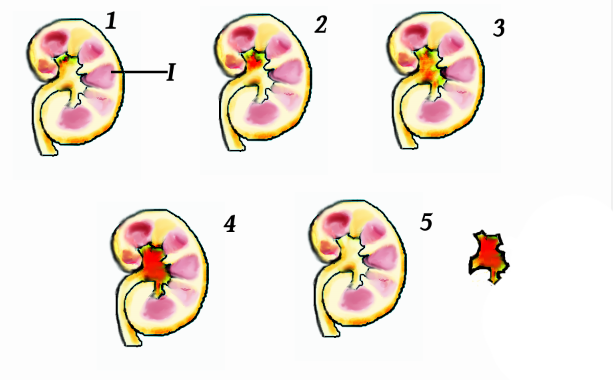
Staghorn may refer to: *The Horn (anatomy) of a stag *Staghorn calculus, a type of kidney stone * Staghorn coral, a branching coral *''Rhus typhina'', a shrub commonly called ''Staghorn sumac'' *''Lycopodium clavatum'', a moss commonly called ''Staghorn moss'' *''Platycerium ''Platycerium'' is a genus of about 18 fern species in the polypod family, Polypodiaceae. Ferns in this genus are widely known as staghorn or elkhorn ferns due to their uniquely shaped fronds. This genus is epiphytic and is native to tropical and ...'', a fern commonly called ''Staghorn fern'' * Pacific staghorn sculpin, a type of fish * Staghorn (He-Man), an action figure from the Mattel * Struvite, a type of kidney stone, also referred to as ''Staghorn calculus'' {{disambiguation, plant ... [...More Info...] [...Related Items...] OR: [Wikipedia] [Google] [Baidu] |
Urinary Bladder
The urinary bladder, or simply bladder, is a hollow organ in humans and other vertebrates that stores urine from the kidneys before disposal by urination. In humans the bladder is a distensible organ that sits on the pelvic floor. Urine enters the bladder via the ureters and exits via the urethra. The typical adult human bladder will hold between 300 and (10.14 and ) before the urge to empty occurs, but can hold considerably more. The Latin phrase for "urinary bladder" is ''vesica urinaria'', and the term ''vesical'' or prefix ''vesico -'' appear in connection with associated structures such as vesical veins. The modern Latin word for "bladder" – ''cystis'' – appears in associated terms such as cystitis (inflammation of the bladder). Structure In humans, the bladder is a hollow muscular organ situated at the base of the pelvis. In gross anatomy, the bladder can be divided into a broad , a body, an apex, and a neck. The apex (also called the vertex) is directed fo ... [...More Info...] [...Related Items...] OR: [Wikipedia] [Google] [Baidu] |
Ureter
The ureters are tubes made of smooth muscle that propel urine from the kidneys to the urinary bladder. In a human adult, the ureters are usually long and around in diameter. The ureter is lined by urothelial cells, a type of transitional epithelium, and has an additional smooth muscle layer that assists with peristalsis in its lowest third. The ureters can be affected by a number of diseases, including urinary tract infections and kidney stone. is when a ureter is narrowed, due to for example chronic inflammation. Congenital abnormalities that affect the ureters can include the development of two ureters on the same side or abnormally placed ureters. Additionally, reflux of urine from the bladder back up the ureters is a condition commonly seen in children. The ureters have been identified for at least two thousand years, with the word "ureter" stemming from the stem relating to urinating and seen in written records since at least the time of Hippocrates. It is, ho ... [...More Info...] [...Related Items...] OR: [Wikipedia] [Google] [Baidu] |
Urinary System
The urinary system, also known as the urinary tract or renal system, consists of the kidneys, ureters, bladder, and the urethra. The purpose of the urinary system is to eliminate waste from the body, regulate blood volume and blood pressure, control levels of electrolytes and metabolites, and regulate blood pH. The urinary tract is the body's drainage system for the eventual removal of urine. The kidneys have an extensive blood supply via the renal arteries which leave the kidneys via the renal vein. Each kidney consists of functional units called nephrons. Following filtration of blood and further processing, wastes (in the form of urine) exit the kidney via the ureters, tubes made of smooth muscle fibres that propel urine towards the urinary bladder, where it is stored and subsequently expelled from the body by urination ( voiding). The female and male urinary system are very similar, differing only in the length of the urethra. Urine is formed in the kidneys through ... [...More Info...] [...Related Items...] OR: [Wikipedia] [Google] [Baidu] |
Renal Pelvis
The renal pelvis or pelvis of the kidney is the funnel-like dilated part of the ureter in the kidney. It is formed by the covnvergence of the major calyces, acting as a funnel for urine flowing from the major calyces to the ureter. It has a mucous membrane and is covered with transitional epithelium and an underlying lamina propria of loose-to-dense connective tissue. The renal pelvis is situated within the renal sinus alongside the other structures of the renal sinus. The renal pelvis is the location of several kinds of kidney cancer and is affected by infection in pyelonephritis. Clinical significance The renal pelvis is the location of several kinds of kidney cancer and is affected by infection in pyelonephritis. A large "staghorn" kidney stone may block all or part of the renal pelvis. The size of the renal pelvis plays a major role in the grading of hydronephrosis. Normally, the anteroposterior diameter of the renal pelvis is less than 4 mm in fetuses up to 32 wee ... [...More Info...] [...Related Items...] OR: [Wikipedia] [Google] [Baidu] |
Renal Papilla
The renal medulla is the innermost part of the kidney. The renal medulla is split up into a number of sections, known as the renal pyramids. Blood enters into the kidney via the renal artery, which then splits up to form the segmental arteries which then branch to form interlobar arteries. The interlobar arteries each in turn branch into arcuate arteries, which in turn branch to form interlobular arteries, and these finally reach the glomeruli. At the glomerulus the blood reaches a highly disfavourable pressure gradient and a large exchange surface area, which forces the serum portion of the blood out of the vessel and into the renal tubules. Flow continues through the renal tubules, including the proximal tubule, the Loop of Henle, through the distal tubule and finally leaves the kidney by means of the collecting duct, leading to the renal pelvis, the dilated portion of the ureter. The renal medulla (Latin: ''medulla renis'' 'marrow of the kidney') contains the structures ... [...More Info...] [...Related Items...] OR: [Wikipedia] [Google] [Baidu] |


