|
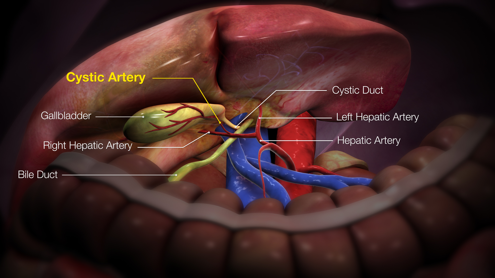
Right Hepatic Artery
The hepatic artery proper (also proper hepatic artery) is the artery that supplies the liver and gallbladder. It raises from the common hepatic artery, a branch of the celiac artery. Structure The hepatic artery proper arises from the common hepatic artery and runs alongside the portal vein and the common bile duct to form the portal triad. A branch of the common hepatic artery –the gastroduodenal artery gives off the small supraduodenal artery to the duodenal bulb. Then the right gastric artery comes off and runs to the left along the lesser curvature of the stomach to meet the left gastric artery, which is a branch of the celiac trunk. It subsequently bifurcates into the right and left hepatic arteries. Variant anatomy Of note, the right and left hepatic arteries may demonstrate variant anatomy. A misplaced right hepatic artery may arise from the superior mesenteric artery (SMA) and a misplaced left hepatic artery may arise from the left gastric artery. The cystic ar ... [...More Info...] [...Related Items...] OR: [Wikipedia] [Google] [Baidu] |
Common Hepatic Artery
The common hepatic artery is a short blood vessel that supplies oxygenated blood to the liver, pylorus of the stomach, duodenum, pancreas, and gallbladder. It arises from the celiac artery and has the following branches: Additional images File:Common hepatic artery.jpg, Common hepatic artery and its branches including hepatic artery proper and right gastric artery (pyloric artery) References External links * - "Stomach, Spleen and Liver: Contents of the Hepatoduodenal ligament The hepatoduodenal ligament is the portion of the lesser omentum extending between the porta hepatis of the liver and the superior part of the duodenum. Running inside it are the following structures collectively known as the portal triad: * hep ..." * {{Authority control Arteries of the abdomen ... [...More Info...] [...Related Items...] OR: [Wikipedia] [Google] [Baidu] |
Duodenal Bulb
The duodenal bulb is the portion of the duodenum closest to the stomach. It normally has a length of about 5 centimeters. The duodenal bulb begins at the pylorus and ends at the neck of the gallbladder. It is located posterior to the liver and the gallbladder and superior to the pancreatic head. The gastroduodenal artery, portal vein, and common bile duct lie just behind it. The distal part of the bulb is located retroperitoneally. It is located immediately distal to the pyloric sphincter. The duodenal bulb is the place where duodenal ulcers occur. Duodenal ulcers are more common than gastric ulcers and unlike gastric ulcers, are caused by increased gastric acid secretion. Duodenal ulcers are commonly located anteriorly, and rarely posteriorly. Anterior ulcers can be complicated by perforation, while the posterior ones bleed. The reason for that is explained by their location. The peritoneal or abdominal cavity is located anterior to the duodenum. Therefore, if the ulcer grows dee ... [...More Info...] [...Related Items...] OR: [Wikipedia] [Google] [Baidu] |
Cystic Artery
The cystic artery (also known as bachelor artery) supplies oxygenated blood to the gallbladder and cystic duct. Most common arrangement In the classic arrangement, occurring with a frequency of approximately 70%, a singular cystic artery originates from the geniculate flexure of the right hepatic artery in the upper portion of the hepatobiliary triangle. A site of origin from a more proximal or distal portion of the right hepatic artery is also considered relatively normal. After separating from the right hepatic artery, the cystic artery travels superiorly to the cystic duct and produces 2 to 4 minor branches, known as ''Calot’s arteries'', that supply part of the cystic duct and cervix of the gallbladder before dividing into the major superficial and deep branches at the superior aspect of the gallbladder neck: * The ''superficial branch'' (or ''anterior branch'') passes subserously over the left aspect of the gallbladder. * The ''deep branch'' (or ''posterior branch'') runs b ... [...More Info...] [...Related Items...] OR: [Wikipedia] [Google] [Baidu] |
Superior Mesenteric Artery
In human anatomy, the superior mesenteric artery (SMA) is an artery which arises from the anterior surface of the abdominal aorta, just inferior to the origin of the celiac trunk, and supplies blood to the intestine from the lower part of the duodenum through two-thirds of the transverse colon, as well as the pancreas. Structure It arises anterior to lower border of vertebra L1 in an adult. It is usually 1 cm lower than the celiac trunk. It initially travels in an anterior/inferior direction, passing behind/under the neck of the pancreas and the splenic vein. Located under this portion of the superior mesenteric artery, between it and the aorta, are the following: * left renal vein - travels between the left kidney and the inferior vena cava (can be compressed between the SMA and the abdominal aorta at this location, leading to nutcracker syndrome). * the third part of the duodenum, a segment of the small intestines (can be compressed by the SMA at this location, lea ... [...More Info...] [...Related Items...] OR: [Wikipedia] [Google] [Baidu] |
Human Body
The human body is the structure of a Human, human being. It is composed of many different types of Cell (biology), cells that together create Tissue (biology), tissues and subsequently organ systems. They ensure homeostasis and the life, viability of the human body. It comprises a human head, head, hair, neck, Trunk (anatomy), trunk (which includes the thorax and abdomen), arms and hands, human leg, legs and feet. The study of the human body involves anatomy, physiology, histology and embryology. The body anatomical variability, varies anatomically in known ways. Physiology focuses on the systems and organs of the human body and their functions. Many systems and mechanisms interact in order to maintain homeostasis, with safe levels of substances such as sugar and oxygen in the blood. The body is studied by health professionals, physiologists, anatomists, and by artists to assist them in their work. Composition The composition of the human body, human body is composed of ... [...More Info...] [...Related Items...] OR: [Wikipedia] [Google] [Baidu] |
Celiac Trunk
The celiac () artery (also spelled ''coeliac''), also known as the celiac trunk or truncus coeliacus, is the first major branch of the abdominal aorta. It is about 1.25 cm in length. Branching from the aorta at thoracic vertebra 12 (T12) in humans, it is one of three anterior/ midline branches of the abdominal aorta (the others are the superior and inferior mesenteric arteries). Structure The celiac artery is the first major branch of the descending abdominal aorta, branching at a 90° angle. This occurs just below the crus of the diaphragm. This is around the first lumbar vertebra. There are three main divisions of the celiac artery, and each in turn has its own named branches: The celiac artery may also give rise to the inferior phrenic arteries. Function The celiac artery supplies oxygenated blood to the liver, stomach, abdominal esophagus, spleen, and the superior half of both the duodenum and the pancreas. These structures correspond to the embryonic foregut. ... [...More Info...] [...Related Items...] OR: [Wikipedia] [Google] [Baidu] |
Left Gastric Artery
In human anatomy, the left gastric artery arises from the celiac artery and runs along the superior portion of the lesser curvature of the stomach. Branches also supply the lower esophagus. The left gastric artery anastomoses with the right gastric artery, which runs right to left. Important to note is that the esophageal branch of the left gastric artery ascends and passes through the esophageal hiatus. Clinical significance In terms of disease, the left gastric artery may be involved in peptic ulcer disease: if an ulcer erodes through the stomach mucosa into a branch of the artery, this can cause massive blood loss into the stomach, which may result in such symptoms as hematemesis or melaena. Additional images File:Stomach blood supply.svg, Blood supply to the stomach: left and right gastric artery, left and right gastro-omental artery and short gastric artery The short gastric arteries consist of from five to seven small branches, which arise from the end of the splen ... [...More Info...] [...Related Items...] OR: [Wikipedia] [Google] [Baidu] |
Right Gastric Artery
The right gastric artery arises, in most cases (53% of cases), from the proper hepatic artery, descends to the pyloric end of the stomach, and passes from right to left along its lesser curvature, supplying it with branches, and anastomosing with the left gastric artery. It can also arise from the region of division of the common hepatic artery (20% of cases), the left branch of the hepatic artery (15% of cases), the gastroduodenal artery (8% of cases), and most rarely, the common hepatic artery itself (4% of cases). Additional images File:Gray532.png, The celiac artery The celiac () artery (also spelled ''coeliac''), also known as the celiac trunk or truncus coeliacus, is the first major branch of the abdominal aorta. It is about 1.25 cm in length. Branching from the aorta at thoracic vertebra 12 (T12) in ... and its branches; the liver has been raised, and the lesser omentum and anterior layer of the greater omentum removed. File:Slide14fff.JPG, Right gastric artery ... [...More Info...] [...Related Items...] OR: [Wikipedia] [Google] [Baidu] |
Supraduodenal Artery
The supraduodenal artery is an artery which usually branches from Gastroduodenal artery In anatomy, the gastroduodenal artery is a small blood vessel in the abdomen. It supplies blood directly to the pylorus (distal part of the stomach) and proximal part of the duodenum. It also indirectly supplies the pancreatic head (via the anterio .... This artery supplies the superior portion of the duodenum. References External links * Arteries of the abdomen {{circulatory-stub ... [...More Info...] [...Related Items...] OR: [Wikipedia] [Google] [Baidu] |
Liver
The liver is a major Organ (anatomy), organ only found in vertebrates which performs many essential biological functions such as detoxification of the organism, and the Protein biosynthesis, synthesis of proteins and biochemicals necessary for digestion and growth. In humans, it is located in the quadrant (anatomy), right upper quadrant of the abdomen, below the thoracic diaphragm, diaphragm. Its other roles in metabolism include the regulation of Glycogen, glycogen storage, decomposition of red blood cells, and the production of hormones. The liver is an accessory digestive organ that produces bile, an alkaline fluid containing cholesterol and bile acids, which helps the fatty acid degradation, breakdown of fat. The gallbladder, a small pouch that sits just under the liver, stores bile produced by the liver which is later moved to the small intestine to complete digestion. The liver's highly specialized biological tissue, tissue, consisting mostly of hepatocytes, regulates a w ... [...More Info...] [...Related Items...] OR: [Wikipedia] [Google] [Baidu] |
Gastroduodenal Artery
In anatomy, the gastroduodenal artery is a small blood vessel in the abdomen. It supplies blood directly to the pylorus (distal part of the stomach) and proximal part of the duodenum. It also indirectly supplies the pancreatic head (via the anterior and posterior superior pancreaticoduodenal arteries). Structure The gastroduodenal artery most commonly arises from either the left hepatic artery or the right hepatic artery instead. It may also arise from the common hepatic artery of the coeliac trunk in a trifork arrangement with the two other arteries, but there are numerous variations of the origin.Bergman RA, Afifi AK, Miyauchi R. Variations in Origin of Gastroduodenal Artery. from Anatomy Atlases. (http://www.anatomyatlases.org/AnatomicVariants/Cardiovascular/Images0001/0017.shtml) It first gives rise to the supraduodenal artery, followed by the posterior superior pancreaticoduodenal artery. It terminates in a bifurcation when it splits into the right gastroepiploic artery a ... [...More Info...] [...Related Items...] OR: [Wikipedia] [Google] [Baidu] |
Portal Triad
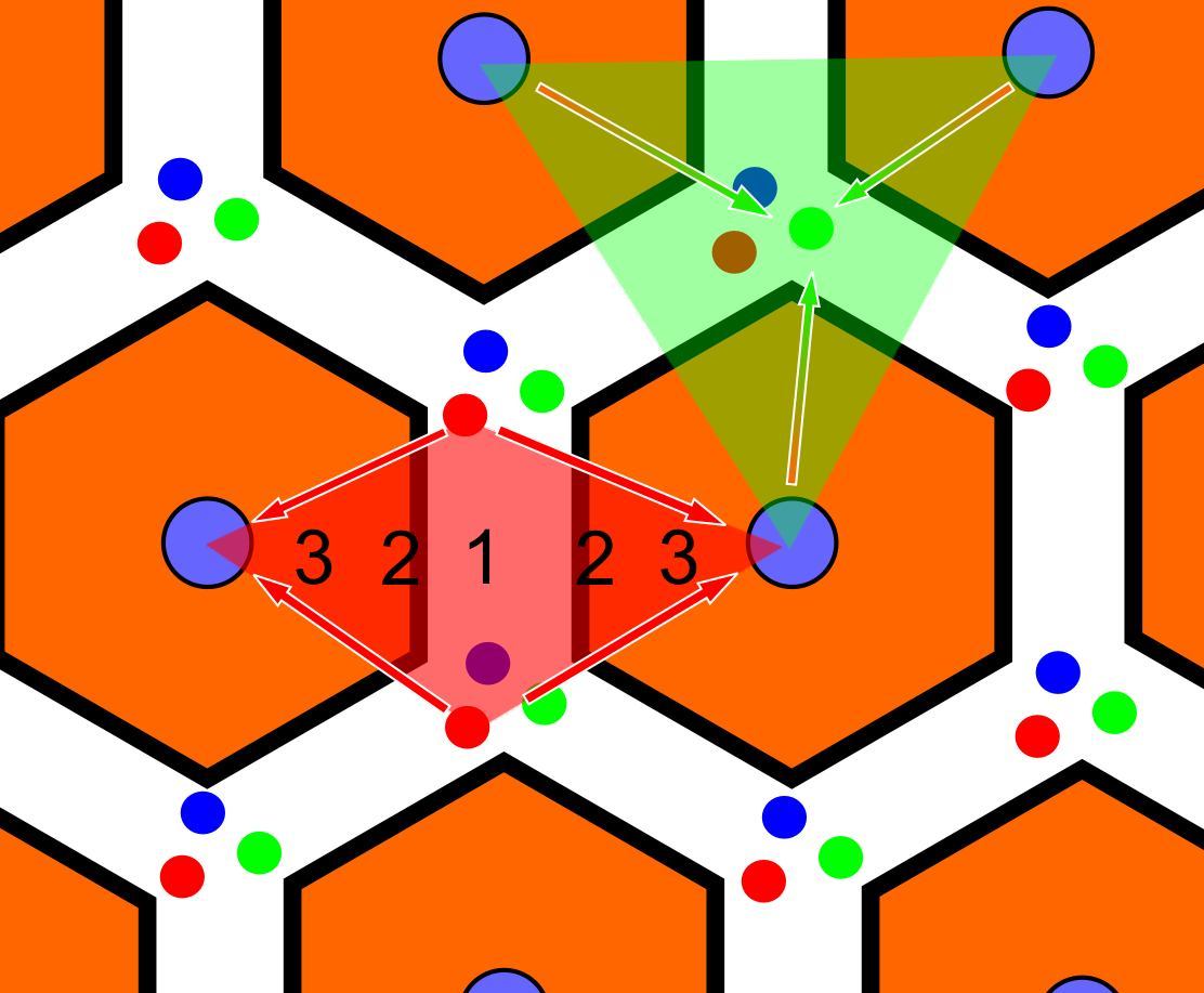
In histology (microscopic anatomy), the lobules of liver, or hepatic lobules, are small divisions of the liver defined at the microscopic scale. The hepatic lobule is a building block of the liver tissue, consisting of a portal triad, hepatocytes arranged in linear cords between a capillary network, and a central vein. Lobules are different from the lobes of liver: they are the smaller divisions of the lobes. The two-dimensional microarchitecture of the liver can be viewed from different perspectives: The term "hepatic lobule", without qualification, typically refers to the classical lobule. Structure The hepatic lobule can be described in terms of metabolic "zones", describing the hepatic acinus (terminal acinus). Each zone is centered on the line connecting two portal triads and extends outwards to the two adjacent central veins. The periportal zone I is nearest to the entering vascular supply and receives the most oxygenated blood, making it least sensitive to ischemic i ... [...More Info...] [...Related Items...] OR: [Wikipedia] [Google] [Baidu] |


