|
Reticuloendothelial System
In anatomy the term "reticuloendothelial system" (abbreviated RES), often associated nowadays with the mononuclear phagocyte system (MPS), was originally launched by the beginning of the 20th century to denote a system of specialised cells that effectively clear colloidal vital stains (so called because they stain living cells) from the blood circulation. The term is still used today, but its meaning has changed over the years, and is used inconsistently in present-day literature. Although RES is commonly associated exclusively with macrophages, recent research has revealed that the cells that accumulate intravenously administrated vital stain belong to a highly specialised group of cells called '' scavenger endothelial cells'' (SECs), that are not macrophages. History In the 1920s, the founder of the term RES, Ludwig Aschoff, reviewed the field of vital staining, and concluded that the cells lining the hepatic sinusoids are by far the most numerous and important cells accumulati ... [...More Info...] [...Related Items...] OR: [Wikipedia] [Google] [Baidu] |
Anatomy
Anatomy () is the branch of biology concerned with the study of the structure of organisms and their parts. Anatomy is a branch of natural science that deals with the structural organization of living things. It is an old science, having its beginnings in prehistoric times. Anatomy is inherently tied to developmental biology, embryology, comparative anatomy, evolutionary biology, and phylogeny, as these are the processes by which anatomy is generated, both over immediate and long-term timescales. Anatomy and physiology, which study the structure and function of organisms and their parts respectively, make a natural pair of related disciplines, and are often studied together. Human anatomy is one of the essential basic sciences that are applied in medicine. The discipline of anatomy is divided into macroscopic and microscopic. Macroscopic anatomy, or gross anatomy, is the examination of an animal's body parts using unaided eyesight. Gross anatomy also includes the br ... [...More Info...] [...Related Items...] OR: [Wikipedia] [Google] [Baidu] |
Mononuclear Phagocyte System
In immunology, the mononuclear phagocyte system or mononuclear phagocytic system (MPS) also known as the reticuloendothelial system or macrophage system is a part of the immune system that consists of the phagocytic cells located in reticular connective tissue. The cells are primarily monocytes and macrophages, and they accumulate in lymph nodes and the spleen. The Kupffer cells of the liver and tissue histiocytes are also part of the MPS. The mononuclear phagocyte system and the monocyte macrophage system refer to two different entities, often mistakenly understood as one. "Reticuloendothelial system" is an older term for the mononuclear phagocyte system, but it is used less commonly now, as it is understood that most endothelial cells are not macrophages. The mononuclear phagocyte system is also a somewhat dated concept trying to combine a broad range of cells, and should be used with caution. Cell types and locations The spleen is the second largest unit of the mononucl ... [...More Info...] [...Related Items...] OR: [Wikipedia] [Google] [Baidu] |
Vital Stain
A vital stain in a casual usage may mean a stain that can be applied on living cells without killing them. Vital stains have been useful for diagnostic and surgical techniques in a variety of medical specialties. In supravital staining, living cells have been removed from an organism, whereas intravital staining is done by injecting or otherwise introducing the stain into the body. The term vital stain is used by some authors to refer to an intravital stain, and by others interchangeably with a supravital stain, the core concept being that the cell being examined is still alive. In a more strict sense, the term vital staining has a meaning contrasting with supravital staining. While in supravital staining the living cells take up the stain, in "vital staining" – the most accepted but apparently paradoxical meaning of this term, the living cells exclude the stain i.e. stain negatively and only the dead cells stain positively and thus viability can be assessed by counting the percen ... [...More Info...] [...Related Items...] OR: [Wikipedia] [Google] [Baidu] |
Scavenger Endothelial Cell
The term scavenger endothelial cell (SEC) was initially coined to describe a specialized sub-group of endothelial cells in vertebrates that express a remarkably high blood clearance activity. The term SEC has now been adopted by several scientists. In vertebrates The term "scavenger endothelial cell", first appearing in the scientific literature in 1999, was coined to distinguish a highly specialized subclass of endothelium in vertebrates that was observed to express a remarkably avid blood clearance activity. Blood borne waste macromolecules are known to be efficiently cleared from the blood circulation via scavenger receptors (stabilin-1, stabilin-2), the mannose receptor, and the Fc gamma receptor IIb2 of the mammalian liver sinusoidal endothelial cells. Ligands that are efficiently cleared from blood by receptor-mediated endocytosis in liver sinusoidal endothelial cells in mammals, are also avidly cleared by liver sinusoidal endothelial cells in birds, reptiles and amphibia, as i ... [...More Info...] [...Related Items...] OR: [Wikipedia] [Google] [Baidu] |
Macrophage
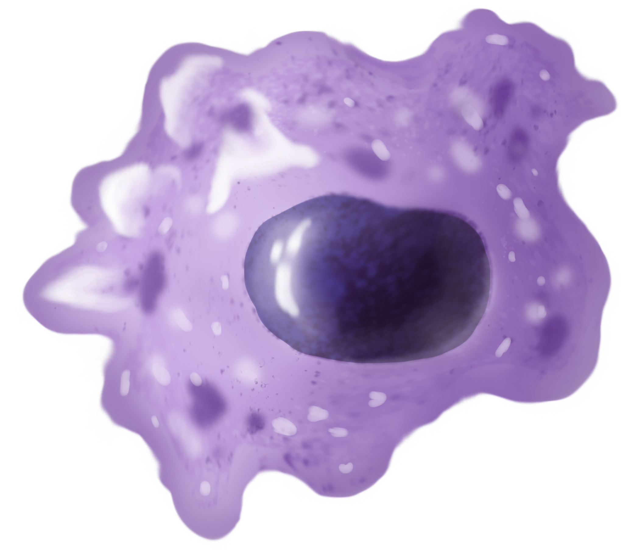
Macrophages (abbreviated as M φ, MΦ or MP) ( el, large eaters, from Greek ''μακρός'' (') = large, ''φαγεῖν'' (') = to eat) are a type of white blood cell of the immune system that engulfs and digests pathogens, such as cancer cells, microbe A microorganism, or microbe,, ''mikros'', "small") and ''organism'' from the el, ὀργανισμός, ''organismós'', "organism"). It is usually written as a single word but is sometimes hyphenated (''micro-organism''), especially in olde ...s, cellular debris, and foreign substances, which do not have proteins that are specific to healthy body cells on their surface. The process is called phagocytosis, which acts to defend the host against infection and injury. These large phagocytes are found in essentially all tissues, where they patrol for potential pathogens by amoeboid movement. They take various forms (with various names) throughout the body (e.g., histiocytes, Kupffer cells, alveolar macrophages, microg ... [...More Info...] [...Related Items...] OR: [Wikipedia] [Google] [Baidu] |
Ludwig Aschoff
Karl Albert Ludwig Aschoff (10 January 1866 – 24 June 1942) was a German physician and pathologist. He is considered to be one of the most influential pathologists of the early 20th century and is regarded as the most important German pathologist after Rudolf Virchow. Early life and education Aschoff was born in Berlin, Prussia on 10 January 1866. He studied medicine at the University of Bonn, University of Strasbourg, and the University of Würzburg. Career After his habilitation in 1894, Ludwig Aschoff was appointed professor for pathology at the University of Göttingen in 1901. Aschoff transferred to the University of Marburg in 1903 to head the department for pathological anatomy. In 1906, he accepted a position as ordinarius at the University of Freiburg, where he remained until his death. Aschoff was interested in the pathology and pathophysiology of the heart. He discovered nodules in the myocardium present during rheumatic fever, the so-called Aschoff bodies. Aschof ... [...More Info...] [...Related Items...] OR: [Wikipedia] [Google] [Baidu] |
Hepatic Sinusoid
A liver sinusoid is a type of capillary known as a sinusoidal capillary, discontinuous capillary or sinusoid, that is similar to a fenestrated capillary, having discontinuous endothelium that serves as a location for mixing of the oxygen-rich blood from the hepatic artery and the nutrient-rich blood from the portal vein. The liver sinusoid has a larger caliber than other types of capillaries and has a lining of specialised endothelial cells known as the liver sinusoidal endothelial cells (LSECs), and Kupffer cells. The cells are porous and have a scavenging function. The LSECs make up around half of the non-parenchymal cells in the liver and are flattened and fenestrated. LSECs have many fenestrae that gives easy communication between the sinusoidal lumen and the space of Disse. They play a part in filtration, endocytosis, and in the regulation of blood flow in the sinusoids. The Kupffer cells can take up and destroy foreign material such as bacteria. Hepatocytes are separate ... [...More Info...] [...Related Items...] OR: [Wikipedia] [Google] [Baidu] |
Lymph
Lymph (from Latin, , meaning "water") is the fluid that flows through the lymphatic system, a system composed of lymph vessels (channels) and intervening lymph nodes whose function, like the venous system, is to return fluid from the tissues to be recirculated. At the origin of the fluid-return process, interstitial fluid—the fluid between the cells in all body tissues—enters the lymph capillaries. This lymphatic fluid is then transported via progressively larger lymphatic vessels through lymph nodes, where substances are removed by tissue lymphocytes and circulating lymphocytes are added to the fluid, before emptying ultimately into the right or the left subclavian vein, where it mixes with central venous blood. Because it is derived from interstitial fluid, with which blood and surrounding cells continually exchange substances, lymph undergoes continual change in composition. It is generally similar to blood plasma, which is the fluid component of blood. Lymph retur ... [...More Info...] [...Related Items...] OR: [Wikipedia] [Google] [Baidu] |
Endothelial Cell
The endothelium is a single layer of squamous endothelial cells that line the interior surface of blood vessels and lymphatic vessels. The endothelium forms an interface between circulating blood or lymph in the lumen and the rest of the vessel wall. Endothelial cells form the barrier between vessels and tissue and control the flow of substances and fluid into and out of a tissue. Endothelial cells in direct contact with blood are called vascular endothelial cells whereas those in direct contact with lymph are known as lymphatic endothelial cells. Vascular endothelial cells line the entire circulatory system, from the heart to the smallest capillaries. These cells have unique functions that include fluid filtration, such as in the glomerulus of the kidney, blood vessel tone, hemostasis, neutrophil recruitment, and hormone trafficking. Endothelium of the interior surfaces of the heart chambers is called endocardium. An impaired function can lead to serious health issu ... [...More Info...] [...Related Items...] OR: [Wikipedia] [Google] [Baidu] |
Liver Sinusoidal Endothelial Cell
Liver sinusoidal endothelial cells (LSECs) form the lining of the smallest blood vessels in the liver, also called the hepatic sinusoids. LSECs are highly specialized endothelial cells with characteristic morphology and function. They constitute an important part of the reticuloendothelial system (RES). Structure Although the LSECs make up only about 3% of the total liver cell volume, their surface in a normal adult human liver is about 210 m2, or nearly the size of a tennis court. The LSEC structure differs from other endothelia. The cells contain many open pores, or fenestrae, with diameters from 100 to 150 nm. The fenestrae occupy 20% of the LSEC surface and are arranged in groups referred to as "sieve plates". Filtering fluid between the sinusoidal lumen and the space of Disse, the fenestrae are crucial for lipoprotein traffic between the hepatocytes and the sinusoidal lumen. The LSECs lack an organized basal lamina. The LSECs contain 45% and 17% of the liver's total mass ... [...More Info...] [...Related Items...] OR: [Wikipedia] [Google] [Baidu] |
Kupffer Cells
Kupffer cells, also known as stellate macrophages and Kupffer–Browicz cells, are specialized cells localized in the liver within the lumen of the liver sinusoids and are adhesive to their endothelial cells which make up the blood vessel walls. Kupffer cells comprise the largest population of tissue-resident macrophages in the body. Gut bacteria, bacterial endotoxins, and microbial debris transported to the liver from the gastrointestinal tract via the portal vein will first come in contact with Kupffer cells, the first immune cells in the liver. It is because of this that any change to Kupffer cell functions can be connected to various liver diseases such as alcoholic liver disease, viral hepatitis, intrahepatic cholestasis, steatohepatitis, activation or rejection of the liver during liver transplantation and liver fibrosis. They form part of the mononuclear phagocyte system. Location and structure Kupffer cells can be found attached to sinusoidal endothelial cells in both th ... [...More Info...] [...Related Items...] OR: [Wikipedia] [Google] [Baidu] |
Mononuclear Phagocyte System
In immunology, the mononuclear phagocyte system or mononuclear phagocytic system (MPS) also known as the reticuloendothelial system or macrophage system is a part of the immune system that consists of the phagocytic cells located in reticular connective tissue. The cells are primarily monocytes and macrophages, and they accumulate in lymph nodes and the spleen. The Kupffer cells of the liver and tissue histiocytes are also part of the MPS. The mononuclear phagocyte system and the monocyte macrophage system refer to two different entities, often mistakenly understood as one. "Reticuloendothelial system" is an older term for the mononuclear phagocyte system, but it is used less commonly now, as it is understood that most endothelial cells are not macrophages. The mononuclear phagocyte system is also a somewhat dated concept trying to combine a broad range of cells, and should be used with caution. Cell types and locations The spleen is the second largest unit of the mononucl ... [...More Info...] [...Related Items...] OR: [Wikipedia] [Google] [Baidu] |


