|
Pseudophakic Bullous Keratopathy
Bullous keratopathy, also known as pseudophakic bullous keratopathy (PBK), is a pathological condition in which small vesicles, or '' bullae'', are formed in the cornea due to endothelial dysfunction. In a healthy cornea, endothelial cells keeps the tissue from excess fluid absorption, pumping it back into the aqueous humor. When affected by some reason, such as Fuchs' dystrophy Fuchs dystrophy, also referred to as Fuchs endothelial corneal dystrophy (FECD) and Fuchs endothelial dystrophy (FED), is a slowly progressing corneal dystrophy that usually affects both eyes and is slightly more common in women than in men. Althou ... or a trauma during cataract removal, endothelial cells suffer mortality or damage. The corneal endothelial cells normally do not undergo mitotic cell division, and cell loss results in permanent loss of function. When endothelial cell counts drop too low, the pump starts failing to function and fluid moves anterior into the stroma and epithelium. The excess ... [...More Info...] [...Related Items...] OR: [Wikipedia] [Google] [Baidu] |
Bulla (dermatology)
A skin condition, also known as cutaneous condition, is any medical condition that affects the integumentary system—the organ system that encloses the body and includes skin, nails, and related muscle and glands. The major function of this system is as a barrier against the external environment. Conditions of the human integumentary system constitute a broad spectrum of diseases, also known as dermatoses, as well as many nonpathologic states (like, in certain circumstances, melanonychia and racquet nails). While only a small number of skin diseases account for most visits to the physician, thousands of skin conditions have been described. Classification of these conditions often presents many nosological challenges, since underlying causes and pathogenetics are often not known. Therefore, most current textbooks present a classification based on location (for example, conditions of the mucous membrane), morphology ( chronic blistering conditions), cause (skin conditions res ... [...More Info...] [...Related Items...] OR: [Wikipedia] [Google] [Baidu] |
Cornea
The cornea is the transparent front part of the eye that covers the iris, pupil, and anterior chamber. Along with the anterior chamber and lens, the cornea refracts light, accounting for approximately two-thirds of the eye's total optical power. In humans, the refractive power of the cornea is approximately 43 dioptres. The cornea can be reshaped by surgical procedures such as LASIK. While the cornea contributes most of the eye's focusing power, its focus is fixed. Accommodation (the refocusing of light to better view near objects) is accomplished by changing the geometry of the lens. Medical terms related to the cornea often start with the prefix "'' kerat-''" from the Greek word κέρας, ''horn''. Structure The cornea has unmyelinated nerve endings sensitive to touch, temperature and chemicals; a touch of the cornea causes an involuntary reflex to close the eyelid. Because transparency is of prime importance, the healthy cornea does not have or need blood vessels with ... [...More Info...] [...Related Items...] OR: [Wikipedia] [Google] [Baidu] |
Corneal Endothelium
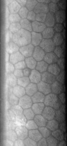
The corneal endothelium is a single layer of endothelial cells on the inner surface of the cornea. It faces the chamber formed between the cornea and the iris. The corneal endothelium are specialized, flattened, mitochondria-rich cells that line the posterior surface of the cornea and face the anterior chamber of the eye. The corneal endothelium governs fluid and solute transport across the posterior surface of the cornea and maintains the cornea in the slightly dehydrated state that is required for optical transparency. Embryology and anatomy The corneal endothelium is embryologically derived from the neural crest. The postnatal total endothelial cellularity of the cornea (approximately 300,000 cells per cornea) is achieved as early as the second trimester of gestation. Thereafter the endothelial cell density (but not the absolute number of cells) rapidly declines, as the fetal cornea grows in surface area, achieving a final adult density of approximately 2400 - 3200 cells ... [...More Info...] [...Related Items...] OR: [Wikipedia] [Google] [Baidu] |
Fuchs' Dystrophy
Fuchs dystrophy, also referred to as Fuchs endothelial corneal dystrophy (FECD) and Fuchs endothelial dystrophy (FED), is a slowly progressing corneal dystrophy that usually affects both eyes and is slightly more common in women than in men. Although early signs of Fuchs dystrophy are sometimes seen in people in their 30s and 40s, the disease rarely affects vision until people reach their 50s and 60s. Signs and symptoms As a progressive, chronic condition, signs and symptoms of Fuchs dystrophy gradually progress over decades of life, starting in middle age. Early symptoms include blurry vision upon wakening which improves during the morning, as fluid retained in the cornea is unable to evaporate through the surface of the eye when the lids are closed overnight. As the disease worsens, the interval of blurry morning vision extends from minutes to hours. In moderate stages of the disease, an increase in guttae and swelling in the cornea can contribute to changes in vision and decreas ... [...More Info...] [...Related Items...] OR: [Wikipedia] [Google] [Baidu] |

