|
Primate Basal Ganglia System
The basal ganglia form a major brain system in all species of vertebrates, but in primates (including humans) there are special features that justify a separate consideration. As in other vertebrates, the primate basal ganglia can be divided into striatal, pallidal, nigral, and subthalamic components. In primates, however, there are two pallidal subdivisions called the external globus pallidus (GPe) and internal globus pallidus (GPi). Also in primates, the dorsal striatum is divided by a large tract called the internal capsule into two masses named the caudate nucleus and the putamen—in most other species no such division exists, and only the striatum as a whole is recognized. Beyond this, there is a complex circuitry of connections between the striatum and cortex that is specific to primates. This complexity reflects the difference in functioning of different cortical areas in the primate brain. Functional imaging studies have been performed mainly using human subjects. ... [...More Info...] [...Related Items...] OR: [Wikipedia] [Google] [Baidu] |
Basal Ganglia Diagram
Basal or basilar is a term meaning ''base'', ''bottom'', or ''minimum''. Science * Basal (anatomy), an anatomical term of location for features associated with the base of an organism or structure * Basal (medicine), a minimal level that is necessary for health or life, such as a minimum insulin dose * Basal (phylogenetics), a sister group relationship in a phylogenetic tree Places * Basal, Hungary, a village in Hungary * Basal, Pakistan, a village in the Attock District Other * Basal plate (other) * Basal sliding, the act of a glacier sliding over the bed before it due to meltwater increasing the water pressure underneath the glacier causing it to be lifted from its bed * Basal conglomerate, see conglomerate (geology) Conglomerate () is a clastic sedimentary rock that is composed of a substantial fraction of rounded to subangular gravel-size clasts. A conglomerate typically contains a matrix of finer-grained sediments, such as sand, silt, or clay, which fill ... ... [...More Info...] [...Related Items...] OR: [Wikipedia] [Google] [Baidu] |
Glutamatergic
Glutamatergic means "related to glutamate". A glutamatergic agent (or drug) is a chemical that directly modulates the excitatory amino acid (glutamate/ aspartate) system in the body or brain. Examples include excitatory amino acid receptor agonists, excitatory amino acid receptor antagonists, and excitatory amino acid reuptake inhibitors. See also * Adenosinergic * Adrenergic * Cannabinoidergic * Cholinergic * Dopaminergic * GABAergic * GHBergic * Glycinergic * Histaminergic * Melatonergic * Monoaminergic * Opioidergic * Serotonergic * Sigmaergic Sigma receptors (σ-receptors) are protein cell surface receptors that bind ligands such as 4-PPBP (4-phenyl-1-(4-phenylbutyl) piperidine), SA 4503 (cutamesine), ditolylguanidine, dimethyltryptamine, and siramesine. There are two subtypes, ... References Neurochemistry Neurotransmitters {{nervous-system-drug-stub ... [...More Info...] [...Related Items...] OR: [Wikipedia] [Google] [Baidu] |
μ-opioid Receptor
The μ-opioid receptors (MOR) are a class of opioid receptors with a high affinity for enkephalins and beta-endorphin, but a low affinity for dynorphins. They are also referred to as μ(''mu'')-opioid peptide (MOP) receptors. The prototypical μ-opioid receptor agonist is morphine, the primary psychoactive alkaloid in opium. It is an inhibitory G-protein coupled receptor that activates the Gi alpha subunit, inhibiting adenylate cyclase activity, lowering cAMP levels. Structure The structure of the μ-opioid receptor has been determined with the antagonist β-FNA, the agonist BU72, and in a complex with DAMGO and Gi protein. Splice variants Three variants of the μ-opioid receptor are well characterized, though RT-PCR has identified up to 10 total splice variants in humans. Location They can exist either presynaptically or postsynaptically depending upon cell types. The μ-opioid receptors exist mostly presynaptically in the periaqueductal gray region, and in the superfi ... [...More Info...] [...Related Items...] OR: [Wikipedia] [Google] [Baidu] |
Striosome
The striosomes (also referred to as ''patches'') are one of two complementary chemical compartments within the striatum (the other compartment is known as the matrix) that can be visualized by staining for immunocytochemical markers such as acetylcholinesterase, enkephalin, substance P, limbic system-associated membrane protein (LAMP), AMPA receptor subunit 1 (GluR1), dopamine receptor subunits, and calcium binding proteins. Striosomal abnormalities have been associated with neurological disorders, such as mood dysfunction in Huntington's disease, though their precise function remains unknown. Striosomes were discovered by Ann Graybiel in 1978 using acetylcholinesterase histochemistry Immunohistochemistry (IHC) is the most common application of immunostaining. It involves the process of selectively identifying antigens (proteins) in cells of a tissue section by exploiting the principle of antibodies binding specifically to ant .... References External links *{{cite web ... [...More Info...] [...Related Items...] OR: [Wikipedia] [Google] [Baidu] |
Staining
Staining is a technique used to enhance contrast in samples, generally at the microscopic level. Stains and dyes are frequently used in histology (microscopic study of biological tissues), in cytology (microscopic study of cells), and in the medical fields of histopathology, hematology, and cytopathology that focus on the study and diagnoses of diseases at the microscopic level. Stains may be used to define biological tissues (highlighting, for example, muscle fibers or connective tissue), cell populations (classifying different blood cells), or organelles within individual cells. In biochemistry, it involves adding a class-specific ( DNA, proteins, lipids, carbohydrates) dye to a substrate to qualify or quantify the presence of a specific compound. Staining and fluorescent tagging can serve similar purposes. Biological staining is also used to mark cells in flow cytometry, and to flag proteins or nucleic acids in gel electrophoresis. Light microscopes are used for viewin ... [...More Info...] [...Related Items...] OR: [Wikipedia] [Google] [Baidu] |
Cerebral Cortex
The cerebral cortex, also known as the cerebral mantle, is the outer layer of neural tissue of the cerebrum of the brain in humans and other mammals. The cerebral cortex mostly consists of the six-layered neocortex, with just 10% consisting of allocortex. It is separated into two cortices, by the longitudinal fissure that divides the cerebrum into the left and right cerebral hemispheres. The two hemispheres are joined beneath the cortex by the corpus callosum. The cerebral cortex is the largest site of neural integration in the central nervous system. It plays a key role in attention, perception, awareness, thought, memory, language, and consciousness. The cerebral cortex is part of the brain responsible for cognition. In most mammals, apart from small mammals that have small brains, the cerebral cortex is folded, providing a greater surface area in the confined volume of the cranium. Apart from minimising brain and cranial volume, cortical folding is crucial for the brain ... [...More Info...] [...Related Items...] OR: [Wikipedia] [Google] [Baidu] |
Motor Cortex
The motor cortex is the region of the cerebral cortex believed to be involved in the planning, control, and execution of voluntary movements. The motor cortex is an area of the frontal lobe located in the posterior precentral gyrus immediately anterior to the central sulcus. Components of the motor cortex The motor cortex can be divided into three areas: 1. The primary motor cortex is the main contributor to generating neural impulses that pass down to the spinal cord and control the execution of movement. However, some of the other motor areas in the brain also play a role in this function. It is located on the anterior paracentral lobule on the medial surface. 2. The premotor cortex is responsible for some aspects of motor control, possibly including the preparation for movement, the sensory guidance of movement, the spatial guidance of reaching, or the direct control of some movements with an emphasis on control of proximal and trunk muscles of the body. Located anterior ... [...More Info...] [...Related Items...] OR: [Wikipedia] [Google] [Baidu] |
Olfactory Tubercle
The olfactory tubercle (OT), also known as the tuberculum olfactorium, is a multi-sensory processing center that is contained within the olfactory cortex and ventral striatum and plays a role in reward cognition. The OT has also been shown to play a role in locomotor and attentional behaviors, particularly in relation to social and sensory responsiveness, and it may be necessary for behavioral flexibility. The OT is interconnected with numerous brain regions, especially the sensory, arousal, and reward centers, thus making it a potentially critical interface between processing of sensory information and the subsequent behavioral responses. The OT is a composite structure that receives direct input from the olfactory bulb and contains the morphological and histochemical characteristics of the ventral pallidum and the striatum of the forebrain. The dopaminergic neurons of the mesolimbic pathway project onto the GABAergic medium spiny neurons of the nucleus accumbens and olfact ... [...More Info...] [...Related Items...] OR: [Wikipedia] [Google] [Baidu] |
Nucleus Accumbens
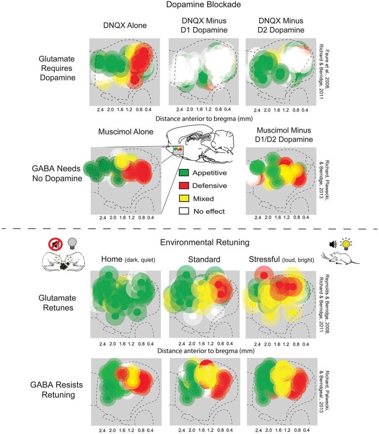
The nucleus accumbens (NAc or NAcc; also known as the accumbens nucleus, or formerly as the ''nucleus accumbens septi'', Latin for "nucleus adjacent to the septum") is a region in the basal forebrain rostral to the preoptic area of the hypothalamus. The nucleus accumbens and the olfactory tubercle collectively form the ventral striatum. The ventral striatum and dorsal striatum collectively form the striatum, which is the main component of the basal ganglia. The dopaminergic neurons of the mesolimbic pathway project onto the GABAergic medium spiny neurons of the nucleus accumbens and olfactory tubercle. Each cerebral hemisphere has its own nucleus accumbens, which can be divided into two structures: the nucleus accumbens core and the nucleus accumbens shell. These substructures have different morphology and functions. Different NAcc subregions (core vs shell) and neuron subpopulations within each region (D1-type vs D2-type medium spiny neurons) are responsible for different ... [...More Info...] [...Related Items...] OR: [Wikipedia] [Google] [Baidu] |
Tonic (physiology)
Tonic in physiology refers to a physiological response which is slow and may be graded. This term is typically used in opposition to a fast response. For instance, tonic muscles are contrasted by the more typical and much faster twitch muscles, while tonic sensory nerve endings are contrasted to the much faster phasic sensory nerve endings. Tonic muscles Tonic muscles are much slower than twitch fibers in terms of time from stimulus to full activation, time to full relaxation upon cessation of stimuli, and maximal shortening velocity.Kardong, K. 2008. Vertebrates: Comparative Anatomy, Function, Evolution. 5th edition. McGraw-Hill Science/Engineering/Math. These muscles are rarely found in mammals (only in the muscles moving the eye and in the middle ear), but are common in reptiles and amphibians. Tonic sensory receptors Tonic sensory input adapts slowly to a stimulushttp://caspar.bgsu.edu/~courses/Glossary.htm and continues to produce action potentials over the duration of th ... [...More Info...] [...Related Items...] OR: [Wikipedia] [Google] [Baidu] |
Cholinergic
Cholinergic agents are compounds which mimic the action of acetylcholine and/or butyrylcholine. In general, the word "choline" describes the various quaternary ammonium salts containing the ''N'',''N'',''N''-trimethylethanolammonium cation. Found in most animal tissues, choline is a primary component of the neurotransmitter acetylcholine and functions with inositol as a basic constituent of lecithin. Choline also prevents fat deposits in the liver and facilitates the movement of fats into cells. The parasympathetic nervous system, which uses acetylcholine almost exclusively to send its messages, is said to be almost entirely cholinergic. Neuromuscular junctions, preganglionic neurons of the sympathetic nervous system, the basal forebrain, and brain stem complexes are also cholinergic, as are the receptor for the merocrine sweat glands. In neuroscience and related fields, the term cholinergic is used in these related contexts: * A substance (or ligand) is cholinergic if it ... [...More Info...] [...Related Items...] OR: [Wikipedia] [Google] [Baidu] |
Dendritic Spine
A dendritic spine (or spine) is a small membranous protrusion from a neuron's dendrite that typically receives input from a single axon at the synapse. Dendritic spines serve as a storage site for synaptic strength and help transmit electrical signals to the neuron's cell body. Most spines have a bulbous head (the spine head), and a thin neck that connects the head of the spine to the shaft of the dendrite. The dendrites of a single neuron can contain hundreds to thousands of spines. In addition to spines providing an anatomical substrate for memory storage and synaptic transmission, they may also serve to increase the number of possible contacts between neurons. It has also been suggested that changes in the activity of neurons have a positive effect on spine morphology. Structure Dendritic spines are small with spine head volumes ranging 0.01 μm3 to 0.8 μm3. Spines with strong synaptic contacts typically have a large spine head, which connects to the dendrite via a ... [...More Info...] [...Related Items...] OR: [Wikipedia] [Google] [Baidu] |


