|
Posterior Cord
The posterior cord is a part of the brachial plexus The brachial plexus is a network () of nerves formed by the anterior rami of the lower four cervical nerves and first thoracic nerve ( C5, C6, C7, C8, and T1). This plexus extends from the spinal cord, through the cervicoaxillary canal in th .... It consists of contributions from all of the roots of the brachial plexus. The posterior cord gives rise to the following nerves: Additional images File:PLEXUS BRACHIALIS.jpg, Brachial plexus File:Slide12OOO.JPG, Posterior cord File:Slide1SSS.JPG, Posterior cord File:Slide1cord.JPG, Brachial plexus.Deep dissection. File:Slide1ecc.JPG, Brachial plexus.Deep dissection.Anterolateral view References MBBS resources http://mbbsbasic.googlepages.com/ External links * - "Axilla, dissection, anterior view" Nerves of the upper limb {{neuroscience-stub ... [...More Info...] [...Related Items...] OR: [Wikipedia] [Google] [Baidu] |
Brachial Plexus
The brachial plexus is a network () of nerves formed by the anterior rami of the lower four cervical nerves and first thoracic nerve ( C5, C6, C7, C8, and T1). This plexus extends from the spinal cord, through the cervicoaxillary canal in the neck, over the first rib, and into the armpit, it supplies afferent and efferent nerve fibers the to chest, shoulder, arm, forearm, and hand. Structure The brachial plexus is divided into five ''roots'', three ''trunks'', six ''divisions'' (three anterior and three posterior), three ''cords'', and five ''branches''. There are five "terminal" branches and numerous other "pre-terminal" or "collateral" branches, such as the subscapular nerve, the thoracodorsal nerve, and the long thoracic nerve, that leave the plexus at various points along its length. A common structure used to identify part of the brachial plexus in cadaver dissections is the M or W shape made by the musculocutaneous nerve, lateral cord, median nerve, medial cord, a ... [...More Info...] [...Related Items...] OR: [Wikipedia] [Google] [Baidu] |
Upper Subscapular Nerve
The upper (superior) subscapular nerve is the first branch of the posterior cord of the brachial plexus. The upper subscapular nerve contains axons from the ventral rami of the C5 and C6 cervical spinal nerves. It innervates the superior portion of the subscapularis muscle. The inferior portion of the subscapularis is innervated by the lower subscapular nerve. Structure The axons which form the upper subscapular nerve travel from the ventral rami of C5 and C6. They join at the upper trunk and move through its posterior division to form the posterior cord, along with the other two posterior divisions of the middle and lower trunks. The axons then branch from the posterior cord and form the upper subscapular nerve. Function The upper subscapular nerve innervates the superior portion of the subscapularis muscle The subscapularis is a large triangular muscle which fills the subscapular fossa and inserts into the lesser tubercle of the humerus and the front of the ca ... [...More Info...] [...Related Items...] OR: [Wikipedia] [Google] [Baidu] |
Lower Subscapular Nerve
The lower subscapular nerve, also known as the inferior subscapular nerve, is the third branch of the posterior cord of the brachial plexus. It innervates the inferior portion of the subscapularis muscle and the teres major muscle. Structure The lower subscapular nerve contains axons from the ventral rami of the C5 and C6 cervical spinal nerves. It is the third branch of the posterior cord of the brachial plexus. It gives branches to 2 muscles: * subscapularis muscle. It usually gives 4 branches to innervate the subscapularis, and can give up to 8 branches. * teres major muscle. Function The lower subscapular nerve innervates the subscapularis muscle and the teres major muscle. These muscles medially rotate and adduct the humerus The humerus (; ) is a long bone in the arm that runs from the shoulder to the elbow. It connects the scapula and the two bones of the lower arm, the radius and ulna, and consists of three sections. The humeral upper extremity consists o ... [...More Info...] [...Related Items...] OR: [Wikipedia] [Google] [Baidu] |
Thoracodorsal Nerve
The thoracodorsal nerve is a nerve present in humans and other animals, also known as the middle subscapular nerve or the long subscapular nerve. It supplies the latissimus dorsi muscle. Structure The thoracodorsal nerve arises from the brachial plexus. It derives its fibers from the sixth, seventh, and eighth cervical nerves. It is derived from their ventral rami, in spite of the fact that the latissimus dorsi is found in the back. The thoracodorsal nerve is a branch of the posterior cord of the brachial plexus, and is made up of fibres from the posterior divisions of all three trunks of the brachial plexus. It follows the course of the subscapular artery, along the posterior wall of the axilla to the latissimus dorsi muscle, in which it may be traced as far as the lower border of the muscle. Function The thoracodorsal nerve innervates the latissimus dorsi muscle on its deep surface. Clinical Significance The latissimus dorsi is occasionally used for transplantation, ... [...More Info...] [...Related Items...] OR: [Wikipedia] [Google] [Baidu] |
Axillary Nerve
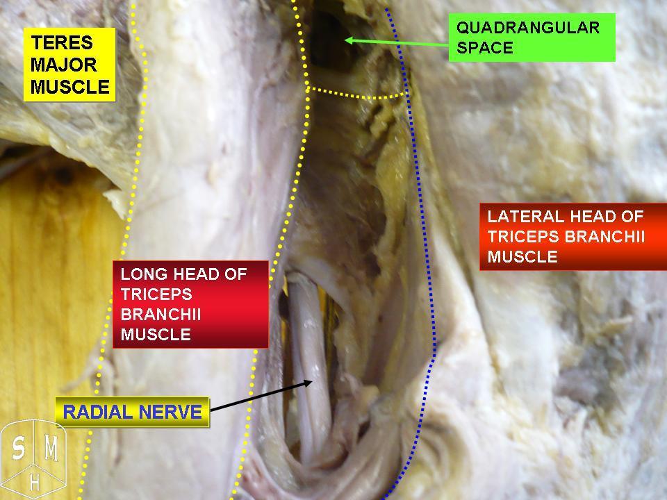
The axillary nerve or the circumflex nerve is a nerve of the human body, that originates from the brachial plexus ( upper trunk, posterior division, posterior cord) at the level of the axilla (armpit) and carries nerve fibers from C5 and C6. The axillary nerve travels through the quadrangular space with the posterior circumflex humeral artery and vein to innervate the deltoid and teres minor. Structure The nerve lies at first behind the axillary artery, and in front of the subscapularis, and passes downward to the lower border of that muscle. It then winds from anterior to posterior around the neck of the humerus, in company with the posterior humeral circumflex artery, through the quadrangular space (bounded above by the teres minor, below by the teres major, medially by the long head of the triceps brachii, and laterally by the surgical neck of the humerus), and divides into an anterior, a posterior, and a collateral branch to the long head of the triceps brachii bra ... [...More Info...] [...Related Items...] OR: [Wikipedia] [Google] [Baidu] |
Radial Nerve
The radial nerve is a nerve in the human body that supplies the posterior portion of the upper limb. It innervates the medial and lateral heads of the triceps brachii muscle of the arm, as well as all 12 muscles in the posterior osteofascial compartment of the forearm and the associated joints and overlying skin. It originates from the brachial plexus, carrying fibers from the ventral roots of spinal nerves C5, C6, C7, C8 & T1. The radial nerve and its branches provide motor innervation to the dorsal arm muscles (the triceps brachii and the anconeus) and the extrinsic extensors of the wrists and hands; it also provides cutaneous sensory innervation to most of the back of the hand, except for the back of the little finger and adjacent half of the ring finger (which are innervated by the ulnar nerve). The radial nerve divides into a deep branch, which becomes the posterior interosseous nerve, and a superficial branch, which goes on to innervate the dorsum (back) of the hand ... [...More Info...] [...Related Items...] OR: [Wikipedia] [Google] [Baidu] |
Subscapularis
The subscapularis is a large triangular muscle which fills the subscapular fossa and inserts into the lesser tubercle of the humerus and the front of the capsule of the shoulder-joint. Structure It arises from its medial two-thirds and Some fibers arise from tendinous laminae, which intersect the muscle and are attached to ridges on the bone; others from an aponeurosis, which separates the muscle from the teres major and the long head of the triceps brachii. The fibers pass laterally and coalesce into a tendon that is inserted into the lesser tubercle of the humerus and the anterior part of the shoulder-joint capsule. Tendinous fibers extend to the greater tubercle with insertions into the bicipital groove. Relations The tendon of the muscle is separated from the neck of the scapula by a large bursa, which communicates with the cavity of the shoulder-joint through an aperture in the capsule. The subscapularis is separated from the serratus anterior books.google.com/books?i ... [...More Info...] [...Related Items...] OR: [Wikipedia] [Google] [Baidu] |
Rotator Cuff
The rotator cuff is a group of muscles and their tendons that act to stabilize the human shoulder and allow for its extensive range of motion. Of the seven scapulohumeral muscles, four make up the rotator cuff. The four muscles are the supraspinatus muscle, the infraspinatus muscle, teres minor muscle, and the subscapularis muscle. Structure Muscles composing rotator cuff The supraspinatus muscle spreads out in a horizontal band to insert on the superior facet of the greater tubercle of the humerus. The greater tubercle projects as the most lateral structure of the humeral head. Medial to this, in turn, is the lesser tubercle of the humeral head. The subscapularis muscle origin is divided from the remainder of the rotator cuff origins as it is deep to the scapula. The four tendons of these muscles converge to form the rotator cuff tendon. These tendinous insertions along with the articular capsule, the coracohumeral ligament, and the glenohumeral ligament complex, ... [...More Info...] [...Related Items...] OR: [Wikipedia] [Google] [Baidu] |
Teres Major
The teres major muscle is a muscle of the upper limb. It attaches to the scapula and the humerus and is one of the seven scapulohumeral muscles. It is a thick but somewhat flattened muscle. The teres major muscle (from Latin ''teres'', meaning "rounded") is positioned above the latissimus dorsi muscle and assists in the extension and medial rotation of the humerus. This muscle is commonly confused as a rotator cuff muscle, but it is not because it does not attach to the capsule of the shoulder joint, unlike the teres minor muscle for example. Structure The teres major muscle originates on the dorsal surface of the inferior angle and the lower part of the lateral border of the scapula. The fibers of teres major insert into the medial lip of the intertubercular sulcus of the humerus. Relations The tendon, at its insertion, lies behind that of the latissimus dorsi, from which it is separated by a bursa, the two tendons being, however, united along their lower borders for a sh ... [...More Info...] [...Related Items...] OR: [Wikipedia] [Google] [Baidu] |
Latissimus Dorsi
The latissimus dorsi () is a large, flat muscle on the back that stretches to the sides, behind the arm, and is partly covered by the trapezius on the back near the midline. The word latissimus dorsi (plural: ''latissimi dorsorum'') comes from Latin and means "broadest uscleof the back", from "latissimus" ( la, broadest)' and "dorsum" ( la, back). The pair of muscles are commonly known as "lats", especially among bodybuilders. The latissimus dorsi is the largest muscle in the upper body. The latissimus dorsi is responsible for extension, adduction, transverse extension also known as horizontal abduction (or horizontal extension), flexion from an extended position, and (medial) internal rotation of the shoulder joint. It also has a synergistic role in extension and lateral flexion of the lumbar spine. Due to bypassing the scapulothoracic joints and attaching directly to the spine, the actions the latissimi dorsi have on moving the arms can also influence the movement of the ... [...More Info...] [...Related Items...] OR: [Wikipedia] [Google] [Baidu] |
Shoulder
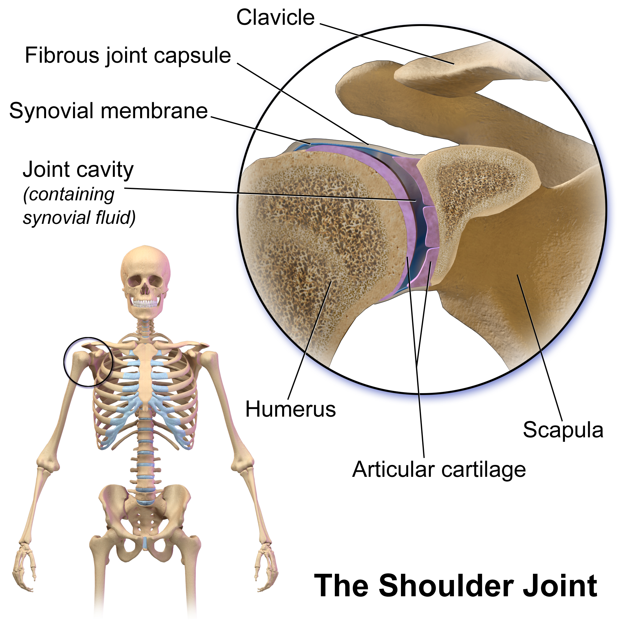
The human shoulder is made up of three bones: the clavicle (collarbone), the scapula (shoulder blade), and the humerus (upper arm bone) as well as associated muscles, ligaments and tendons. The articulations between the bones of the shoulder make up the shoulder joints. The shoulder joint, also known as the glenohumeral joint, is the major joint of the shoulder, but can more broadly include the acromioclavicular joint. In human anatomy, the shoulder joint comprises the part of the body where the humerus attaches to the scapula, and the head sits in the glenoid cavity. The shoulder is the group of structures in the region of the joint. The shoulder joint is the main joint of the shoulder. It is a ball and socket joint that allows the arm to rotate in a circular fashion or to hinge out and up away from the body. The joint capsule is a soft tissue envelope that encircles the glenohumeral joint and attaches to the scapula, humerus, and head of the biceps. It is lined by a thin, ... [...More Info...] [...Related Items...] OR: [Wikipedia] [Google] [Baidu] |
Deltoid Muscle
The deltoid muscle is the muscle forming the rounded contour of the human shoulder. It is also known as the 'common shoulder muscle', particularly in other animals such as the domestic cat. Anatomically, the deltoid muscle appears to be made up of three distinct sets of muscle fibers, namely the # anterior or clavicular part (pars clavicularis) # posterior or scapular part (pars scapularis) # intermediate or acromial part (pars acromialis) However, electromyography suggests that it consists of at least seven groups that can be independently coordinated by the nervous system. It was previously called the deltoideus (plural ''deltoidei'') and the name is still used by some anatomists. It is called so because it is in the shape of the Greek capital letter delta (Δ). Deltoid is also further shortened in slang as "delt". A study of 30 shoulders revealed an average mass of in humans, ranging from to . Structure Previous studies showed that the insertions of the tendons of the delt ... [...More Info...] [...Related Items...] OR: [Wikipedia] [Google] [Baidu] |


