|
Petrosquamous Suture
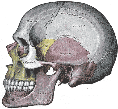
The petrosquamous suture is a cranial suture between the petrous portion and the squama of the temporal bone. It forms the Koerner's septum. The petrous portion forms the medial component of the osseous margin, while the squama forms the lateral component. The anterolateral portion (squama) arises from the mesenchyme at 8 weeks of embryogenesis while the petromastoid portion develops later from a cartilaginous center at 6 months of fetal development. In certain people, it can contain an emissary vein, referred to as the ''petrosquamosal sinus''. Being aware of this anatomic variant with preoperative CT scanning can be important to prevent bleeding in certain types of otolaryngological surgeries. Some authors have theorized that a persistent venous sinus reflects an arrest in embryologic development. See also * Petrotympanic fissure The petrotympanic fissure (also known as the squamotympanic fissure or the glaserian fissure) is a fissure in the temporal bone that runs from th ... [...More Info...] [...Related Items...] OR: [Wikipedia] [Google] [Baidu] |
Temporal Bone
The temporal bones are situated at the sides and base of the skull, and lateral to the temporal lobes of the cerebral cortex. The temporal bones are overlaid by the sides of the head known as the temples, and house the structures of the ears. The lower seven cranial nerves and the major vessels to and from the brain traverse the temporal bone. Structure The temporal bone consists of four parts— the squamous, mastoid, petrous and tympanic parts. The squamous part is the largest and most superiorly positioned relative to the rest of the bone. The zygomatic process is a long, arched process projecting from the lower region of the squamous part and it articulates with the zygomatic bone. Posteroinferior to the squamous is the mastoid part. Fused with the squamous and mastoid parts and between the sphenoid and occipital bones lies the petrous part, which is shaped like a pyramid. The tympanic part is relatively small and lies inferior to the squamous part, anterior to the mast ... [...More Info...] [...Related Items...] OR: [Wikipedia] [Google] [Baidu] |
Cranial Suture
In anatomy, fibrous joints are joints connected by Fibrous connective tissue, fibrous tissue, consisting mainly of collagen. These are fixed joints where bones are united by a layer of white fibrous tissue of varying thickness. In the skull the joints between the bones are called Suture (anatomy), sutures. Such immovable joints are also referred to as synarthrosis, synarthroses. Types Most fibrous joints are also called "fixed" or "immovable". These joints have no joint cavity and are connected via fibrous connective tissue. The skull bones are connected by fibrous joints called ''#Sutures, sutures''. In fetus, fetal skulls the sutures are wide to allow slight movement during birth. They later become rigid (synarthrosis, synarthrodial). Some of the long bones in the body such as the radius (bone), radius and ulna in the forearm are joined by a ''#Syndesmosis, syndesmosis'' (along the interosseous membrane of forearm, interosseous membrane). Syndemoses are slightly moveable (amph ... [...More Info...] [...Related Items...] OR: [Wikipedia] [Google] [Baidu] |
Petrous Portion Of The Temporal Bone
The petrous part of the temporal bone is pyramid-shaped and is wedged in at the base of the skull between the sphenoid and occipital bones. Directed medially, forward, and a little upward, it presents a base, an apex, three surfaces, and three angles, and houses in its interior, the components of the inner ear. The petrous portion is among the most basal elements of the skull and forms part of the endocranium. Petrous comes from the Latin word ''petrosus'', meaning "stone-like, hard". It is one of the densest bones in the body. The petrous bone is important for studies of ancient DNA from skeletal remains, as it tends to contain extremely well-preserved DNA. Base The base is fused with the internal surfaces of the squamous and mastoid parts. Apex The apex, which is rough and uneven, is received into the angular interval between the posterior border of the great wing of the sphenoid bone and the basilar part of the occipital bone; it presents the anterior or internal openin ... [...More Info...] [...Related Items...] OR: [Wikipedia] [Google] [Baidu] |
Squama Temporalis
The squamous part of temporal bone, or temporal squama, forms the front and upper part of the temporal bone, and is scale-like, thin, and translucent. Surfaces Its outer surface is smooth and convex; it affords attachment to the temporal muscle, and forms part of the temporal fossa; on its hinder part is a vertical groove for the middle temporal artery. A curved line, the ''temporal line'', or ''supramastoid crest'', runs backward and upward across its posterior part; it serves for the attachment of the temporal fascia, and limits the origin of the temporalis muscle. The boundary between the squamous part and the mastoid portion of the bone, as indicated by traces of the original suture, lies about 1 cm. below this line. Projecting from the lower part of the squamous part is a long, arched process, the ''zygomatic process''. This process is at first directed lateralward, its two surfaces looking upward and downward; it then appears as if twisted inward upon itself, and runs ... [...More Info...] [...Related Items...] OR: [Wikipedia] [Google] [Baidu] |
Temporal Bone
The temporal bones are situated at the sides and base of the skull, and lateral to the temporal lobes of the cerebral cortex. The temporal bones are overlaid by the sides of the head known as the temples, and house the structures of the ears. The lower seven cranial nerves and the major vessels to and from the brain traverse the temporal bone. Structure The temporal bone consists of four parts— the squamous, mastoid, petrous and tympanic parts. The squamous part is the largest and most superiorly positioned relative to the rest of the bone. The zygomatic process is a long, arched process projecting from the lower region of the squamous part and it articulates with the zygomatic bone. Posteroinferior to the squamous is the mastoid part. Fused with the squamous and mastoid parts and between the sphenoid and occipital bones lies the petrous part, which is shaped like a pyramid. The tympanic part is relatively small and lies inferior to the squamous part, anterior to the mast ... [...More Info...] [...Related Items...] OR: [Wikipedia] [Google] [Baidu] |
Koerner's Septum
Koerner's septum is an anatomic boundary in the temporal bone formed by the petrosquamous suture between the petrous and squamosal portions of the mastoid air cells, at the anatomic level of the antrum. Along with the middle ear ossicles, it is usually eroded in middle ear cholesteatomas. Superiorly, this continues as the petrosquamous suture, a normal anatomic structure that can be mistaken for fractures on temporal bone CT. It is surgically important as it may cause difficulty in locating the antrum and the deeper cells and thus may lead to incomplete removal of disease at mastoidectomy. See also *Temporal bone *Temporal fenestrae *Temporal muscle *Temporomandibular joint In anatomy, the temporomandibular joints (TMJ) are the two joints connecting the jawbone to the skull. It is a bilateral synovial articulation between the temporal bone of the skull above and the mandible below; it is from these bones that it ... References External links * {{Cranium Bones of the ... [...More Info...] [...Related Items...] OR: [Wikipedia] [Google] [Baidu] |
Mesenchyme
Mesenchyme () is a type of loosely organized animal embryonic connective tissue of undifferentiated cells that give rise to most tissues, such as skin, blood or bone. The interactions between mesenchyme and epithelium help to form nearly every organ in the developing embryo. Vertebrates Structure Mesenchyme is characterized morphologically by a prominent ground substance matrix containing a loose aggregate of reticular fibers and unspecialized mesenchymal stem cells. Mesenchymal cells can migrate easily (in contrast to epithelial cells, which lack mobility), are organized into closely adherent sheets, and are polarized in an apical-basal orientation. Development The mesenchyme originates from the mesoderm. From the mesoderm, the mesenchyme appears as an embryologically primitive "soup". This "soup" exists as a combination of the mesenchymal cells plus serous fluid plus the many different tissue proteins. Serous fluid is typically stocked with the many serous elements, such a ... [...More Info...] [...Related Items...] OR: [Wikipedia] [Google] [Baidu] |
Embryogenesis
An embryo is an initial stage of development of a multicellular organism. In organisms that reproduce sexually, embryonic development is the part of the life cycle that begins just after fertilization of the female egg cell by the male sperm cell. The resulting fusion of these two cells produces a single-celled zygote that undergoes many cell divisions that produce cells known as blastomeres. The blastomeres are arranged as a solid ball that when reaching a certain size, called a morula, takes in fluid to create a cavity called a blastocoel. The structure is then termed a blastula, or a blastocyst in mammals. The mammalian blastocyst hatches before implantating into the endometrial lining of the womb. Once implanted the embryo will continue its development through the next stages of gastrulation, neurulation, and organogenesis. Gastrulation is the formation of the three germ layers that will form all of the different parts of the body. Neurulation forms the nervous sys ... [...More Info...] [...Related Items...] OR: [Wikipedia] [Google] [Baidu] |
Emissary Vein
The emissary veins connect the extracranial venous system with the intracranial venous sinuses. They connect the veins outside the cranium to the venous sinuses inside the cranium. They drain from the scalp, through the human skull, skull, into the larger meningeal veins and dural venous sinuses. Emissary veins have an important role in selective cooling of the head. They also serve as routes where infections are carried into the cranial cavity from the extracranial veins to the intracranial veins. There are several types of emissary veins including posterior condyloid, mastoid, occipital and parietal emissary vein. Structure There are also emissary veins passing through the Foramen ovale (skull), foramen ovale, jugular foramen, foramen lacerum, and hypoglossal canal. Function Because the emissary veins are valveless, they are an important part in selective brain cooling through bidirectional flow of cooler blood from the evaporating surface of the head. In general, blood flow ... [...More Info...] [...Related Items...] OR: [Wikipedia] [Google] [Baidu] |
Otolaryngology
Otorhinolaryngology ( , abbreviated ORL and also known as otolaryngology, otolaryngology–head and neck surgery (ORL–H&N or OHNS), or ear, nose, and throat (ENT)) is a surgical subspeciality within medicine that deals with the surgical and medical management of conditions of the head and neck. Doctors who specialize in this area are called otorhinolaryngologists, otolaryngologists, head and neck surgeons, or ENT surgeons or physicians. Patients seek treatment from an otorhinolaryngologist for diseases of the ear, nose, throat, base of skull, base of the skull, head, and neck. These commonly include functional diseases that affect the senses and activities of eating, drinking, speaking, breathing, swallowing, and hearing. In addition, ENT surgery encompasses the surgical management of cancers and benign tumors and reconstruction of the head and neck as well as plastic surgery of the face and neck. Etymology The term is a combination of New Latin classical compound, combini ... [...More Info...] [...Related Items...] OR: [Wikipedia] [Google] [Baidu] |
Petrotympanic Fissure
The petrotympanic fissure (also known as the squamotympanic fissure or the glaserian fissure) is a fissure in the temporal bone that runs from the temporomandibular joint to the tympanic cavity. The mandibular fossa is bounded, in front, by the articular tubercle; behind, by the tympanic part of the bone, which separates it from the external acoustic meatus; it is divided into two parts by a narrow slit, the petrotympanic fissure. It opens just above and in front of the ring of bone into which the tympanic membrane is inserted; in this situation it is a mere slit about 2 mm. in length. It lodges the anterior process and anterior ligament of the malleus, and gives passage to the anterior tympanic branch of the internal maxillary artery. Eponym It is also known as the "Glaserian fissure", after Johann Glaser. Contents The contents of the fissure include communications of cranial nerve VII to the infratemporal fossa. A branch of cranial nerve VII, the chorda tympani, runs th ... [...More Info...] [...Related Items...] OR: [Wikipedia] [Google] [Baidu] |


