|
Panuveitis
Panuveitis also known as Diffuse uveitis or Total uveitis is an eye disease affecting the internal structures of the eye. In this inflammation occurs throughout the uveal tract, with no specific areas of predominant inflammation. In most cases, along with the uvea, the retina, vitreous humor, optic nerve or lens are also involved. Definition Panuitis is a type of uveitis in which inflammation occurs throughout the uveal tract, with no specific areas of predominant inflammation. In most cases, along with the uvea, the retina, vitreous humor, optic nerve or lens are involved. International Uveitis Study Group (IUSG) defines panuveitis as generalized inflammation of all three parts (iris, ciliary body and choroid) of the uvea. Causes In many cases the cause of panuveitis is unknown. Possible causes include exogenous or endogenous infection, injury, or an autoimmune disease. Endogenous infections caused by syphilis, tuberculosis, mumps, smallpox, influenza, toxoplasmosis, lupus, sar ... [...More Info...] [...Related Items...] OR: [Wikipedia] [Google] [Baidu] |
Cyclitis
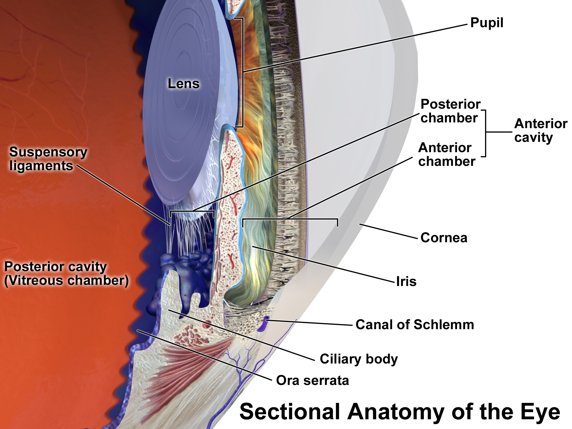
Uveitis () is inflammation of the uvea, the pigmented layer of the eye between the inner retina and the outer fibrous layer composed of the sclera and cornea. The uvea consists of the middle layer of pigmented vascular structures of the eye and includes the iris, ciliary body, and choroid. Uveitis is described anatomically, by the part of the eye affected, as anterior, intermediate or posterior, or panuveitic if all parts are involved. Anterior uveitis ( iridocyclytis) is the most common, with the incidence of uveitis overall affecting approximately 1:4500, most commonly those between the ages of 20-60. Symptoms include eye pain, eye redness, floaters and blurred vision, and ophthalmic examination may show dilated ciliary blood vessels and the presence of cells in the anterior chamber. Uveitis may arise spontaneously, have a genetic component, or be associated with an autoimmune disease or infection. While the eye is a relatively protected environment, its immune mechanisms m ... [...More Info...] [...Related Items...] OR: [Wikipedia] [Google] [Baidu] |
Iridocyclitis
Uveitis () is inflammation of the uvea, the pigmented layer of the eye between the inner retina and the outer fibrous layer composed of the sclera and cornea. The uvea consists of the middle layer of pigmented vascular structures of the eye and includes the iris, ciliary body, and choroid. Uveitis is described anatomically, by the part of the eye affected, as anterior, intermediate or posterior, or panuveitic if all parts are involved. Anterior uveitis ( iridocyclytis) is the most common, with the incidence of uveitis overall affecting approximately 1:4500, most commonly those between the ages of 20-60. Symptoms include eye pain, eye redness, floaters and blurred vision, and ophthalmic examination may show dilated ciliary blood vessels and the presence of cells in the anterior chamber. Uveitis may arise spontaneously, have a genetic component, or be associated with an autoimmune disease or infection. While the eye is a relatively protected environment, its immune mechanisms may ... [...More Info...] [...Related Items...] OR: [Wikipedia] [Google] [Baidu] |
Uveitis
Uveitis () is inflammation of the uvea, the pigmented layer of the eye between the inner retina and the outer fibrous layer composed of the sclera and cornea. The uvea consists of the middle layer of pigmented vascular structures of the eye and includes the iris, ciliary body, and choroid. Uveitis is described anatomically, by the part of the eye affected, as anterior, intermediate or posterior, or panuveitic if all parts are involved. Anterior uveitis ( iridocyclytis) is the most common, with the incidence of uveitis overall affecting approximately 1:4500, most commonly those between the ages of 20-60. Symptoms include eye pain, eye redness, floaters and blurred vision, and ophthalmic examination may show dilated ciliary blood vessels and the presence of cells in the anterior chamber. Uveitis may arise spontaneously, have a genetic component, or be associated with an autoimmune disease or infection. While the eye is a relatively protected environment, its immune mechanisms ... [...More Info...] [...Related Items...] OR: [Wikipedia] [Google] [Baidu] |
Vogt–Koyanagi–Harada Disease
Vogt–Koyanagi–Harada disease (VKH) is a multisystem disease of presumed autoimmune disease, autoimmune cause that affects melanin-pigmented tissues. The most significant manifestation is bilateral, diffuse uveitis, which affects the eyes. VKH may variably also involve the inner ear, with effects on hearing, the skin, and the meninges of the central nervous system. Signs and symptoms Overview The disease is characterised by bilateral diffuse uveitis, with pain, redness and vision loss, blurring of vision. The eye symptoms may be accompanied by a varying constellation of systemic symptoms, such as auditory (tinnitus, vertigo, and hypoacusis), neurological (meningismus, with malaise, fever, headache, nausea, abdominal pain, stiffness of the neck and back, or a combination of these factors; meningitis, Cerebrospinal fluid, CSF pleocytosis, cranial nerve palsy, palsies, hemiparesis, transverse myelitis and ciliary ganglionitis), and cutaneous manifestations, including poli ... [...More Info...] [...Related Items...] OR: [Wikipedia] [Google] [Baidu] |
Choroiditis
Chorioretinitis is an inflammation of the choroid (thin pigmented vascular coat of the eye) and retina of the eye. It is a form of posterior uveitis. If only the choroid is inflamed, not the retina, the condition is termed choroiditis. The ophthalmologist's goal in treating these potentially blinding conditions is to eliminate the inflammation and minimize the potential risk of therapy to the patient. Symptoms Symptoms may include the presence of floating black spots, blurred vision, pain or redness in the eye, sensitivity to light, or excessive tearing. Causes Chorioretinitis is often caused by toxoplasmosis and cytomegalovirus infections (mostly seen in immunodeficient subjects such as people with HIV/AIDS or on immunosuppressant drugs). Congenital toxoplasmosis via transplacental transmission can also lead to sequelae such as chorioretinitis along with hydrocephalus and cerebral calcifications. Other possible causes of chorioretinitis are syphilis, sarcoidosis, tuberculosis, Be ... [...More Info...] [...Related Items...] OR: [Wikipedia] [Google] [Baidu] |
Ophthalmology
Ophthalmology ( ) is a surgical subspecialty within medicine that deals with the diagnosis and treatment of eye disorders. An ophthalmologist is a physician who undergoes subspecialty training in medical and surgical eye care. Following a medical degree, a doctor specialising in ophthalmology must pursue additional postgraduate residency training specific to that field. This may include a one-year integrated internship that involves more general medical training in other fields such as internal medicine or general surgery. Following residency, additional specialty training (or fellowship) may be sought in a particular aspect of eye pathology. Ophthalmologists prescribe medications to treat eye diseases, implement laser therapy, and perform surgery when needed. Ophthalmologists provide both primary and specialty eye care - medical and surgical. Most ophthalmologists participate in academic research on eye diseases at some point in their training and many include research as part ... [...More Info...] [...Related Items...] OR: [Wikipedia] [Google] [Baidu] |
Influenza
Influenza, commonly known as "the flu", is an infectious disease caused by influenza viruses. Symptoms range from mild to severe and often include fever, runny nose, sore throat, muscle pain, headache, coughing, and fatigue. These symptoms begin from one to four days after exposure to the virus (typically two days) and last for about 2–8 days. Diarrhea and vomiting can occur, particularly in children. Influenza may progress to pneumonia, which can be caused by the virus or by a subsequent bacterial infection. Other complications of infection include acute respiratory distress syndrome, meningitis, encephalitis, and worsening of pre-existing health problems such as asthma and cardiovascular disease. There are four types of influenza virus, termed influenza viruses A, B, C, and D. Aquatic birds are the primary source of Influenza A virus (IAV), which is also widespread in various mammals, including humans and pigs. Influenza B virus (IBV) and Influenza C virus (ICV) pri ... [...More Info...] [...Related Items...] OR: [Wikipedia] [Google] [Baidu] |
Keratic Precipitate
Keratic precipitate (KP) is an inflammatory cellular deposit seen on corneal endothelium. Acute KPs are white and round in shape whereas old KPs are faded and irregular in shape. Mutton-fat KPs are large in shape and are greasy-white in color and are formed from macrophages and epithelioid cell According to a common point of view epithelioid cells (also called epithelioid histiocytes) are derivatives of activated macrophages resembling epithelial cells. Structure and function Structurally, epithelioid cells (when examined by light mi ...s. They are indicative of inflammatory disease. Mutton fat KPs are due to granulomatous iridocyclitis. Another variant called red KPs may be seen in hemorrhagic uveitis. References Disorders of sclera and cornea Inflammations {{eye-stub ... [...More Info...] [...Related Items...] OR: [Wikipedia] [Google] [Baidu] |
Slit Lamp
A slit lamp is an instrument consisting of a high-intensity light source that can be focused to shine a thin sheet of light into the eye. It is used in conjunction with a biomicroscope. The lamp facilitates an examination of the anterior segment and posterior segment of the human eye, which includes the eyelid, sclera, conjunctiva, iris, natural crystalline lens, and cornea. The binocular slit-lamp examination provides a stereoscopic magnified view of the eye structures in detail, enabling anatomical diagnoses to be made for a variety of eye conditions. A second, hand-held lens is used to examine the retina. History Two conflicting trends emerged in the development of the slit lamp. One trend originated from clinical research and aimed to apply the increasingly complex and advanced technology of the time. [...More Info...] [...Related Items...] OR: [Wikipedia] [Google] [Baidu] |
Floater
Floaters or eye floaters are sometimes visible deposits (e.g., the shadows of tiny structures of protein or other cell debris projected onto the retina) within the eye's vitreous humour ("the vitreous"), which is normally transparent, or between the vitreous and retina.Cline D; Hofstetter HW; Griffin JR. ''Dictionary of Visual Science''. 4th ed. Butterworth-Heinemann, Boston 1997. They can become particularly noticeable when looking at a blank surface or an open monochromatic space, such as blue sky. Each floater can be measured by its size, shape, consistency, refractive index, and motility. They are also called ''muscae volitantes'' (Latin for 'flying flies'), or ''mouches volantes'' (from the same phrase in French). The vitreous usually starts out transparent, but imperfections may gradually develop as one ages. The common type of floater, present in most people's eyes, is due to these degenerative changes of the vitreous. The perception of floaters, which may be annoying ... [...More Info...] [...Related Items...] OR: [Wikipedia] [Google] [Baidu] |
Glaucoma Surgery
Glaucoma is a group of diseases affecting the optic nerve that results in vision loss and is frequently characterized by raised intraocular pressure (IOP). There are many glaucoma surgeries, and variations or combinations of those surgeries, that facilitate the escape of excess aqueous humor from the eye to lower intraocular pressure, and a few that lower IOP by decreasing the production of aqueous humor. Procedures that facilitate outflow of aqueous humor Laser trabeculoplasty A trabeculoplasty is a modification of the trabecular meshwork. Laser trabeculoplasty (LTP) is the application of a laser beam to burn areas of the trabecular meshwork, located near the base of the iris, to increase fluid outflow. LTP is used in the treatment of various open-angle glaucomas. The two types of laser trabeculoplasty are argon laser trabeculoplasty (ALT) and selective laser trabeculoplasty (SLT). As its name suggests, argon laser trabeculoplasty uses an argon laser to create tiny burns on the t ... [...More Info...] [...Related Items...] OR: [Wikipedia] [Google] [Baidu] |
Cataract Surgery
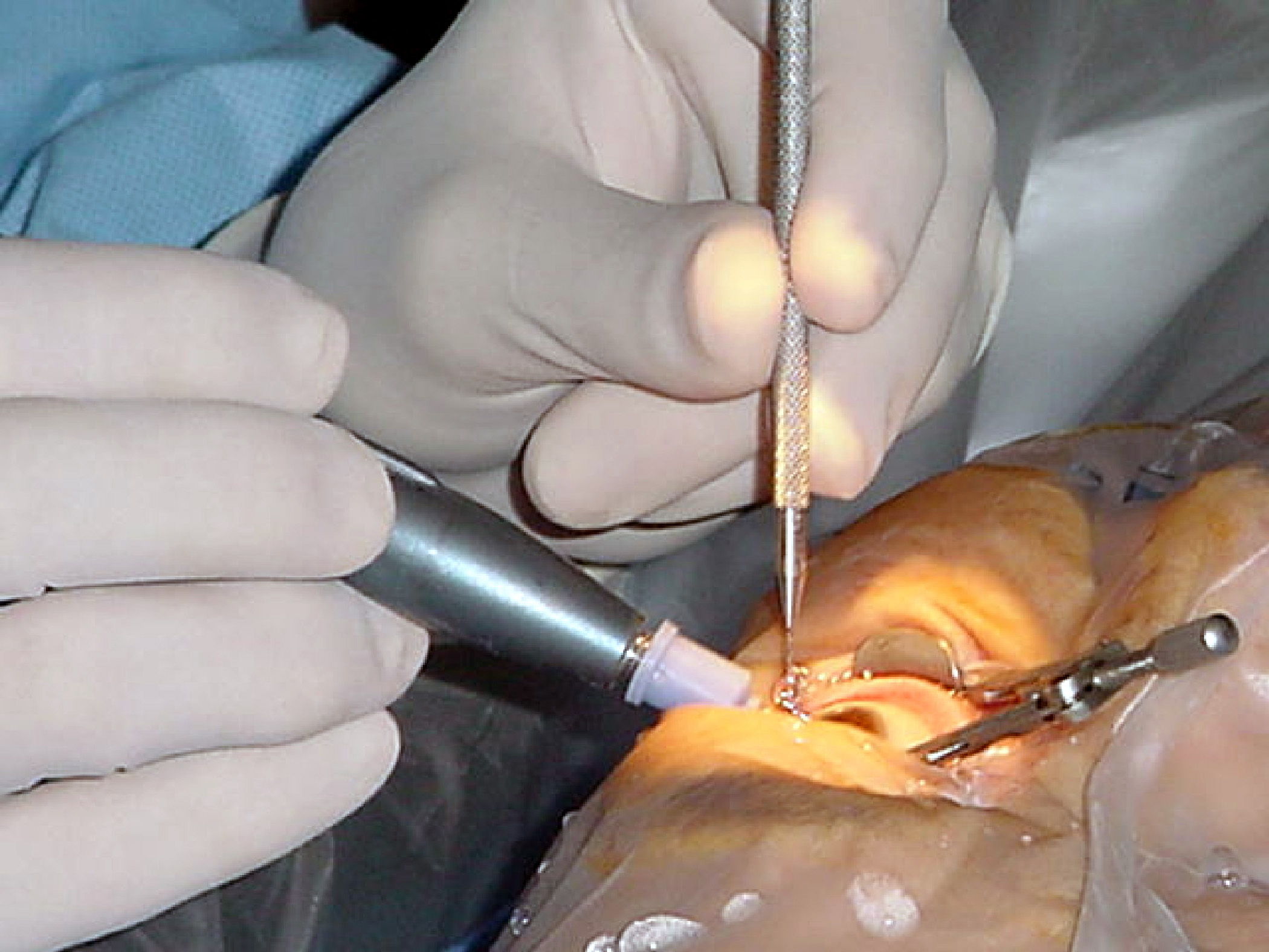
Cataract surgery, also called lens replacement surgery, is the removal of the natural lens of the eye (also called "crystalline lens") that has developed an opacification, which is referred to as a cataract, and its replacement with an intraocular lens. Metabolic changes of the crystalline lens fibers over time lead to the development of the cataract, causing impairment or loss of vision. Some infants are born with congenital cataracts, and certain environmental factors may also lead to cataract formation. Early symptoms may include strong glare from lights and small light sources at night, and reduced acuity at low light levels. During cataract surgery, a patient's cloudy natural cataract lens is removed, either by emulsification in place or by cutting it out. An artificial intraocular lens (IOL) is implanted in its place. Cataract surgery is generally performed by an ophthalmologist in an ambulatory setting at a surgical center or hospital rather than an inpatient setting. Eit ... [...More Info...] [...Related Items...] OR: [Wikipedia] [Google] [Baidu] |


