|
Polar Body Biopsy
Polar body biopsy is the sampling of a polar body of an oocyte. It was first applied clinically in humans in 1987 after extensive animal studies. A ''polar body'' is a small haploid cell that is formed concomitantly as an egg cell during oogenesis, but which generally does not have the ability to be fertilized. After sampling of a polar body, subsequent analysis can be used to predict viability and pregnancy chance of the oocyte, as well as the future health of a person resulting from such a pregnancy. The latter use makes it a form of preimplantation genetic screening (PGS). Compared to a blastocyst biopsy, a polar body biopsy can potentially be of lower costs, less harmful side-effects, and more sensitive in detecting abnormalities."Delivery of a chromosomally normal child from an oocyte with reciprocal aneuploid polar bodies". Scott Jr, Richard T., Nathan R. Treff, John Stevens, Eric J. Forman, Kathleen H. Hong, Mandy G. Katz-Jaffe, William B. Schoolcraft. Journal of Assisted R ... [...More Info...] [...Related Items...] OR: [Wikipedia] [Google] [Baidu] |
Sampling (medicine)
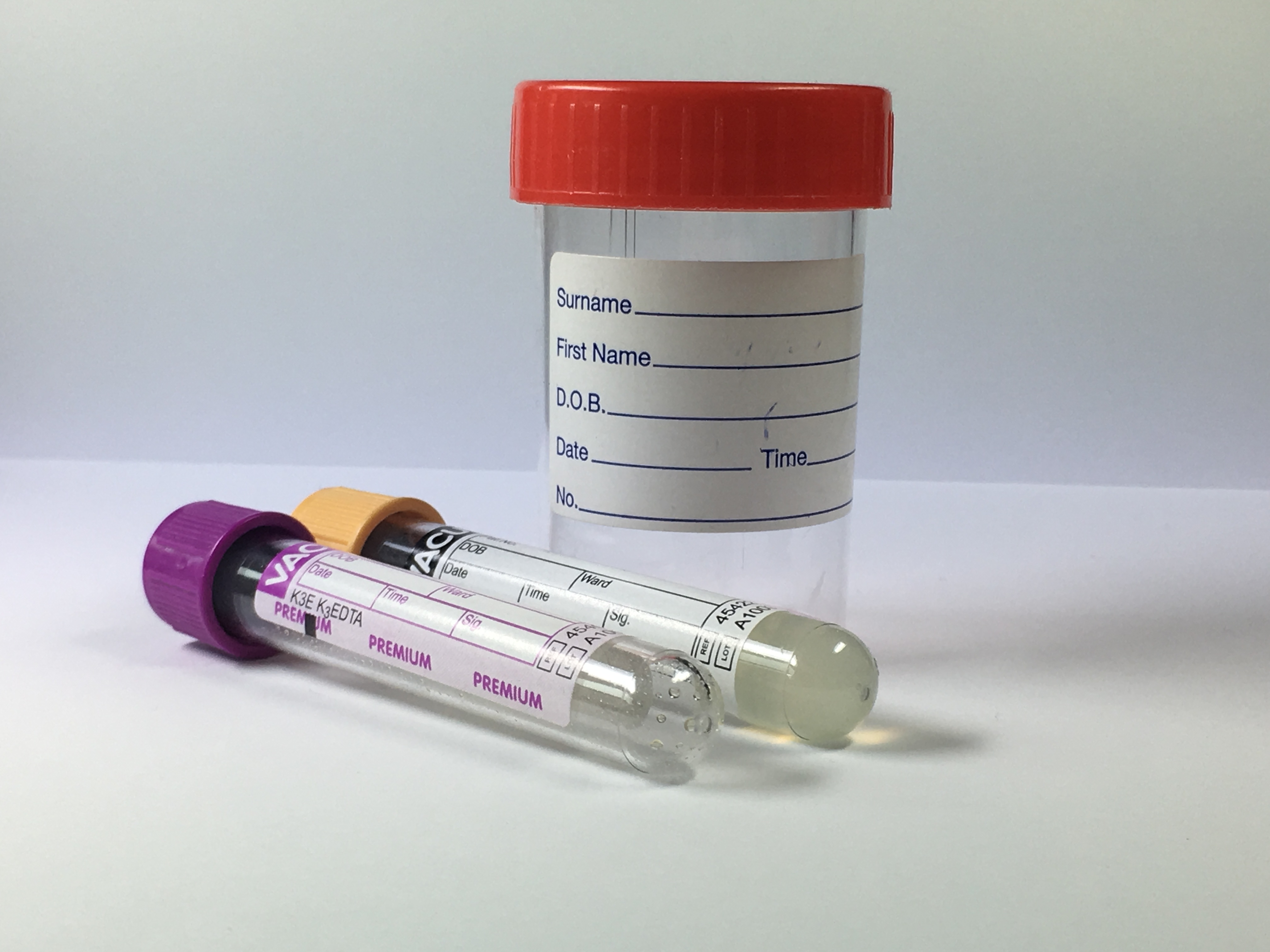
In medicine, sampling is gathering of matter from the body to aid in the process of a medical diagnosis and/or evaluation of an indication for treatment, further medical tests or other procedures. In this sense, the sample is the gathered matter, and the sampling tool or sampler is the person or material to collect the sample. Sampling is a prerequisite for many medical tests, but generally not for medical history, physical examination and radiologic tests. By sampling technique *Obtaining excretions or materials that leave the body anyway, such as urine, stool, sputum, or vomitus, by direct collection as they exit. A sample of saliva can also be collected from the mouth. Excision (cutting out), a surgical method for the removal of solid or soft tissue samples. *Puncture (also called ''centesis'') followed by aspiration is the main method used for sampling of many types of tissues and body fluids. Examples are thoracocentesis to sample pleural fluid, and amniocentesis to ... [...More Info...] [...Related Items...] OR: [Wikipedia] [Google] [Baidu] |
Fluorescence In Situ Hybridization
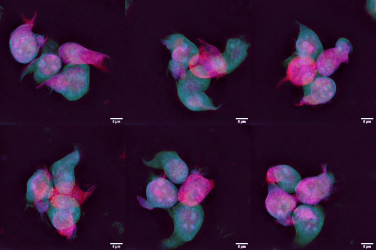
Fluorescence ''in situ'' hybridization (FISH) is a molecular cytogenetic technique that uses fluorescent probes that bind to only particular parts of a nucleic acid sequence with a high degree of sequence complementarity. It was developed by biomedical researchers in the early 1980s to detect and localize the presence or absence of specific DNA sequences on chromosomes. Fluorescence microscopy can be used to find out where the fluorescent probe is bound to the chromosomes. FISH is often used for finding specific features in DNA for use in genetic counseling, medicine, and species identification. FISH can also be used to detect and localize specific RNA targets (mRNA, lncRNA and miRNA) in cells, circulating tumor cells, and tissue samples. In this context, it can help define the spatial-temporal patterns of gene expression within cells and tissues. Probes – RNA and DNA In biology, a probe is a single strand of DNA or RNA that is complementary to a nucleotide sequence o ... [...More Info...] [...Related Items...] OR: [Wikipedia] [Google] [Baidu] |
Euploid
Ploidy () is the number of complete sets of chromosomes in a cell, and hence the number of possible alleles for autosomal and pseudoautosomal genes. Sets of chromosomes refer to the number of maternal and paternal chromosome copies, respectively, in each homologous chromosome pair, which chromosomes naturally exist as. Somatic cells, tissues, and individual organisms can be described according to the number of sets of chromosomes present (the "ploidy level"): monoploid (1 set), diploid (2 sets), triploid (3 sets), tetraploid (4 sets), pentaploid (5 sets), hexaploid (6 sets), heptaploid or septaploid (7 sets), etc. The generic term polyploid is often used to describe cells with three or more chromosome sets. Virtually all sexually reproducing organisms are made up of somatic cells that are diploid or greater, but ploidy level may vary widely between different organisms, between different tissues within the same organism, and at different stages in an organism's life cycle. Half ... [...More Info...] [...Related Items...] OR: [Wikipedia] [Google] [Baidu] |
Chromosomes
A chromosome is a long DNA molecule with part or all of the genetic material of an organism. In most chromosomes the very long thin DNA fibers are coated with packaging proteins; in eukaryotic cells the most important of these proteins are the histones. These proteins, aided by chaperone proteins, bind to and condense the DNA molecule to maintain its integrity. These chromosomes display a complex three-dimensional structure, which plays a significant role in transcriptional regulation. Chromosomes are normally visible under a light microscope only during the metaphase of cell division (where all chromosomes are aligned in the center of the cell in their condensed form). Before this happens, each chromosome is duplicated (S phase), and both copies are joined by a centromere, resulting either in an X-shaped structure (pictured above), if the centromere is located equatorially, or a two-arm structure, if the centromere is located distally. The joined copies are now called sis ... [...More Info...] [...Related Items...] OR: [Wikipedia] [Google] [Baidu] |
Chromatids
A chromatid (Greek ''khrōmat-'' 'color' + ''-id'') is one half of a duplicated chromosome. Before replication, one chromosome is composed of one DNA molecule. In replication, the DNA molecule is copied, and the two molecules are known as chromatids. During the later stages of cell division these chromatids separate longitudinally to become individual chromosomes. Chromatid pairs are normally genetically identical, and said to be homozygous. However, if mutations occur, they will present slight differences, in which case they are heterozygous. The pairing of chromatids should not be confused with the ploidy of an organism, which is the number of homologous versions of a chromosome. Sister chromatids Chromatids may be sister or non-sister chromatids. A sister chromatid is either one of the two chromatids of the same chromosome joined together by a common centromere. A pair of sister chromatids is called a dyad. Once sister chromatids have separated (during the anaphase of m ... [...More Info...] [...Related Items...] OR: [Wikipedia] [Google] [Baidu] |
Aneuploidy
Aneuploidy is the presence of an abnormal number of chromosomes in a cell, for example a human cell having 45 or 47 chromosomes instead of the usual 46. It does not include a difference of one or more complete sets of chromosomes. A cell with any number of complete chromosome sets is called a ''euploid'' cell. An extra or missing chromosome is a common cause of some genetic disorders. Some cancer cells also have abnormal numbers of chromosomes. About 68% of human solid tumors are aneuploid. Aneuploidy originates during cell division when the chromosomes do not separate properly between the two cells (nondisjunction). Most cases of aneuploidy in the autosomes result in miscarriage, and the most common extra autosomal chromosomes among live births are 21, 18 and 13. Chromosome abnormalities are detected in 1 of 160 live human births. Autosomal aneuploidy is more dangerous than sex chromosome aneuploidy, as autosomal aneuploidy is almost always lethal to embryos that cease develo ... [...More Info...] [...Related Items...] OR: [Wikipedia] [Google] [Baidu] |
Epigenetic
In biology, epigenetics is the study of stable phenotypic changes (known as ''marks'') that do not involve alterations in the DNA sequence. The Greek prefix '' epi-'' ( "over, outside of, around") in ''epigenetics'' implies features that are "on top of" or "in addition to" the traditional genetic basis for inheritance. Epigenetics most often involves changes that affect the regulation of gene expression, but the term can also be used to describe any heritable phenotypic change. Such effects on cellular and physiological phenotypic traits may result from external or environmental factors, or be part of normal development. The term also refers to the mechanism of changes: functionally relevant alterations to the genome that do not involve mutation of the nucleotide sequence. Examples of mechanisms that produce such changes are DNA methylation and histone modification, each of which alters how genes are expressed without altering the underlying DNA sequence. Gene expression can ... [...More Info...] [...Related Items...] OR: [Wikipedia] [Google] [Baidu] |
Microarrays
A microarray is a multiplex lab-on-a-chip. Its purpose is to simultaneously detect the expression of thousands of genes from a sample (e.g. from a tissue). It is a two-dimensional array on a solid substrate—usually a glass slide or silicon thin-film cell—that assays (tests) large amounts of biological material using high-throughput screening miniaturized, multiplexed and parallel processing and detection methods. The concept and methodology of microarrays was first introduced and illustrated in antibody microarrays (also referred to as antibody matrix) by Tse Wen Chang in 1983 in a scientific publication and a series of patents. The "gene chip" industry started to grow significantly after the 1995 ''Science Magazine'' article by the Ron Davis and Pat Brown labs at Stanford University. With the establishment of companies, such as Affymetrix, Agilent, Applied Microarrays, Arrayjet, Illumina, and others, the technology of DNA microarrays has become the most sophisticated and ... [...More Info...] [...Related Items...] OR: [Wikipedia] [Google] [Baidu] |
Metaphase
Metaphase ( and ) is a stage of mitosis in the eukaryotic cell cycle in which chromosomes are at their second-most condensed and coiled stage (they are at their most condensed in anaphase). These chromosomes, carrying genetic information, align in the equator of the cell before being separated into each of the two daughter cells. Metaphase accounts for approximately 4% of the cell cycle's duration. Preceded by events in prometaphase and followed by anaphase, microtubules formed in prophase have already found and attached themselves to kinetochores in metaphase. In metaphase, the centromeres of the chromosomes convene themselves on the ''metaphase plate'' (or ''equatorial plate''), an imaginary line that is equidistant from the two centrosome poles. This even alignment is due to the counterbalance of the pulling powers generated by the opposing kinetochore microtubules, analogous to a tug-of-war between two people of equal strength, ending with the destruction of B cyclin. In ce ... [...More Info...] [...Related Items...] OR: [Wikipedia] [Google] [Baidu] |
Lymphocytes
A lymphocyte is a type of white blood cell (leukocyte) in the immune system of most vertebrates. Lymphocytes include natural killer cells (which function in cell-mediated, cytotoxic innate immunity), T cells (for cell-mediated, cytotoxic adaptive immunity), and B cells (for humoral, antibody-driven adaptive immunity). They are the main type of cell found in lymph, which prompted the name "lymphocyte". Lymphocytes make up between 18% and 42% of circulating white blood cells. Types The three major types of lymphocyte are T cells, B cells and natural killer (NK) cells. Lymphocytes can be identified by their large nucleus. T cells and B cells T cells (thymus cells) and B cells ( bone marrow- or bursa-derived cells) are the major cellular components of the adaptive immune response. T cells are involved in cell-mediated immunity, whereas B cells are primarily responsible for humoral immunity (relating to antibodies). The function of T cells and B cells is to recognize specif ... [...More Info...] [...Related Items...] OR: [Wikipedia] [Google] [Baidu] |
Zygote
A zygote (, ) is a eukaryotic cell formed by a fertilization event between two gametes. The zygote's genome is a combination of the DNA in each gamete, and contains all of the genetic information of a new individual organism. In multicellular organisms, the zygote is the earliest developmental stage. In humans and most other anisogamous organisms, a zygote is formed when an egg cell and sperm cell come together to create a new unique organism. In single-celled organisms, the zygote can divide asexually by mitosis to produce identical offspring. German zoologists Oscar and Richard Hertwig made some of the first discoveries on animal zygote formation in the late 19th century. Humans In human fertilization, a released ovum (a haploid secondary oocyte with replicate chromosome copies) and a haploid sperm cell (male gamete) combine to form a single diploid cell called the zygote. Once the single sperm fuses with the oocyte, the latter completes the division of the second ... [...More Info...] [...Related Items...] OR: [Wikipedia] [Google] [Baidu] |


