|
Piriformis
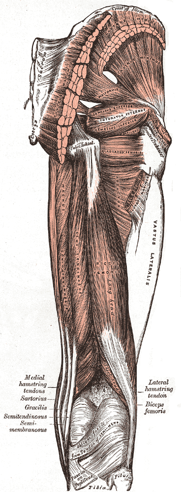
The piriformis muscle () is a flat, pyramidally-shaped muscle in the gluteal region of the lower limbs. It is one of the six muscles in the lateral rotator group. The piriformis muscle has its origin upon the front surface of the sacrum, and inserts onto the greater trochanter of the femur. Depending upon the given position of the leg, it acts either as external (lateral) rotator of the thigh or as abductor of the thigh. It is innervated by the piriformis nerve. Structure The piriformis is a flat muscle, and is pyramidal in shape. Origin The piriformis muscle originates from the anterior (front) surface of the sacrum by three fleshy digitations attached to the second, third, and fourth sacral vertebra. It also arises from the superior margin of the greater sciatic notch, the gluteal surface of the ilium (near the posterior inferior iliac spine), the sacroiliac joint capsule, and (sometimes) the sacrotuberous ligament (more specifically, the superior part of the pelvic su ... [...More Info...] [...Related Items...] OR: [Wikipedia] [Google] [Baidu] |
Piriformis Nerve
The piriformis nerve, also known as the nerve to piriformis, is the peripheral nerve that innervates the piriformis muscle. Structure The piriformis nerve to piriformis originates from the ventral rami of S1 and S2 in the sacral plexus. It enters the anterior surface of the piriformis muscle. This nerve may be double. This nerve is not to be confused with the inferior gluteal nerve, which also arises from posterior divisions of the first and second sacral ventral rami (L5, S1, S2). Function The piriformis nerve innervates the piriformis muscle. See also * Piriformis * Sacral plexus In human anatomy, the sacral plexus is a nerve plexus which provides motor and sensory nerves for the posterior thigh, most of the lower leg and foot, and part of the pelvis. It is part of the lumbosacral plexus and emerges from the lumbar ver ... References 홍재기 Nerves of the lower limb and lower torso {{neuroanatomy-stub ... [...More Info...] [...Related Items...] OR: [Wikipedia] [Google] [Baidu] |
Inferior Gluteal Artery
The inferior gluteal artery (sciatic artery), the smaller of the two terminal branches of the anterior trunk of the internal iliac artery, is distributed chiefly to the buttock and back of the thigh. It passes down on the sacral plexus of nerves and the piriformis muscle, behind the internal pudendal artery. It passes through the lower part of the greater sciatic foramen. It escapes from the pelvis between piriformis muscle and coccygeus muscle. It then descends in the interval between the greater trochanter of the femur and tuberosity of the ischium. It is accompanied by the sciatic nerve and the posterior femoral cutaneous nerves The posterior cutaneous nerve of the thigh (also called the posterior femoral cutaneous nerve) is a sensory nerve in the thigh. It supplies the skin of the posterior surface of the thigh, leg, buttock, and also the perineum. Structure ..., and covered by the gluteus maximus. It continues down the back of the thigh, supplying the sk ... [...More Info...] [...Related Items...] OR: [Wikipedia] [Google] [Baidu] |
Superior Gluteal Artery
The superior gluteal artery is the largest and final branch of the internal iliac artery. It is the continuation of the posterior division of that vessel. It is a short artery which runs backward between the lumbosacral trunk and the first sacral nerve. It divides into a superficial and a deep branch after passing out of the pelvis above the upper border of the piriformis muscle. Within the pelvis, it gives off branches to the iliacus, piriformis, and obturator internus muscles. Just previous to exiting the pelvic cavity, it also gives off a nutrient artery which enters the ilium. Structure The superior gluteal artery is the largest and final branch of the internal iliac artery. It branches from the posterior division of the internal iliac artery. It exits the pelvis through the greater sciatic foramen. It divides into a superficial and a deep branch. Superficial branch The superficial branch passes over the piriformis muscle. It enters the deep surface of the gluteus maximus mu ... [...More Info...] [...Related Items...] OR: [Wikipedia] [Google] [Baidu] |
Lateral Rotator Group
The lateral rotator group is a group of six small muscles of the hip which all externally (laterally) rotate the femur in the hip joint. It consists of the following muscles: piriformis, gemellus superior, obturator internus, gemellus inferior, quadratus femoris and the obturator externus. All muscles in the lateral rotator group originate from the hip bone and insert on to the upper extremity of the femur. The muscles are innervated by the sacral plexus ( L4- S2), except the obturator externus muscle, which is innervated by the lumbar plexus. Individual muscles Other lateral rotators This group does not include all muscles which aid in lateral rotation of the hip joint: rather it is a collection of ones which are known for primarily performing this action. Other muscles that contribute to lateral rotation of the hip include: *Gluteus maximus muscle (lower fibres) *Gluteus medius muscle and gluteus minimus muscle when the hip is flexed (become medial rotators when hip is ... [...More Info...] [...Related Items...] OR: [Wikipedia] [Google] [Baidu] |
Greater Trochanter
The greater trochanter of the femur is a large, irregular, quadrilateral eminence and a part of the skeletal system. It is directed lateral and medially and slightly posterior. In the adult it is about 2–4 cm lower than the femoral head.Standring, Susan, editor. ''Gray’s Anatomy: The Anatomical Basis of Clinical Practice''. Forty-First edition, Elsevier Limited, 2016, p. 1327. Because the pelvic outlet in the female is larger than in the male, there is a greater distance between the greater trochanters in the female. It has two surfaces and four borders. It is a traction epiphysis. Surfaces The ''lateral surface'', quadrilateral in form, is broad, rough, convex, and marked by a diagonal impression, which extends from the postero-superior to the antero-inferior angle, and serves for the insertion of the tendon of the gluteus medius. Above the impression is a triangular surface, sometimes rough for part of the tendon of the same muscle, sometimes smooth for the interpo ... [...More Info...] [...Related Items...] OR: [Wikipedia] [Google] [Baidu] |
Greater Sciatic Notch
The greater sciatic notch is a notch in the ilium, one of the bones that make up the human pelvis. It lies between the posterior inferior iliac spine (above), and the ischial spine (below). The sacrospinous ligament changes this notch into an opening, the greater sciatic foramen. The notch holds the piriformis, the superior gluteal vein and artery, and the superior gluteal nerve; the inferior gluteal vein and artery and the inferior gluteal nerve; the sciatic and posterior femoral cutaneous nerves; the internal pudendal artery and veins, and the nerves to the internal obturator and quadratus femoris muscles. Of these, the superior gluteal vessels and nerve pass out above the piriformis, and the other structures below it. The greater sciatic notch is wider in women (about 74.4 degrees on average) than in men (about 50.4 degrees). [...More Info...] [...Related Items...] OR: [Wikipedia] [Google] [Baidu] |
Sacral Spinal Nerve 1
The sacral spinal nerve 1 (S1) is a spinal nerve of the sacral segment. Nervous System -- Groups of Nerves It originates from the from below the 1st body of the sacrum
The sacrum (plural: ''sacra'' or ''sacrums''), in human anatomy, is a large, triangular bone at the base of the spine that forms by the fusing of the sacral vertebrae (S1S5) between ages 18 and 30.
The sacrum situates at the upper, back part ... [...More Info...] [...Related Items...] OR: [Wikipedia] [Google] [Baidu] |
Sacral Spinal Nerve 2
The sacral spinal nerve 2 (S2) is a spinal nerve of the sacral segment. Nervous System -- Groups of Nerves It originates from the spinal column from below the 2nd body of the sacrum 
Muscles S2 supplies many muscles, either directly or through nerves originating from S2. They are not innervated with S2 as single origin, but partly by S2 and partly by other spinal nerves. They are most commonly known to govern the toes. The muscles are: * Sphincter urethrae, sphincter urethrae membranaceae * gluteus maximus muscle * piriformis muscle, piriformis * obturator internus muscle * superior gemellus muscle, superior ge ...[...More Info...] [...Related Items...] OR: [Wikipedia] [Google] [Baidu] |
Greater Trochanter
The greater trochanter of the femur is a large, irregular, quadrilateral eminence and a part of the skeletal system. It is directed lateral and medially and slightly posterior. In the adult it is about 2–4 cm lower than the femoral head.Standring, Susan, editor. ''Gray’s Anatomy: The Anatomical Basis of Clinical Practice''. Forty-First edition, Elsevier Limited, 2016, p. 1327. Because the pelvic outlet in the female is larger than in the male, there is a greater distance between the greater trochanters in the female. It has two surfaces and four borders. It is a traction epiphysis. Surfaces The ''lateral surface'', quadrilateral in form, is broad, rough, convex, and marked by a diagonal impression, which extends from the postero-superior to the antero-inferior angle, and serves for the insertion of the tendon of the gluteus medius. Above the impression is a triangular surface, sometimes rough for part of the tendon of the same muscle, sometimes smooth for the interpo ... [...More Info...] [...Related Items...] OR: [Wikipedia] [Google] [Baidu] |
Lower Limbs
The human leg, in the general word sense, is the entire lower limb of the human body, including the foot, thigh or sometimes even the hip or gluteal region. However, the definition in human anatomy refers only to the section of the lower limb extending from the knee to the ankle, also known as the crus or, especially in non-technical use, the shank. Legs are used for standing, and all forms of locomotion including recreational such as dancing, and constitute a significant portion of a person's mass. Female legs generally have greater hip anteversion and tibiofemoral angles, but shorter femur and tibial lengths than those in males. Structure In human anatomy, the lower leg is the part of the lower limb that lies between the knee and the ankle. Anatomists restrict the term ''leg'' to this use, rather than to the entire lower limb. The thigh is between the hip and knee and makes up the rest of the lower limb. The term ''lower limb'' or ''lower extremity'' is commonly used to de ... [...More Info...] [...Related Items...] OR: [Wikipedia] [Google] [Baidu] |
Pelvic Surface Of Sacrum
The sacrum (plural: ''sacra'' or ''sacrums''), in human anatomy, is a large, triangular bone at the base of the spine that forms by the fusing of the sacral vertebrae (S1S5) between ages 18 and 30. The sacrum situates at the upper, back part of the pelvic cavity, between the two wings of the pelvis. It forms joints with four other bones. The two projections at the sides of the sacrum are called the alae (wings), and articulate with the ilium at the L-shaped sacroiliac joints. The upper part of the sacrum connects with the last lumbar vertebra (L5), and its lower part with the coccyx (tailbone) via the sacral and coccygeal cornua. The sacrum has three different surfaces which are shaped to accommodate surrounding pelvic structures. Overall it is concave (curved upon itself). The base of the sacrum, the broadest and uppermost part, is tilted forward as the sacral promontory internally. The central part is curved outward toward the posterior, allowing greater room for the pelvi ... [...More Info...] [...Related Items...] OR: [Wikipedia] [Google] [Baidu] |


