|
Superior Gluteal Artery
The superior gluteal artery is the largest and final branch of the internal iliac artery. It is the continuation of the posterior division of that vessel. It is a short artery which runs backward between the lumbosacral trunk and the first sacral nerve. It divides into a superficial and a deep branch after passing out of the pelvis above the upper border of the piriformis muscle. Within the pelvis, it gives off branches to the iliacus, piriformis, and obturator internus muscles. Just previous to exiting the pelvic cavity, it also gives off a nutrient artery which enters the ilium. Structure The superior gluteal artery is the largest and final branch of the internal iliac artery. It branches from the posterior division of the internal iliac artery. It exits the pelvis through the greater sciatic foramen. It divides into a superficial and a deep branch. Superficial branch The superficial branch passes over the piriformis muscle. It enters the deep surface of the gluteus maximus mus ... [...More Info...] [...Related Items...] OR: [Wikipedia] [Google] [Baidu] |
Sciatic Nerve
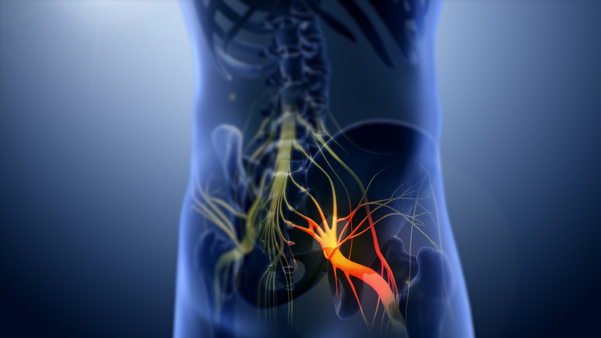
The sciatic nerve, also called the ischiadic nerve, is a large nerve in humans and other vertebrate animals which is the largest branch of the sacral plexus and runs alongside the hip joint and down the lower limb. It is the longest and widest single nerve in the human body, going from the top of the leg to the foot on the posterior aspect. The sciatic nerve has no cutaneous branches for the thigh. This nerve provides the connection to the nervous system for the skin of the lateral leg and the whole foot, the muscles of the back of the thigh, and those of the leg and foot. It is derived from spinal nerves L4 to S3. It contains fibers from both the anterior and posterior divisions of the lumbosacral plexus. Structure In humans, the sciatic nerve is formed from the L4 to S3 segments of the sacral plexus, a collection of nerve fibres that emerge from the sacral part of the spinal cord. The lumbosacral trunk from the L4 and L5 roots descends between the sacral promontory and ala and ... [...More Info...] [...Related Items...] OR: [Wikipedia] [Google] [Baidu] |
Greater Sciatic Foramen
The greater sciatic foramen is an opening (foramen) in the posterior human pelvis. It is formed by the sacrotuberous and sacrospinous ligaments. The piriformis muscle passes through the foramen and occupies most of its volume. The greater sciatic foramen is wider in women than in men. Structure It is bounded as follows: * anterolaterally by the greater sciatic notch of the ilium. * posteromedially by the sacrotuberous ligament. * inferiorly by the sacrospinous ligament and the ischial spine. * superiorly by the anterior sacroiliac ligament. Function The piriformis, which exits the pelvis through the foramen, occupies most of its volume. The following structures also exit the pelvis through the greater sciatic foramen: See also *Lesser sciatic foramen The lesser sciatic foramen is an opening (foramen) between the pelvis and the back of the thigh. The foramen is formed by the sacrotuberous ligament which runs between the sacrum and the ischial tuberosity and the sacrospino ... [...More Info...] [...Related Items...] OR: [Wikipedia] [Google] [Baidu] |
Hip-joint
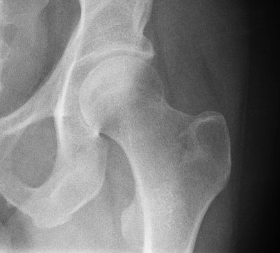
In vertebrate anatomy, hip (or "coxa"Latin ''coxa'' was used by Celsus in the sense "hip", but by Pliny the Elder in the sense "hip bone" (Diab, p 77) in medical terminology) refers to either an anatomical region or a joint. The hip region is located lateral and anterior to the gluteal region, inferior to the iliac crest, and overlying the greater trochanter of the femur, or "thigh bone". In adults, three of the bones of the pelvis have fused into the hip bone or acetabulum which forms part of the hip region. The hip joint, scientifically referred to as the acetabulofemoral joint (''art. coxae''), is the joint between the head of the femur and acetabulum of the pelvis and its primary function is to support the weight of the body in both static (e.g., standing) and dynamic (e.g., walking or running) postures. The hip joints have very important roles in retaining balance, and for maintaining the pelvic inclination angle. Pain of the hip may be the result of numerous causes, ... [...More Info...] [...Related Items...] OR: [Wikipedia] [Google] [Baidu] |
Gluteal Muscles
The gluteal muscles, often called glutes are a group of three muscles which make up the gluteal region commonly known as the buttocks: the gluteus maximus, gluteus medius and gluteus minimus. The three muscles originate from the ilium and sacrum and insert on the femur. The functions of the muscles include extension, abduction, external rotation, and internal rotation of the hip joint. Structure The gluteus maximus is the largest and most superficial of the three gluteal muscles. It makes up a large part of the shape and appearance of the hips. It is a narrow and thick fleshy mass of a quadrilateral shape, and forms the prominence of the buttocks. The gluteus medius is a broad, thick, radiating muscle, situated on the outer surface of the pelvis. It lies profound to the gluteus maximus and its posterior third is covered by the gluteus maximus, its anterior two-thirds by the gluteal aponeurosis, which separates it from the superficial fascia and skin. The gluteus minimus is ... [...More Info...] [...Related Items...] OR: [Wikipedia] [Google] [Baidu] |
Lateral Femoral Circumflex Artery
The lateral circumflex femoral artery, also known as the lateral femoral circumflex artery, or the external circumflex artery, is an artery in the upper thigh. It is usually a branch of the profunda femoris artery, and produces three branches. It is mostly distributed to the muscles of the lateral thigh. Structure Origin The lateral femoral circumflex artery usually arises from the lateral side of the profunda femoris artery, but may occasionally arise directly from the femoral artery. It is the largest branch of the profunda femoris artery. Course and relations The lateral circumflex femoral artery usually courses anterior to the femoral neck. It passes horizontally between the divisions of the femoral nerve. It passes posterior to the sartorius muscle and rectus femoris muscle. It passes laterally across the hip joint capsule. It divides into ascending, transverse, and descending branches. Branches The lateral circumflex femoral artery has three branches: # The ascending ... [...More Info...] [...Related Items...] OR: [Wikipedia] [Google] [Baidu] |
Deep Iliac Circumflex Artery
Deep or The Deep may refer to: Places United States * Deep Creek (Appomattox River tributary), Virginia * Deep Creek (Great Salt Lake), Idaho and Utah * Deep Creek (Mahantango Creek tributary), Pennsylvania * Deep Creek (Mojave River tributary), California * Deep Creek (Pine Creek tributary), Pennsylvania * Deep Creek (Soque River tributary), Georgia * Deep Creek (Texas), a tributary of the Colorado River * Deep Creek (Washington), a tributary of the Spokane River * Deep River (Indiana), a tributary of the Little Calumet River * Deep River (Iowa), a minor tributary of the English River * Deep River (North Carolina) * Deep River (Washington), a minor tributary of the Columbia River * Deep Voll Brook, New Jersey, also known as Deep Brook Elsewhere * Deep Creek (Bahamas) * Deep Creek (Melbourne, Victoria), Australia, a tributary of the Maribyrnong River * Deep River (Western Australia) People * Deep (given name) * Deep (rapper), Punjabi rapper from Houston, Texas * Ravi Deep ... [...More Info...] [...Related Items...] OR: [Wikipedia] [Google] [Baidu] |
Anterior Superior Iliac Spine
The anterior superior iliac spine (abbreviated: ASIS) is a bony projection of the iliac bone, and an important landmark of surface anatomy. It refers to the anterior extremity of the iliac crest of the pelvis. It provides attachment for the inguinal ligament, and the sartorius muscle. The tensor fasciae latae muscle attaches to the lateral aspect of the superior anterior iliac spine, and also about 5 cm away at the iliac tubercle. Structure The anterior superior iliac spine refers to the anterior extremity of the iliac crest of the pelvis. This is a key surface landmark, and easily palpated. It provides attachment for the inguinal ligament, the sartorius muscle, and the tensor fasciae latae muscle. A variety of structures lie close to the anterior superior iliac spine, including the subcostal nerve, the femoral artery (which passes between it and the pubic symphysis), and the iliohypogastric nerve. Clinical significance The anterior superior iliac spine provides a c ... [...More Info...] [...Related Items...] OR: [Wikipedia] [Google] [Baidu] |
Lateral Sacral Arteries
The lateral sacral arteries is an artery in the pelvis that arises from the posterior division of the internal iliac artery. It later splits into two smaller branches, a superior and an inferior. Structure The lateral sacral artery is the second branch of the posterior division of the internal iliac artery. It is a parietal branch. Superior The superior, of large size, passes medialward, and, after anastomosing with branches from the middle sacral, enters the first or second anterior sacral foramen, supplies branches to the contents of the sacral canal, and, escaping by the corresponding posterior sacral foramen, is distributed to the skin and muscles on the dorsum of the sacrum, anastomosing with the superior gluteal. Inferior The inferior runs obliquely across the front of the piriformis and the sacral nerves to the medial side of the anterior sacral foramina, descends on the front of the sacrum, and anastomoses over the coccyx with the middle sacral and opposite lateral ... [...More Info...] [...Related Items...] OR: [Wikipedia] [Google] [Baidu] |
Inferior Gluteal Artery
The inferior gluteal artery (sciatic artery), the smaller of the two terminal branches of the anterior trunk of the internal iliac artery, is distributed chiefly to the buttock and back of the thigh. It passes down on the sacral plexus of nerves and the piriformis muscle, behind the internal pudendal artery. It passes through the lower part of the greater sciatic foramen. It escapes from the pelvis between piriformis muscle and coccygeus muscle. It then descends in the interval between the greater trochanter of the femur and tuberosity of the ischium. It is accompanied by the sciatic nerve and the posterior femoral cutaneous nerves, and covered by the gluteus maximus. It continues down the back of the thigh, supplying the skin, and anastomosing with branches of the perforating arteries. Additional images File:Gray544.png, The arteries of the gluteal and posterior femoral regions. File:Gray829.png, Dissection of side wall of pelvis showing sacral and pudendal plexuses. See a ... [...More Info...] [...Related Items...] OR: [Wikipedia] [Google] [Baidu] |
Gluteus Maximus
The gluteus maximus is the main extensor muscle of the hip. It is the largest and outermost of the three gluteal muscles and makes up a large part of the shape and appearance of each side of the hips. It is the single largest muscle in the human body. Its thick fleshy mass, in a quadrilateral shape, forms the prominence of the buttocks. The other gluteal muscles are the medius and minimus, and sometimes informally these are collectively referred to as the glutes. Its large size is one of the most characteristic features of the muscular system in humans,Norman Eizenberg et al., ''General Anatomy: Principles and Applications'' (2008), p. 17. connected as it is with the power of maintaining the trunk in the erect posture. Other primates have much flatter hips and cannot sustain standing erectly. The muscle is made up of muscle fascicles lying parallel with one another, and are collected together into larger bundles separated by fibrous septa. Structure The gluteus maximus is the ... [...More Info...] [...Related Items...] OR: [Wikipedia] [Google] [Baidu] |
Obturator Internus
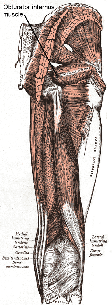
The internal obturator muscle or obturator internus muscle originates on the medial surface of the obturator membrane, the ischium near the membrane, and the rim of the pubis. It exits the pelvic cavity through the lesser sciatic foramen. The internal obturator is situated partly within the lesser pelvis, and partly at the back of the hip-joint. It functions to help laterally rotate femur with hip extension and abduct femur with hip flexion, as well as to steady the femoral head in the acetabulum. Structure Origin The internal obturator muscle arises from the inner surface of the antero-lateral wall of the pelvis. It surrounds the obturator foramen. It is attached to the inferior pubic ramus and ischium, and at the side to the inner surface of the hip bone below and behind the pelvic brim. It reaches from the upper part of the greater sciatic foramen above and behind to the obturator foramen below and in front. It also arises from the pelvic surface of the obturator membra ... [...More Info...] [...Related Items...] OR: [Wikipedia] [Google] [Baidu] |
Internal Iliac Artery
The internal iliac artery (formerly known as the hypogastric artery) is the main artery of the pelvis. Structure The internal iliac artery supplies the walls and viscera of the pelvis, the buttock, the reproductive organs, and the medial compartment of the thigh. The vesicular branches of the internal iliac arteries supply the bladder. It is a short, thick vessel, smaller than the external iliac artery, and about 3 to 4 cm in length. Course The internal iliac artery arises at the bifurcation of the common iliac artery, opposite the lumbosacral articulation, and, passing downward to the upper margin of the greater sciatic foramen, divides into two large trunks, an anterior and a posterior. It is posterior to the ureter, anterior to the internal iliac vein, anterior to the lumbosacral trunk, and anterior to the piriformis muscle. Near its origin, it is medial to the external iliac vein, which lies between it and the psoas major muscle. It is above the obturator nerve. Bra ... [...More Info...] [...Related Items...] OR: [Wikipedia] [Google] [Baidu] |


