|
Phlebography
Venography (also called phlebography or ascending phlebography) is a procedure in which an x-ray of the veins, a venogram, is taken after a special dye is injected into the bone marrow or veins. The dye has to be injected constantly via a catheter, making it an invasive procedure. Normally the catheter is inserted by the groin and moved to the appropriate site by navigating through the vascular system. Contrast venography is the gold standard for judging diagnostic imaging methods for deep venous thrombosis; although, because of its cost, invasiveness, and other limitations this test is rarely performed. Venography can also be used to distinguish blood clots from obstructions in the veins, to evaluate congenital vein problems, to see how the deep leg vein valves are working, or to identify a vein for arterial bypass grafting. Areas of the venous system that can be investigated include the lower extremities, the inferior vena cava, and the upper extremities. The United States Na ... [...More Info...] [...Related Items...] OR: [Wikipedia] [Google] [Baidu] |
Vein
Veins are blood vessels in humans and most other animals that carry blood towards the heart. Most veins carry deoxygenated blood from the tissues back to the heart; exceptions are the pulmonary and umbilical veins, both of which carry oxygenated blood to the heart. In contrast to veins, arteries carry blood away from the heart. Veins are less muscular than arteries and are often closer to the skin. There are valves (called ''pocket valves'') in most veins to prevent backflow. Structure Veins are present throughout the body as tubes that carry blood back to the heart. Veins are classified in a number of ways, including superficial vs. deep, pulmonary vs. systemic, and large vs. small. * Superficial veins are those closer to the surface of the body, and have no corresponding arteries. *Deep veins are deeper in the body and have corresponding arteries. *Perforator veins drain from the superficial to the deep veins. These are usually referred to in the lower limbs and feet. *Communic ... [...More Info...] [...Related Items...] OR: [Wikipedia] [Google] [Baidu] |
Vein Valve
Veins are blood vessels in humans and most other animals that carry blood towards the heart. Most veins carry deoxygenated blood from the tissues back to the heart; exceptions are the pulmonary and umbilical veins, both of which carry oxygenated blood to the heart. In contrast to veins, arteries carry blood away from the heart. Veins are less muscular than arteries and are often closer to the skin. There are valves (called ''pocket valves'') in most veins to prevent backflow. Structure Veins are present throughout the body as tubes that carry blood back to the heart. Veins are classified in a number of ways, including superficial vs. deep, pulmonary vs. systemic, and large vs. small. *Superficial veins are those closer to the surface of the body, and have no corresponding arteries. *Deep veins are deeper in the body and have corresponding arteries. *Perforator veins drain from the superficial to the deep veins. These are usually referred to in the lower limbs and feet. *Communica ... [...More Info...] [...Related Items...] OR: [Wikipedia] [Google] [Baidu] |
Deep Venous Thrombosis
Deep vein thrombosis (DVT) is a type of venous thrombosis involving the formation of a blood clot in a deep vein, most commonly in the legs or pelvis. A minority of DVTs occur in the arms. Symptoms can include pain, swelling, redness, and enlarged veins in the affected area, but some DVTs have no symptoms. The most common life-threatening concern with DVT is the potential for a clot to embolize (detach from the veins), travel as an embolus through the right side of the heart, and become lodged in a pulmonary artery that supplies blood to the lungs. This is called a pulmonary embolism (PE). DVT and PE comprise the cardiovascular disease of venous thromboembolism (VTE). About two-thirds of VTE manifests as DVT only, with one-third manifesting as PE with or without DVT. The most frequent long-term DVT complication is post-thrombotic syndrome, which can cause pain, swelling, a sensation of heaviness, itching, and in severe cases, ulcers. Recurrent VTE occurs in about 30% of those i ... [...More Info...] [...Related Items...] OR: [Wikipedia] [Google] [Baidu] |
Deep Vein Thrombosis
Deep vein thrombosis (DVT) is a type of venous thrombosis involving the formation of a blood clot in a deep vein, most commonly in the legs or pelvis. A minority of DVTs occur in the arms. Symptoms can include pain, swelling, redness, and enlarged veins in the affected area, but some DVTs have no symptoms. The most common life-threatening concern with DVT is the potential for a clot to embolize (detach from the veins), travel as an embolus through the right side of the heart, and become lodged in a pulmonary artery that supplies blood to the lungs. This is called a pulmonary embolism (PE). DVT and PE comprise the cardiovascular disease of venous thromboembolism (VTE). About two-thirds of VTE manifests as DVT only, with one-third manifesting as PE with or without DVT. The most frequent long-term DVT complication is post-thrombotic syndrome, which can cause pain, swelling, a sensation of heaviness, itching, and in severe cases, ulcers. Recurrent VTE occurs in about 30% of those i ... [...More Info...] [...Related Items...] OR: [Wikipedia] [Google] [Baidu] |
Phlebitis
Phlebitis (or Venitis) is inflammation of a vein, usually in the legs. It most commonly occurs in superficial veins. Phlebitis often occurs in conjunction with thrombosis and is then called thrombophlebitis or superficial thrombophlebitis. Unlike deep vein thrombosis, the probability that superficial thrombophlebitis will cause a clot to break up and be transported in pieces to the lung is very low. Signs and symptoms * Localized redness and swelling * Pain or burning along the length of the vein * Vein being hard and cord-like There is usually a slow onset of a tender red area along the superficial veins on the skin. A long, thin red area may be seen as the inflammation follows a superficial vein. This area may feel hard, warm, and tender. The skin around the vein may be itchy and swollen. The area may begin to throb or burn. Symptoms may be worse when the leg is lowered, especially when first getting out of bed in the morning. A low-grade fever may occur. Sometimes phlebitis m ... [...More Info...] [...Related Items...] OR: [Wikipedia] [Google] [Baidu] |
Femoral Vein
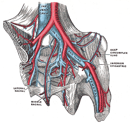
In the human body, the femoral vein is a blood vessel that accompanies the femoral artery in the femoral sheath. It begins at the adductor hiatus (an opening in the adductor magnus muscle) as the continuation of the popliteal vein. It ends at the inferior margin of the inguinal ligament where it becomes the external iliac vein. The femoral vein bears valves which are mostly bicuspid and whose number is variable between individuals and often between left and right leg. Structure Segments *The common femoral vein is the segment of the femoral vein between the branching point of the deep femoral vein and the inferior margin of the inguinal ligament.Page 590 in: *The subsartorial vein or superficial femoral vein are designations ... [...More Info...] [...Related Items...] OR: [Wikipedia] [Google] [Baidu] |
Valsalva Maneuver
The Valsalva maneuver is performed by a forceful attempt of exhalation against a closed airway, usually done by closing one's mouth and pinching one's nose shut while expelling air out as if blowing up a balloon. Variations of the maneuver can be used either in medical examination as a test of cardiac function and autonomic nervous control of the heart, or to clear the ears and sinuses (that is, to equalize pressure between them) when ambient pressure changes, as in scuba diving, hyperbaric oxygen therapy, or air travel. A modified version is done by expiring against a closed glottis. This will elicit the cardiovascular responses described below but will not force air into the Eustachian tubes. History The technique is named after Antonio Maria Valsalva, a 17th-century physician and anatomist from Bologna whose principal scientific interest was the human ear. He described the Eustachian tube and the maneuver to test its patency (openness). He also described the use of this ... [...More Info...] [...Related Items...] OR: [Wikipedia] [Google] [Baidu] |
Anterior Tibial Vein
The anterior tibial vein is a vein in the lower leg. In human anatomy, there are two anterior tibial veins. They originate and receive blood from the dorsal venous arch, on the back of the foot and empties into the popliteal vein. The anterior tibial veins drain the ankle joint, knee joint, tibiofibular joint, and the anterior portion of the lower leg. The two anterior tibial veins ascend in the interosseous membrane between the tibia and fibula and unite with the posterior tibial veins to form the popliteal vein. Like most deep veins in legs, anterior tibial veins are accompanied by the homonym artery, the anterior tibial artery The anterior tibial artery is an artery of the leg. It carries blood to the anterior compartment of the leg and dorsum (biology), dorsal surface of the foot, from the popliteal artery. Structure Course The anterior tibial artery is a branch o ..., along its course. References Veins of the lower limb {{Circulatory-stub ... [...More Info...] [...Related Items...] OR: [Wikipedia] [Google] [Baidu] |
Superficial Vein
Superficial veins are veins that are close to the surface of the body, as opposed to deep veins, which are far from the surface. Superficial veins are not paired with an artery, unlike the deep veins, which are typically associated with an artery of the same name. Superficial veins are important physiologically for cooling of the body. When the body is too hot, the body shunts blood from the deep veins to the superficial veins to facilitate heat transfer to the body's surroundings. Superficial veins are often visible underneath the skin. Those below the level of the heart tend to bulge out, which can be readily witnessed in the hand, where the veins bulge significantly less after the arm has been raised above the head for a short time. Veins become more visually prominent when lifting heavy weight, especially after a period of proper strength training. Physiologically, the superficial veins are not as important as the deep veins (as they carry less blood) and are sometimes r ... [...More Info...] [...Related Items...] OR: [Wikipedia] [Google] [Baidu] |
Sepsis
Sepsis, formerly known as septicemia (septicaemia in British English) or blood poisoning, is a life-threatening condition that arises when the body's response to infection causes injury to its own tissues and organs. This initial stage is followed by suppression of the immune system. Common signs and symptoms include fever, tachycardia, increased heart rate, hyperventilation, increased breathing rate, and mental confusion, confusion. There may also be symptoms related to a specific infection, such as a cough with pneumonia, or dysuria, painful urination with a pyelonephritis, kidney infection. The very young, old, and people with a immunodeficiency, weakened immune system may have no symptoms of a specific infection, and the hypothermia, body temperature may be low or normal instead of having a fever. Severe sepsis causes organ dysfunction, poor organ function or blood flow. The presence of Hypotension, low blood pressure, high blood Lactic acid, lactate, or Oliguria, low urine o ... [...More Info...] [...Related Items...] OR: [Wikipedia] [Google] [Baidu] |
Axillary Vein
In human anatomy, the axillary vein is a large blood vessel that conveys blood from the lateral aspect of the thorax, axilla (armpit) and upper limb toward the heart. There is one axillary vein on each side of the body. Structure Its origin is at the lower margin of the teres major muscle and a continuation of the brachial vein. This large vein is formed by the brachial vein and the basilic vein. At its terminal part, it is also joined by the cephalic vein. Other tributaries include the subscapular vein, circumflex humeral vein, lateral thoracic vein and thoraco-acromial vein. It terminates at the lateral margin of the first rib, at which it becomes the subclavian vein. It is accompanied along its course by a similarly named artery, the axillary artery In human anatomy, the axillary artery is a large blood vessel that conveys oxygenated blood to the lateral aspect of the thorax, the axilla (armpit) and the upper limb. Its origin is at the lateral margin of the first ... [...More Info...] [...Related Items...] OR: [Wikipedia] [Google] [Baidu] |
Digital Subtraction Angiography
Digital subtraction angiography (DSA) is a fluoroscopy technique used in interventional radiology to clearly visualize blood vessels in a bony or dense soft tissue environment. Images are produced using contrast medium by subtracting a "pre-contrast image" or ''mask'' from subsequent images, once the contrast medium has been introduced into a structure. Hence the term "digital ''subtraction'' angiography. Subtraction angiography was first described in 1935 and in English sources in 1962 as a manual technique. Digital technology made DSA practical starting in the 1970s. Procedure DSA and fluoroscopy In traditional angiography, images are acquired by exposing an area of interest with time-controlled x-rays while injecting a contrast medium into the blood vessels. The image obtained includes the blood vessels, together with all overlying and underlying structures. The images are useful for determining anatomical position and variations, but unhelpful for visualizing blood vessels acc ... [...More Info...] [...Related Items...] OR: [Wikipedia] [Google] [Baidu] |


