|
Oral Mucocele
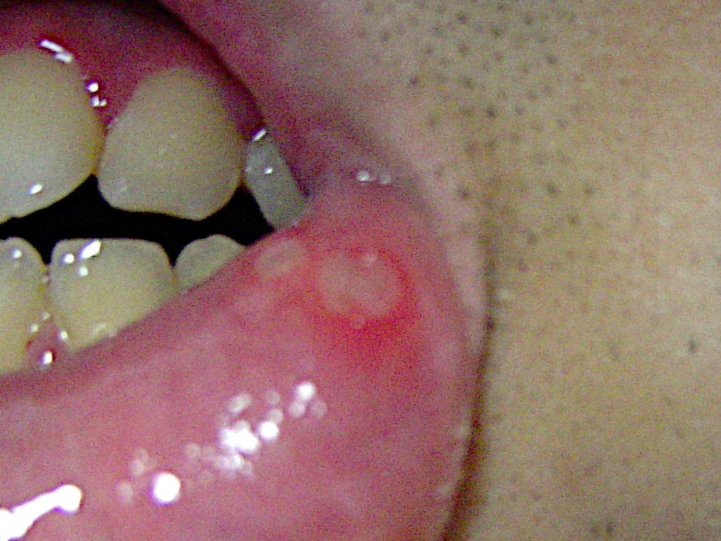
Oral mucocele (also mucous extravasation cyst, mucous cyst of the oral mucosa, and mucous retention and extravasation phenomena.) is a condition caused by two related phenomena - mucus extravasation phenomenon and mucous retention cyst. Mucous extravasation phenomenon is a swelling of connective tissue consisting of a collection of fluid called mucus. This occurs because of a ruptured salivary gland duct usually caused by local trauma (damage) in the case of mucous extravasation phenomenon and an obstructed or ruptured salivary duct in the case of a mucus retention cyst. The mucocele has a bluish, translucent color, and is more commonly found in children and young adults. Although these lesions are often called cysts, mucoceles are not true cysts because they have no epithelial lining. Rather, they are polyps. Signs and symptoms The size of oral mucoceles vary from 1 mm to several centimeters and they usually are slightly transparent with a blue tinge. On palpation, muco ... [...More Info...] [...Related Items...] OR: [Wikipedia] [Google] [Baidu] |
Swelling (medical)
Edema, also spelled oedema, and also known as fluid retention, dropsy, hydropsy and swelling, is the build-up of fluid in the body's tissue. Most commonly, the legs or arms are affected. Symptoms may include skin which feels tight, the area may feel heavy, and joint stiffness. Other symptoms depend on the underlying cause. Causes may include venous insufficiency, heart failure, kidney problems, low protein levels, liver problems, deep vein thrombosis, infections, angioedema, certain medications, and lymphedema. It may also occur after prolonged sitting or standing and during menstruation or pregnancy. The condition is more concerning if it starts suddenly, or pain or shortness of breath is present. Treatment depends on the underlying cause. If the underlying mechanism involves sodium retention, decreased salt intake and a diuretic may be used. Elevating the legs and support stockings may be useful for edema of the legs. Older people are more commonly affected. The word i ... [...More Info...] [...Related Items...] OR: [Wikipedia] [Google] [Baidu] |
Frontoethmoidal Suture
The frontoethmoidal suture is the suture between the ethmoid bone and the frontal bone. It is located in the anterior cranial fossa The anterior cranial fossa is a depression in the floor of the cranial base which houses the projecting frontal lobes of the brain. It is formed by the orbital part of frontal bone, orbital plates of the Frontal bone, frontal, the cribriform plate .... References External links * Bones of the head and neck Cranial sutures Human head and neck Joints Joints of the head and neck Skeletal system Skull {{musculoskeletal-stub ... [...More Info...] [...Related Items...] OR: [Wikipedia] [Google] [Baidu] |
Histiocyte
A histiocyte is a vertebrate cell that is part of the mononuclear phagocyte system (also known as the reticuloendothelial system or lymphoreticular system). The mononuclear phagocytic system is part of the organism's immune system. The histiocyte is a tissue macrophage or a dendritic cell (histio, diminutive of histo, meaning ''tissue'', and cyte, meaning ''cell''). Part of their job is to clear out neutrophils once they've reached the end of their lifespan. Development Histiocytes are derived from the bone marrow by multiplication from a stem cell. The derived cells migrate from the bone marrow to the blood as monocytes. They circulate through the body and enter various organs, where they undergo differentiation into histiocytes, which are part of the mononuclear phagocytic system (MPS). However, the term ''histiocyte'' has been used for multiple purposes in the past, and some cells called "histocytes" do not appear to derive from monocytic-macrophage lines. The term Histioc ... [...More Info...] [...Related Items...] OR: [Wikipedia] [Google] [Baidu] |
Neutrophil
Neutrophils (also known as neutrocytes or heterophils) are the most abundant type of granulocytes and make up 40% to 70% of all white blood cells in humans. They form an essential part of the innate immune system, with their functions varying in different animals. They are formed from stem cells in the bone marrow and Cellular differentiation, differentiated into #Subpopulations, subpopulations of neutrophil-killers and neutrophil-cagers. They are short-lived and highly mobile, as they can enter parts of tissue where other cells/molecules cannot. Neutrophils may be subdivided into segmented neutrophils and banded neutrophils (or Band cell, bands). They form part of the polymorphonuclear cells family (PMNs) together with basophils and eosinophils. The name ''neutrophil'' derives from staining characteristics on hematoxylin and eosin (H&E stain, H&E) histology, histological or cell biology, cytological preparations. Whereas basophilic white blood cells stain dark blue and eosinoph ... [...More Info...] [...Related Items...] OR: [Wikipedia] [Google] [Baidu] |
Inflammation
Inflammation (from la, wikt:en:inflammatio#Latin, inflammatio) is part of the complex biological response of body tissues to harmful stimuli, such as pathogens, damaged cells, or Irritation, irritants, and is a protective response involving immune cells, blood vessels, and molecular mediators. The function of inflammation is to eliminate the initial cause of cell injury, clear out necrotic cells and tissues damaged from the original insult and the inflammatory process, and initiate tissue repair. The five cardinal signs are heat, pain, redness, swelling, and Functio laesa, loss of function (Latin ''calor'', ''dolor'', ''rubor'', ''tumor'', and ''functio laesa''). Inflammation is a generic response, and therefore it is considered as a mechanism of innate immune system, innate immunity, as compared to adaptive immune system, adaptive immunity, which is specific for each pathogen. Too little inflammation could lead to progressive tissue destruction by the harmful stimulus (e.g. b ... [...More Info...] [...Related Items...] OR: [Wikipedia] [Google] [Baidu] |
Granulation Tissue
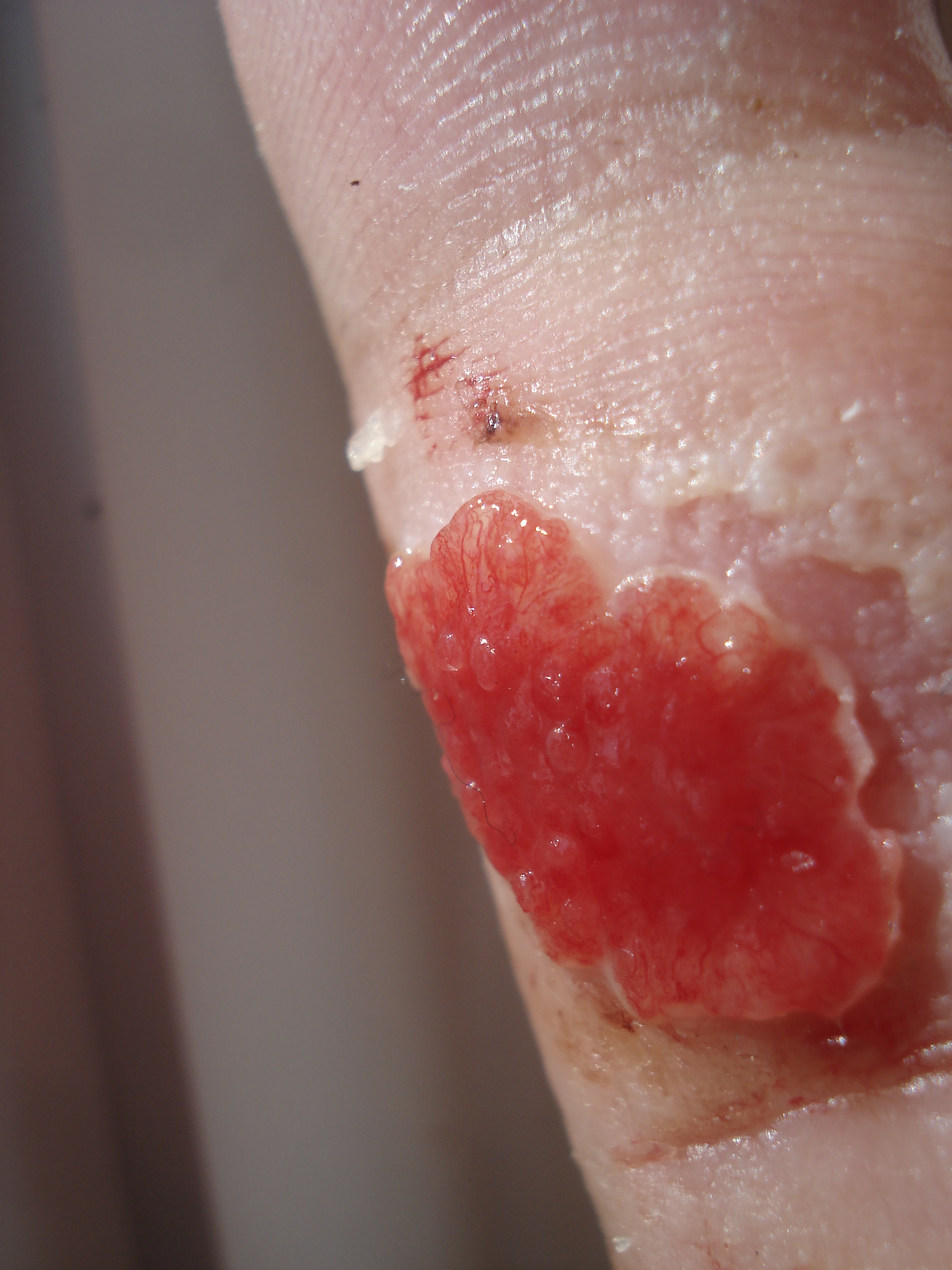
Granulation tissue is new connective tissue and microscopic blood vessels that form on the surfaces of a wound during the healing process. Granulation tissue typically grows from the base of a wound and is able to fill wounds of almost any size. Examples of granulation tissue can be seen in pyogenic granulomas and pulp polyps. Its histological appearance is characterized by proliferation of fibroblasts and new thin-walled, delicate capillaries (angiogenesis), infiltrated inflammatory cells in a loose extracellular matrix. Appearance During the migratory phase of wound healing, granulation tissue is: * light red or dark pink, being perfused with new capillary loops or "buds"; * soft to the touch; * moist; * bumpy (granular) in appearance, due to punctate hemorrhages; * pulsatile on palpation; * painless when healthy; Structure Granulation tissue is composed of tissue matrix supporting a variety of cell types, most of which can be associated with one of the following functions: ... [...More Info...] [...Related Items...] OR: [Wikipedia] [Google] [Baidu] |
Mucocele Of Lower Lip (1)
A mucocele is a distension of a hollow organ or cavity because of mucus buildup. By location Oral Oral mucocele is the most common benign lesion of the salivary glands generally conceded to be of traumatic origin. It is characterized by the pooling of mucus in a cavity due to the rupture of salivary ducts or acini. It can occur in the lower lip, palate, cheeks, tongue and the floor of the mouth. Appendix Appendiceal mucocele is found in 0.3 to 0.7% of the appendectomies.Creative Commons Attribution 4.0 Internationallicense It is characterized by the dilation of the organ lumen with mucus accumulation. Appendix mucocele may come as a consequence of obstructive or inflammatory processes, cystadenomas or cystadenocarcinomas. Its main complication is pseudomyxoma peritonei. File:Gross pathology of mucocele of the appendix.jpg, Gross pathology of mucocele of the appendix File:Pie chart of histological types of mucocele of the appendix.jpg, Pie chart of histological types of mucocele ... [...More Info...] [...Related Items...] OR: [Wikipedia] [Google] [Baidu] |
Lipoma
A lipoma is a benign tumor made of fat tissue. They are generally soft to the touch, movable, and painless. They usually occur just under the skin, but occasionally may be deeper. Most are less than in size. Common locations include upper back, shoulders, and abdomen. It is possible to have a number of lipomas. The cause is generally unclear. Risk factors include family history, obesity, and lack of exercise. Diagnosis is typically based on a physical exam. Occasionally medical imaging or tissue biopsy is used to confirm the diagnosis. Treatment is typically by observation or surgical removal. Rarely, the condition may recur following removal, but this can generally be managed with repeat surgery. They are not generally associated with a future risk of cancer. Lipomas have a prevalence of roughly 2 out of every 100 people. Lipomas typically occur in adults between 40 and 60 years of age. Males are more often affected than females. They are the most common noncancerous soft-t ... [...More Info...] [...Related Items...] OR: [Wikipedia] [Google] [Baidu] |
Aphthous Stomatitis
Aphthous stomatitis, or recurrent aphthous stomatitis (RAS), is a common condition characterized by the repeated formation of benign and non- contagious mouth ulcers (aphthae) in otherwise healthy individuals. The informal term ''canker sore'' is also used, mainly in North America, although it may also refer to other types of mouth ulcers. The cause is not completely understood but involves a T cell-mediated immune response triggered by a variety of factors which may include nutritional deficiencies, local trauma, stress, hormonal influences, allergies, genetic predisposition, certain foods, dehydration, some food additives, or some hygienic chemical additives like SDS (common in toothpaste). These ulcers occur periodically and heal completely between attacks. In the majority of cases, the individual ulcers last about 7–10 days, and ulceration episodes occur 3–6 times per year. Most appear on the non-keratinizing epithelial surfaces in the mouth – i.e. anywhere exce ... [...More Info...] [...Related Items...] OR: [Wikipedia] [Google] [Baidu] |
Vesicle (biology)
In cell biology, a vesicle is a structure within or outside a cell, consisting of liquid or cytoplasm enclosed by a lipid bilayer. Vesicles form naturally during the processes of secretion (exocytosis), uptake (endocytosis) and transport of materials within the plasma membrane. Alternatively, they may be prepared artificially, in which case they are called liposomes (not to be confused with lysosomes). If there is only one phospholipid bilayer, the vesicles are called ''unilamellar liposomes''; otherwise they are called ''multilamellar liposomes''. The membrane enclosing the vesicle is also a lamellar phase, similar to that of the plasma membrane, and intracellular vesicles can fuse with the plasma membrane to release their contents outside the cell. Vesicles can also fuse with other organelles within the cell. A vesicle released from the cell is known as an extracellular vesicle. Vesicles perform a variety of functions. Because it is separated from the cytosol, the inside of th ... [...More Info...] [...Related Items...] OR: [Wikipedia] [Google] [Baidu] |
Retromolar Pad
The retromolar space or retromolar gap is a space at the rear of the mandible, between the back of the last molar and the anterior edge of the ascending ramus where it crosses the alveolar margin. This gap is generally small or absent in modern humans, but it was more often present in Neanderthals, and it was common among some prehistoric Amerindians, such as Arikara and Mandan. Retromolar pad The retromolar area of a human mandible is covered by the retromolar pad (also known as the piriformis papilla), an elevated triangular area of mucosa A mucous membrane or mucosa is a membrane that lines various cavities in the body of an organism and covers the surface of internal organs. It consists of one or more layers of epithelial cells overlying a layer of loose connective tissue. It is .... It is composed of non-keratinized loose alveolar tissue covering glandular tissues and muscle fibers. It is important for supporting lower complete and partial dentures as well as landmarkin ... [...More Info...] [...Related Items...] OR: [Wikipedia] [Google] [Baidu] |
Palate
The palate () is the roof of the mouth in humans and other mammals. It separates the oral cavity from the nasal cavity. A similar structure is found in crocodilians, but in most other tetrapods, the oral and nasal cavities are not truly separated. The palate is divided into two parts, the anterior, bony hard palate and the posterior, fleshy soft palate (or velum). Structure Innervation The maxillary nerve branch of the trigeminal nerve supplies sensory innervation to the palate. Development The hard palate forms before birth. Variation If the fusion is incomplete, a cleft palate results. Function When functioning in conjunction with other parts of the mouth, the palate produces certain sounds, particularly velar, palatal, palatalized, postalveolar, alveolopalatal, and uvular consonants. History Etymology The English synonyms palate and palatum, and also the related adjective palatine (as in palatine bone), are all from the Latin ''palatum'' via Old French ''palat ... [...More Info...] [...Related Items...] OR: [Wikipedia] [Google] [Baidu] |


