|
Operculum (brain)
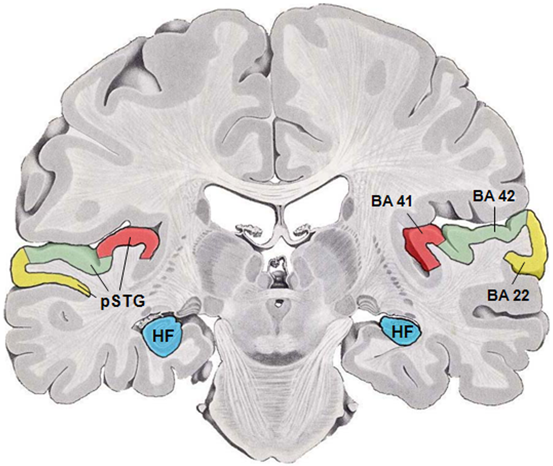
In human brain anatomy, an operculum (Latin, meaning "little lid") (pl. opercula), may refer to the frontal, temporal, or parietal operculum, which together cover the insula as the opercula of insula. It can also refer to the occipital operculum, part of the occipital lobe. The insular lobe is a portion of the cerebral cortex that has invaginated to lie deep within the lateral sulcus. It sits like an island (the meaning of ''insular'') almost surrounded by the groove of the circular sulcus and covered over and obscured by the insular opercula. A part of the parietal lobe, the frontoparietal operculum, covers the upper part of the insular lobe from the front to the back. The opercula lie on the precentral and postcentral gyri (on either side of the central sulcus). The part of the parietal operculum that forms the ceiling of the lateral sulcus functions as the secondary somatosensory cortex. Development Normally, the insular opercula begin to develop between the 20th and ... [...More Info...] [...Related Items...] OR: [Wikipedia] [Google] [Baidu] |
Human Temporal Lobe Areas
Humans (''Homo sapiens'') are the most abundant and widespread species of primate, characterized by bipedality, bipedalism and exceptional cognitive skills due to a large and complex Human brain, brain. This has enabled the development of advanced tools, culture, and language. Humans are highly social and tend to live in complex social structures composed of many cooperating and competing groups, from family, families and kinship networks to political state (polity), states. Social interactions between humans have established a wide variety of values, norm (sociology), social norms, and rituals, which bolster human society. Its intelligence and its desire to understand and influence the environment and to explain and manipulate Phenomenon, phenomena have motivated humanity's development of science, philosophy, mythology, religion, and other fields of study. Although some scientists equate the term ''humans'' with all members of the genus ''Homo'', in common usage, it generall ... [...More Info...] [...Related Items...] OR: [Wikipedia] [Google] [Baidu] |
Central Sulcus
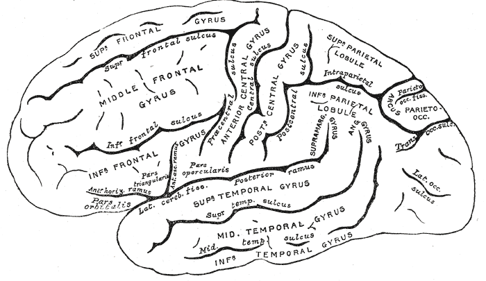
In neuroanatomy, the central sulcus (also central fissure, fissure of Rolando, or Rolandic fissure, after Luigi Rolando) is a sulcus, or groove, in the cerebral cortex in the brains of vertebrates. It is sometimes confused with the longitudinal fissure. The central sulcus is a prominent landmark of the brain, separating the parietal lobe from the frontal lobe and the primary motor cortex from the primary somatosensory cortex. Evolution of the central sulcus The evolution of the central sulcus is theorized to have occurred in mammals when the complete dissociation of the original somatosensory cortex from its mirror duplicate developed in placental mammals such as primates, though the development did not stop there as time progressed the distinction between the two cortices grew. Evolution in primates The central sulcus is more prominent in apes as a result of fine-tuning of the motor system in apes. Hominins (bipedal apes) continued this trend through increased use of thei ... [...More Info...] [...Related Items...] OR: [Wikipedia] [Google] [Baidu] |
Sulcus Lateralis
In neuroanatomy, the lateral sulcus (also called Sylvian fissure, after Franciscus Sylvius, or lateral fissure) is one of the most prominent features of the human brain. The lateral sulcus is a deep fissure in each hemisphere that separates the frontal and parietal lobes from the temporal lobe. The insular cortex lies deep within the lateral sulcus. Anatomy The lateral sulcus divides both the frontal lobe and parietal lobe above from the temporal lobe below. It is in both hemispheres of the brain. The lateral sulcus is one of the earliest-developing sulci of the human brain. It first appears around the fourteenth gestational week. The insular cortex lies deep within the lateral sulcus. The lateral sulcus has a number of side branches. Two of the most prominent and most regularly found are the ascending (also called vertical) ramus and the horizontal ramus of the lateral fissure, which subdivide the inferior frontal gyrus. The lateral sulcus also contains the transverse tempor ... [...More Info...] [...Related Items...] OR: [Wikipedia] [Google] [Baidu] |
Albert Einstein’s Brain
The brain of Albert Einstein has been a subject of much research and speculation. Albert Einstein's brain was removed within seven and a half hours of his death. His apparent regularities or irregularities in the brain have been used to support various ideas about correlations in neuroanatomy with general or mathematical intelligence. Studies have suggested an increased number of glial cells in Einstein's brain. Fate of the brain Einstein's autopsy was conducted in the lab of Thomas Stoltz Harvey. Shortly after Einstein's death in 1955, Harvey removed and weighed the brain at 1230g. Harvey then took the brain to a lab at the University of Pennsylvania where he dissected it into several pieces. Some of the pieces he kept to himself while others were given to leading pathologists. He hoped that cytoarchitectonics, the study of brain cells under a microscope, would reveal useful information. Harvey injected 50% formalin through the internal carotid arteries and afterward suspended th ... [...More Info...] [...Related Items...] OR: [Wikipedia] [Google] [Baidu] |
Full Term
Pregnancy is the time during which one or more offspring develops ( gestates) inside a woman's uterus (womb). A multiple pregnancy involves more than one offspring, such as with twins. Pregnancy usually occurs by sexual intercourse, but can also occur through assisted reproductive technology procedures. A pregnancy may end in a live birth, a miscarriage, an induced abortion, or a stillbirth. Childbirth typically occurs around 40 weeks from the start of the last menstrual period (LMP), a span known as the gestational age. This is just over nine months. Counting by fertilization age, the length is about 38 weeks. Pregnancy is "the presence of an implanted human embryo or fetus in the uterus"; implantation occurs on average 8–9 days after fertilization. An ''embryo'' is the term for the developing offspring during the first seven weeks following implantation (i.e. ten weeks' gestational age), after which the term ''fetus'' is used until birth. Signs and sym ... [...More Info...] [...Related Items...] OR: [Wikipedia] [Google] [Baidu] |
Invagination
Invagination is the process of a surface folding in on itself to form a cavity, pouch or tube. In developmental biology, invagination is a mechanism that takes place during gastrulation. This mechanism or cell movement happens mostly in the vegetal pole. Invagination consists of the folding of an area of the exterior sheet of cells towards the inside of the blastula. In each organism, the complexity will be different depending on the number of cells. Invagination can be referenced as one of the steps of the establishment of the body plan. The term, originally used in embryology, has been adopted in other disciplines as well. There is more than one type of movement for invagination. Two common types are axial and orthogonal. The difference between the production of the tube formed in the cytoskeleton and extracellular matrix. Axial can be formed at a single point along the axis of a surface. Orthogonal is linear and trough. Biology * Invagination is the morphogenetic processes by wh ... [...More Info...] [...Related Items...] OR: [Wikipedia] [Google] [Baidu] |
Prenatal Development
Prenatal development () includes the development of the embryo and of the fetus during a viviparous animal's gestation. Prenatal development starts with fertilization, in the germinal stage of embryonic development, and continues in fetal development until birth. In human pregnancy, prenatal development is also called antenatal development. The development of the human embryo follows fertilization, and continues as fetal development. By the end of the tenth week of gestational age the embryo has acquired its basic form and is referred to as a fetus. The next period is that of fetal development where many organs become fully developed. This fetal period is described both topically (by organ) and chronologically (by time) with major occurrences being listed by gestational age. The very early stages of embryonic development are the same in all mammals, but later stages of development, and the length of gestation varies. Terminology In the human: Different terms are used to ... [...More Info...] [...Related Items...] OR: [Wikipedia] [Google] [Baidu] |
Secondary Somatosensory Cortex
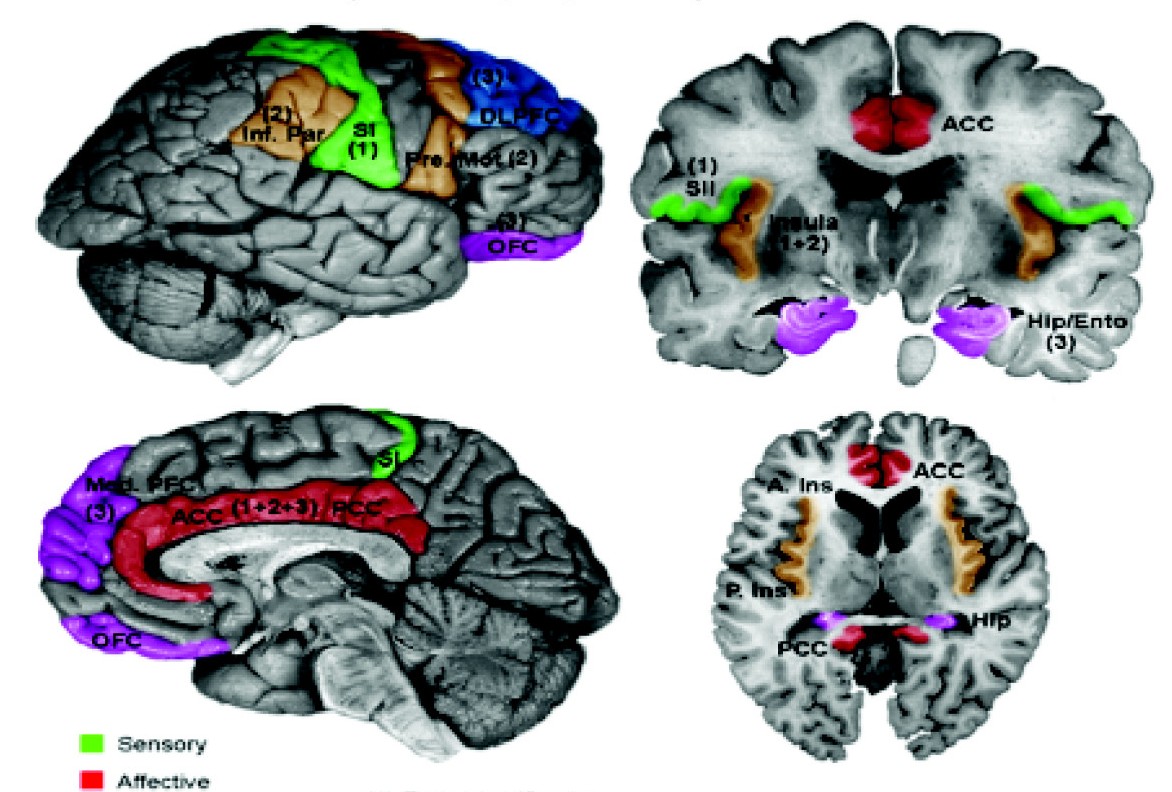
The human secondary somatosensory cortex (S2, SII) is a region of cortex in the parietal operculum on the ceiling of the lateral sulcus. Region S2 was first described by Adrian in 1940, who found that feeling in cats' feet was not only represented in the primary somatosensory cortex (S1) but also in a second region adjacent to S1. In 1954, Penfield and Jasper evoked somatosensory sensations in human patients during neurosurgery by electrically stimulating the ceiling of the lateral sulcus, which lies adjacent to S1, and their findings were confirmed in 1979 by Woolsey et al. using evoked potentials and electrical stimulation. Experiments involving ablation of the second somatosensory cortex in primates indicate that this cortical area is involved in remembering the differences between tactile shapes and textures. Functional neuroimaging studies have found S2 activation in response to light touch, pain, visceral sensation, and tactile attention. In monkeys, apes and hominids, inclu ... [...More Info...] [...Related Items...] OR: [Wikipedia] [Google] [Baidu] |
Gyrus
In neuroanatomy, a gyrus (pl. gyri) is a ridge on the cerebral cortex. It is generally surrounded by one or more sulci (depressions or furrows; sg. ''sulcus''). Gyri and sulci create the folded appearance of the brain in humans and other mammals. Structure The gyri are part of a system of folds and ridges that create a larger surface area for the human brain and other mammalian brains. Because the brain is confined to the skull, brain size is limited. Ridges and depressions create folds allowing a larger cortical surface area, and greater cognitive function, to exist in the confines of a smaller cranium. Development The human brain undergoes gyrification during fetal and neonatal development. In embryonic development, all mammalian brains begin as smooth structures derived from the neural tube. A cerebral cortex without surface convolutions is lissencephalic, meaning 'smooth-brained'. As development continues, gyri and sulci begin to take shape on the fetal brain, with ... [...More Info...] [...Related Items...] OR: [Wikipedia] [Google] [Baidu] |
Human Brain
The human brain is the central organ of the human nervous system, and with the spinal cord makes up the central nervous system. The brain consists of the cerebrum, the brainstem and the cerebellum. It controls most of the activities of the body, processing, integrating, and coordinating the information it receives from the sense organs, and making decisions as to the instructions sent to the rest of the body. The brain is contained in, and protected by, the skull bones of the head. The cerebrum, the largest part of the human brain, consists of two cerebral hemispheres. Each hemisphere has an inner core composed of white matter, and an outer surface – the cerebral cortex – composed of grey matter. The cortex has an outer layer, the neocortex, and an inner allocortex. The neocortex is made up of six neuronal layers, while the allocortex has three or four. Each hemisphere is conventionally divided into four lobes – the frontal, temporal, parietal, and occipital lo ... [...More Info...] [...Related Items...] OR: [Wikipedia] [Google] [Baidu] |
Postcentral Gyrus
In neuroanatomy, the postcentral gyrus is a prominent gyrus in the lateral parietal lobe of the human brain. It is the location of the primary somatosensory cortex, the main sensory receptive area for the somatosensory system, sense of touch. Like other sensory areas, there is a map of sensory space in this location, called the ''sensory homunculus''. The primary somatosensory cortex was initially defined from surface stimulation studies of Wilder Penfield, and parallel surface potential studies of Bard, Woolsey, and Marshall. Although initially defined to be roughly the same as Brodmann areas Brodmann area 3, 3, Brodmann area 1, 1 and Brodmann area 2, 2, more recent work by Jon Kaas, Kaas has suggested that for homogeny with other sensory fields only area 3 should be referred to as "primary somatosensory cortex", as it receives the bulk of the Thalamocortical radiations, thalamocortical projections from the sensory input fields. Structure The lateral postcentral gyrus is bounded ... [...More Info...] [...Related Items...] OR: [Wikipedia] [Google] [Baidu] |
Precentral Gyrus
The precentral gyrus is a prominent gyrus on the surface of the posterior frontal lobe of the brain. It is the site of the primary motor cortex that in humans is cytoarchitecturally defined as Brodmann area 4. Structure The precentral gyrus lies in front of the postcentral gyrus - mostly on the lateral (convex) side of each cerebral hemisphere - from which it is separated by the central sulcus. Its anterior border is represented by the precentral sulcus, while inferiorly it borders to the lateral sulcus (Sylvian fissure). Medially, it is contiguous with the paracentral lobule. The internal pyramidal layer (layer V) of the precentral cortex contains giant (70-100 micrometers) pyramidal neurons called Betz cells, which send long axons to the contralateral motor nuclei of the cranial nerves and to the lower motor neurons in the ventral horn of the spinal cord. These axons form the corticospinal tract. The Betz cells along with their long axons are referred to as upper motor n ... [...More Info...] [...Related Items...] OR: [Wikipedia] [Google] [Baidu] |


