|
Nerve To Subclavius
The subclavian nerve, also known as the nerve to the subclavius, is small branch of the upper trunk of the brachial plexus. It contains axons from C5 and C6. The subclavian nerve innervates the subclavius muscle. Structure The subclavian nerve is a branch of the upper trunk of the brachial plexus. It contains axons derived from the ventral rami of the C5 and C6 cervical spinal nerves. Variation The subclavian nerve can variably give rise to a branch which innervates the diaphragm called the accessory phrenic nerve. The accessory phrenic nerve may rather branch from the C4 or C6 segments or ansa cervicalis. This nerve usually joins with the phrenic nerve before innervating the diaphragm, ventral to the subclavian vein. Function The subclavian nerve innervates the subclavius muscle The subclavius is a small triangular muscle, placed between the clavicle and the first rib. Along with the pectoralis major and pectoralis minor muscles, the subclavius muscle makes up ... [...More Info...] [...Related Items...] OR: [Wikipedia] [Google] [Baidu] |
Brachial Plexus
The brachial plexus is a network () of nerves formed by the anterior rami of the lower four cervical nerves and first thoracic nerve ( C5, C6, C7, C8, and T1). This plexus extends from the spinal cord, through the cervicoaxillary canal in the neck, over the first rib, and into the armpit, it supplies afferent and efferent nerve fibers the to chest, shoulder, arm, forearm, and hand. Structure The brachial plexus is divided into five ''roots'', three ''trunks'', six ''divisions'' (three anterior and three posterior), three ''cords'', and five ''branches''. There are five "terminal" branches and numerous other "pre-terminal" or "collateral" branches, such as the subscapular nerve, the thoracodorsal nerve, and the long thoracic nerve, that leave the plexus at various points along its length. A common structure used to identify part of the brachial plexus in cadaver dissections is the M or W shape made by the musculocutaneous nerve, lateral cord, median nerve, medial cord, and ... [...More Info...] [...Related Items...] OR: [Wikipedia] [Google] [Baidu] |
Subclavius Muscle
The subclavius is a small triangular muscle, placed between the clavicle and the first rib. Along with the pectoralis major and pectoralis minor muscles, the subclavius muscle makes up the anterior axioappendicular muscles, also known as anterior wall of the axilla. Structure It arises by a short, thick tendon from the first rib and its cartilage at their junction, in front of the costoclavicular ligament. The fleshy fibers proceed obliquely superolaterally, to be inserted into the groove on the under surface of the clavicle. Innervation The nerve to subclavius (or subclavian nerve) innervates the muscle. This arises from the junction of the fifth and sixth cervical nerves, from the superior/upper trunk of the brachial plexus. Variation Insertion into coracoid process instead of clavicle or into both clavicle and coracoid process. Sternoscapular fasciculus to the upper border of scapula. Sternoclavicularis from manubrium to clavicle between pectoralis major and coracoclavicular ... [...More Info...] [...Related Items...] OR: [Wikipedia] [Google] [Baidu] |
Upper Trunk
The upper (superior) trunk is part of the brachial plexus. It is formed by joining of the ventral rami of the fifth (C5) and sixth (C6) cervical nerves. The upper trunk divides into an anterior and posterior division. The branches of the upper trunk from proximal to distal are: * subclavian nerve (C5-C6) *suprascapular nerve (C5-C6) *anterior division of upper trunk (C5-C6, forms part of lateral cord) *posterior division of upper trunk (C5-C6, forms part of posterior cord) The axillary, radial, musculocutaneous and median nerve The median nerve is a nerve in humans and other animals in the upper limb. It is one of the five main nerves originating from the brachial plexus. The median nerve originates from the lateral and medial cords of the brachial plexus, and has contr ...s all contain axons derived from the upper trunk. Additional images File:Slide1cord.JPG, Brachial plexus.Deep dissection. File:Slide1ecc.JPG, Brachial plexus.Deep dissection.Anterolateral view {{Authori ... [...More Info...] [...Related Items...] OR: [Wikipedia] [Google] [Baidu] |
Axon
An axon (from Greek ἄξων ''áxōn'', axis), or nerve fiber (or nerve fibre: see spelling differences), is a long, slender projection of a nerve cell, or neuron, in vertebrates, that typically conducts electrical impulses known as action potentials away from the nerve cell body. The function of the axon is to transmit information to different neurons, muscles, and glands. In certain sensory neurons (pseudounipolar neurons), such as those for touch and warmth, the axons are called afferent nerve fibers and the electrical impulse travels along these from the periphery to the cell body and from the cell body to the spinal cord along another branch of the same axon. Axon dysfunction can be the cause of many inherited and acquired neurological disorders that affect both the peripheral and central neurons. Nerve fibers are classed into three typesgroup A nerve fibers, group B nerve fibers, and group C nerve fibers. Groups A and B are myelinated, and group C are unmyelinated. ... [...More Info...] [...Related Items...] OR: [Wikipedia] [Google] [Baidu] |
Cervical Spinal Nerve 5
The cervical spinal nerve 5 (C5) is a spinal nerve of the cervical segment. Nervous System -- Groups of Nerves It originates from the spinal column from above the (C5). It contributes to the phrenic nerve, , and before joining |
Cervical Spinal Nerve 6
The cervical spinal nerve 6 (C6) is a spinal nerve of the cervical segment. It originates from the spinal column from above the cervical vertebra 6 (C6). The C6 nerve root shares a common branch from C5, and has a role in innervating many muscles of the rotator cuff and distal arm, including: *Subclavius *Supraspinatus *Infraspinatus * Biceps Brachii * Brachialis * Deltoid *Teres Minor *Brachioradialis *Serratus Anterior *Subscapularis *Pectoralis Major *Coracobrachialis *Teres Major * Supinator *Extensor Carpi Radialis Brevis * Extensor Carpi Radialis Longus * Latissimus Dorsi Damage to the C6 motor neuron, by way of impingement, ischemia, trauma, or degeneration of nerve tissue, can cause denervation Denervation is any loss of nerve supply regardless of the cause. If the nerves lost to denervation are part of the neuronal communication to a specific function in the body then altered or a loss of physiological functioning can occur. Denervati ... of one or more of the asso ... [...More Info...] [...Related Items...] OR: [Wikipedia] [Google] [Baidu] |
Ventral Ramus Of Spinal Nerve
The ventral ramus (pl. ''rami'') (Latin for ''branch'') is the anterior division of a spinal nerve. The ventral rami supply the antero-lateral parts of the trunk and the limbs. They are mainly larger than the dorsal rami. Shortly after a spinal nerve exits the intervertebral foramen, it branches into the dorsal ramus, the ventral ramus, and the ramus communicans. Each of these three structures carries both sensory and motor information. Each spinal nerve carries both sensory and motor information, via efferent and afferent nerve fibers - ultimately via the motor cortex in the frontal lobe and to somatosensory cortex in the parietal lobe - but also through the phenomenon of reflex. Spinal nerves are referred to as "mixed nerves". In the thoracic region they remain distinct from each other and each innervates a narrow strip of muscle and skin along the sides, chest, ribs, and abdominal wall. These rami are called the intercostal nerves. In regions other than the thoracic, ventr ... [...More Info...] [...Related Items...] OR: [Wikipedia] [Google] [Baidu] |
Spinal Nerve
A spinal nerve is a mixed nerve, which carries motor, sensory, and autonomic signals between the spinal cord and the body. In the human body there are 31 pairs of spinal nerves, one on each side of the vertebral column. These are grouped into the corresponding cervical, thoracic, lumbar, sacral and coccygeal regions of the spine. There are eight pairs of cervical nerves, twelve pairs of thoracic nerves, five pairs of lumbar nerves, five pairs of sacral nerves, and one pair of coccygeal nerves. The spinal nerves are part of the peripheral nervous system. Structure Each spinal nerve is a mixed nerve, formed from the combination of nerve fibers from its dorsal and ventral roots. The dorsal root is the afferent sensory root and carries sensory information to the brain. The ventral root is the efferent motor root and carries motor information from the brain. The spinal nerve emerges from the spinal column through an opening (intervertebral foramen) between adjacent vertebrae. ... [...More Info...] [...Related Items...] OR: [Wikipedia] [Google] [Baidu] |
Thoracic Diaphragm
The thoracic diaphragm, or simply the diaphragm ( grc, διάφραγμα, diáphragma, partition), is a sheet of internal Skeletal striated muscle, skeletal muscle in humans and other mammals that extends across the bottom of the thoracic cavity. The diaphragm is the most important Muscles of respiration, muscle of respiration, and separates the thoracic cavity, containing the heart and lungs, from the abdominal cavity: as the diaphragm contracts, the volume of the thoracic cavity increases, creating a negative pressure there, which draws air into the lungs. Its high oxygen consumption is noted by the many mitochondria and capillaries present; more than in any other skeletal muscle. The term ''diaphragm'' in anatomy, created by Gerard of Cremona, can refer to other flat structures such as the urogenital diaphragm or Pelvic floor, pelvic diaphragm, but "the diaphragm" generally refers to the thoracic diaphragm. In humans, the diaphragm is slightly asymmetric—its right half is h ... [...More Info...] [...Related Items...] OR: [Wikipedia] [Google] [Baidu] |
Ansa Cervicalis
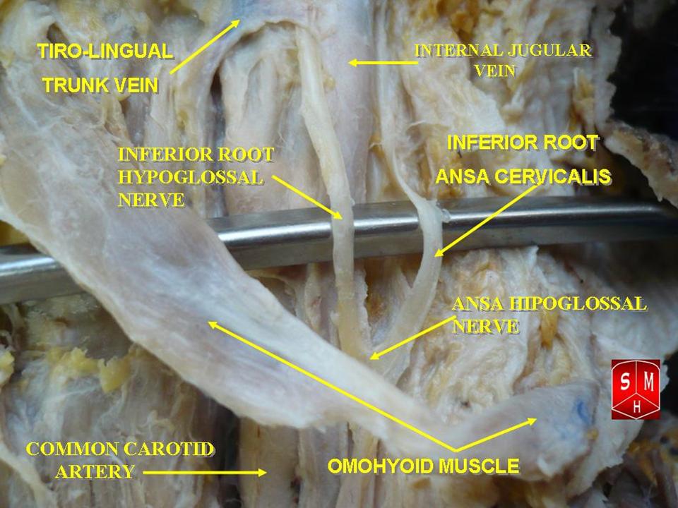
The ansa cervicalis (or ansa hypoglossi in older literature) is a loop of nerves that are part of the cervical plexus. It lies superficial to the internal jugular vein in the carotid triangle. Its name means "handle of the neck" in Latin. Branches from the ansa cervicalis innervate most of the infrahyoid muscles, including the sternothyroid muscle, sternohyoid muscle and the omohyoid muscle. Note that the thyrohyoid muscle, which is also an infrahyoid muscle and the geniohyoid muscle which is a suprahyoid muscle are innervated by cervical spinal nerve 1 via the hypoglossal nerve. Roots Two roots make up the ansa cervicalis, a superior root, and an inferior root. The superior root of the ansa cervicalis is formed from cervical spinal nerve 1 of the cervical plexus. These nerve fibers travel in the hypoglossal nerve before separating in the carotid triangle to form the superior root. The superior root goes around the occipital artery and then descends on the carotid sheath. I ... [...More Info...] [...Related Items...] OR: [Wikipedia] [Google] [Baidu] |
Phrenic Nerve
The phrenic nerve is a mixed motor/sensory nerve which originates from the C3-C5 spinal nerves in the neck. The nerve is important for breathing because it provides exclusive motor control of the diaphragm, the primary muscle of respiration. In humans, the right and left phrenic nerves are primarily supplied by the C4 spinal nerve, but there is also contribution from the C3 and C5 spinal nerves. From its origin in the neck, the nerve travels downward into the chest to pass between the heart and lungs towards the diaphragm. In addition to motor fibers, the phrenic nerve contains sensory fibers, which receive input from the central tendon of the diaphragm and the mediastinal pleura, as well as some sympathetic nerve fibers. Although the nerve receives contributions from nerves roots of the cervical plexus and the brachial plexus, it is usually considered separate from either plexus. The name of the nerve comes from Ancient Greek ''phren'' 'diaphragm'. Structure The phrenic n ... [...More Info...] [...Related Items...] OR: [Wikipedia] [Google] [Baidu] |
Subclavian Vein
The subclavian vein is a paired large vein, one on either side of the body, that is responsible for draining blood from the upper extremities, allowing this blood to return to the heart. The left subclavian vein plays a key role in the absorption of lipids, by allowing products that have been carried by lymph in the thoracic duct to enter the bloodstream. The diameter of the subclavian veins is approximately 1–2 cm, depending on the individual. Structure Each subclavian vein is a continuation of the axillary vein and runs from the outer border of the first rib to the medial border of anterior scalene muscle. From here it joins with the internal jugular vein to form the brachiocephalic vein (also known as "innominate vein"). The angle of union is termed the venous angle. The subclavian vein follows the subclavian artery and is separated from the subclavian artery by the insertion of anterior scalene. Thus, the subclavian vein lies anterior to the anterior scalene while the su ... [...More Info...] [...Related Items...] OR: [Wikipedia] [Google] [Baidu] |


