|
Nectin-1
Poliovirus receptor-related 1 (PVRL1), also known as nectin-1 and CD111 (formerly herpesvirus entry mediator C, HVEC) is a human protein of the immunoglobulin superfamily (IgSF), also considered a member of the nectins. It is a membrane protein with three extracellular immunoglobulin domains, a single transmembrane helix and a cytoplasmic tail. The protein can mediate Calcium in biology, Ca2+-independent cellular adhesion further characterizing it as IgSF cell adhesion molecule (IgSF CAM). Function PVRL1 is an adhesion molecule found in a wide range of tissues where it localizes in various junctions such as the adherens junction of epithelial tissue or the chemical synapse of neurons. The cytoplasmic tail of PVRL1 can bind the protein MLLT4, afadin which is a scaffolding protein that binds actin. In the chemical synapse PVRL1 interacts with PVRL3 (nectin-3) and both proteins can be found in neuronal tissue already in early stages of brain development as well as in aging brains ... [...More Info...] [...Related Items...] OR: [Wikipedia] [Google] [Baidu] |
Nectin
Nectins and Nectin-like molecules (Necl) are families of cellular adhesion molecules involved in Ca2+-independent cellular adhesion. Nectins are ubiquitously expressed and have adhesive roles in a wide range of tissues such as the adherens junction of epithelia or the chemical synapse of the neuronal tissue. Diversity So far four nectins have been identified in humans, namely nectin-1, nectin-2, nectin-3 and nectin-4. These four family members have also been found in most other well studied mammals. Also, five Necls have been identified, these are: Necl-1, Necl-2, Necl-3, Necl-4 and Necl-5. Structure All nectins and all Necls share the same overall structure defined by three extra cellular immunoglobulin domains, a single transmembrane helix and an intracellular domain. For all nectins the intracellular domain can bind a scaffold protein named afadin (the product of the MLLT4 gene). All nectins and Necls can form homo-cis dimers, meaning a dimer of two alike molecules o ... [...More Info...] [...Related Items...] OR: [Wikipedia] [Google] [Baidu] |
PVRL3
Nectin-3, also known as nectin cell adhesion molecule 3, is a protein that in humans is encoded by the ''NECTIN3'' gene. Nectin-3 belongs to the family of immunoglobulin(Ig)-like cellular adhesion molecules involved in Ca2+-independent cellular adhesion in several tissues during the development and was firstly isolated at the turn of 20th and 21st century. Structure and localization Nectin-3 has three splicing variants, nectin-3α, which is the biggest one, nectin-3β and the smallest variant nectin-3γ. Nectin-3α (same as the other splicing variants) is abundately expressed in testis, on slightly level it is also expressed in heart, brain, liver or kidney. It has been also proved that nectin-3α is together with nectin-2 localize at the junctional complex regions in small intestina absorptive epitelia. Nectin-3γ is also detectable in lung, liver and kidney. Nectin-3 is expressed not only on epithelial cells as another nectins, but there was shown that, as the only membe ... [...More Info...] [...Related Items...] OR: [Wikipedia] [Google] [Baidu] |
Immunoglobulin Superfamily
The immunoglobulin superfamily (IgSF) is a large protein superfamily of cell surface and soluble proteins that are involved in the recognition, binding, or adhesion processes of cells. Molecules are categorized as members of this superfamily based on shared structural features with immunoglobulins (also known as antibodies); they all possess a domain known as an immunoglobulin domain or fold. Members of the IgSF include cell surface antigen receptors, co-receptors and co-stimulatory molecules of the immune system, molecules involved in antigen presentation to lymphocytes, cell adhesion molecules, certain cytokine receptors and intracellular muscle proteins. They are commonly associated with roles in the immune system. Otherwise, the sperm-specific protein IZUMO1, a member of the immunoglobulin superfamily, has also been identified as the only sperm membrane protein essential for sperm-egg fusion. Immunoglobulin domains Proteins of the IgSF possess a structural domain known as ... [...More Info...] [...Related Items...] OR: [Wikipedia] [Google] [Baidu] |
Calcium In Biology
Calcium ions (Ca2+) contribute to the physiology and biochemistry of organisms' cell (biology), cells. They play an important role in signal transduction pathways, where they act as a second messenger, in neurotransmitter release from neurons, in contraction of all muscle cell types, and in fertilization. Many enzymes require calcium ions as a Cofactor (biochemistry), cofactor, including several of the coagulation factors. Extracellular calcium is also important for maintaining the potential difference across excitable cell cell membrane, membranes, as well as proper bone formation. Plasma calcium levels in mammals are tightly regulated, electronic-book electronic- with bone acting as the major mineral storage site. Calcium ions, Ca2+, are released from bone into the bloodstream under controlled conditions. Calcium is transported through the bloodstream as dissolved ions or bound to proteins such as serum albumin. Parathyroid hormone secreted by the parathyroid gland regulates ... [...More Info...] [...Related Items...] OR: [Wikipedia] [Google] [Baidu] |
Cellular Adhesion
Cell adhesion is the process by which cells interact and attach to neighbouring cells through specialised molecules of the cell surface. This process can occur either through direct contact between cell surfaces such as cell junctions or indirect interaction, where cells attach to surrounding extracellular matrix, a gel-like structure containing molecules released by cells into spaces between them. Cells adhesion occurs from the interactions between cell-adhesion molecules (CAMs), transmembrane proteins located on the cell surface. Cell adhesion links cells in different ways and can be involved in signal transduction for cells to detect and respond to changes in the surroundings. Other cellular processes regulated by cell adhesion include cell migration and tissue development in multicellular organisms. Alterations in cell adhesion can disrupt important cellular processes and lead to a variety of diseases, including cancer and arthritis. Cell adhesion is also essential for infec ... [...More Info...] [...Related Items...] OR: [Wikipedia] [Google] [Baidu] |
Adherens Junction
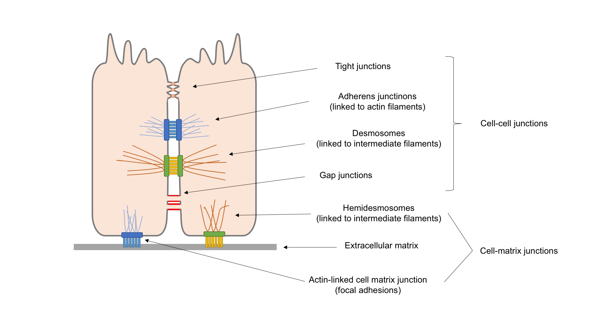
Adherens junctions (or zonula adherens, intermediate junction, or "belt desmosome") are protein complexes that occur at cell–cell junctions, cell–matrix junctions in epithelial and endothelial tissues, usually more basal than tight junctions. An adherens junction is defined as a cell junction whose cytoplasmic face is linked to the actin cytoskeleton. They can appear as bands encircling the cell (zonula adherens) or as spots of attachment to the extracellular matrix (focal adhesion). Adherens junctions uniquely disassemble in uterine epithelial cells to allow the blastocyst to penetrate between epithelial cells. A similar cell junction in non-epithelial, non-endothelial cells is the fascia adherens. It is structurally the same, but appears in ribbonlike patterns that do not completely encircle the cells. One example is in cardiomyocytes. Proteins Adherens junctions are composed of the following proteins: * cadherins. The cadherins are a family of transmembrane proteins tha ... [...More Info...] [...Related Items...] OR: [Wikipedia] [Google] [Baidu] |
Epithelial Tissue
Epithelium or epithelial tissue is one of the four basic types of animal tissue, along with connective tissue, muscle tissue and nervous tissue. It is a thin, continuous, protective layer of compactly packed cells with a little intercellular matrix. Epithelial tissues line the outer surfaces of organs and blood vessels throughout the body, as well as the inner surfaces of cavities in many internal organs. An example is the epidermis, the outermost layer of the skin. There are three principal shapes of epithelial cell: squamous (scaly), columnar, and cuboidal. These can be arranged in a singular layer of cells as simple epithelium, either squamous, columnar, or cuboidal, or in layers of two or more cells deep as stratified (layered), or ''compound'', either squamous, columnar or cuboidal. In some tissues, a layer of columnar cells may appear to be stratified due to the placement of the nuclei. This sort of tissue is called pseudostratified. All glands are made up of epith ... [...More Info...] [...Related Items...] OR: [Wikipedia] [Google] [Baidu] |
Chemical Synapse
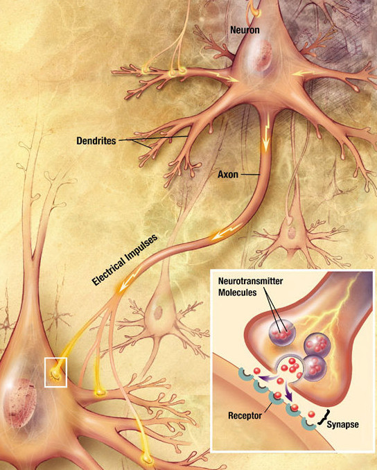
Chemical synapses are biological junctions through which neurons' signals can be sent to each other and to non-neuronal cells such as those in muscles or glands. Chemical synapses allow neurons to form circuits within the central nervous system. They are crucial to the biological computations that underlie perception and thought. They allow the nervous system to connect to and control other systems of the body. At a chemical synapse, one neuron releases neurotransmitter molecules into a small space (the synaptic cleft) that is adjacent to another neuron. The neurotransmitters are contained within small sacs called synaptic vesicles, and are released into the synaptic cleft by exocytosis. These molecules then bind to neurotransmitter receptors on the postsynaptic cell. Finally, the neurotransmitters are cleared from the synapse through one of several potential mechanisms including enzymatic degradation or re-uptake by specific transporters either on the presynaptic cell o ... [...More Info...] [...Related Items...] OR: [Wikipedia] [Google] [Baidu] |
Neurons
A neuron, neurone, or nerve cell is an electrically excitable cell that communicates with other cells via specialized connections called synapses. The neuron is the main component of nervous tissue in all animals except sponges and placozoa. Non-animals like plants and fungi do not have nerve cells. Neurons are typically classified into three types based on their function. Sensory neurons respond to stimuli such as touch, sound, or light that affect the cells of the sensory organs, and they send signals to the spinal cord or brain. Motor neurons receive signals from the brain and spinal cord to control everything from muscle contractions to glandular output. Interneurons connect neurons to other neurons within the same region of the brain or spinal cord. When multiple neurons are connected together, they form what is called a neural circuit. A typical neuron consists of a cell body (soma), dendrites, and a single axon. The soma is a compact structure, and the axon and dend ... [...More Info...] [...Related Items...] OR: [Wikipedia] [Google] [Baidu] |
MLLT4
Afadin is a protein that in humans is encoded by the ''AFDN'' gene. Function Afadin is a Ras (see HRAS; MIM 190020) target that regulates cell–cell adhesions downstream of Ras activation. It is fused with MLL (MIM 159555) in leukemias caused by t(6;11) translocations (Taya et al., 1998). upplied by OMIMref name="entrez"> Interactions Afadin has been shown to interact with: * BCR gene, * EPHB3, * F11 receptor, * HRAS and * LMO2, * PVRL1, * PVRL3, * Profilin 1, * RAP1A, * RAP1GAP, * SORBS1, * SSX2IP, * Tight junction protein 1 Zonula occludens-1 ZO-1, also known as Tight junction protein-1 is a 220-kD peripheral membrane protein that is encoded by the ''TJP1'' gene in humans. It belongs to the family of ''zonula occludens proteins'' (ZO-1, ZO-2, and ZO-3), which are ti ..., and * USP9X. References Further reading * * * * * * * * * * * * * * * * * * * {{PDB Gallery, geneid=4301 ... [...More Info...] [...Related Items...] OR: [Wikipedia] [Google] [Baidu] |
Actin
Actin is a family of globular multi-functional proteins that form microfilaments in the cytoskeleton, and the thin filaments in muscle fibrils. It is found in essentially all eukaryotic cells, where it may be present at a concentration of over 100 μM; its mass is roughly 42 kDa, with a diameter of 4 to 7 nm. An actin protein is the monomeric subunit of two types of filaments in cells: microfilaments, one of the three major components of the cytoskeleton, and thin filaments, part of the contractile apparatus in muscle cells. It can be present as either a free monomer called G-actin (globular) or as part of a linear polymer microfilament called F-actin (filamentous), both of which are essential for such important cellular functions as the mobility and contraction of cells during cell division. Actin participates in many important cellular processes, including muscle contraction, cell motility, cell division and cytokinesis, vesicle and organelle movement, cell sign ... [...More Info...] [...Related Items...] OR: [Wikipedia] [Google] [Baidu] |
Axon
An axon (from Greek ἄξων ''áxōn'', axis), or nerve fiber (or nerve fibre: see spelling differences), is a long, slender projection of a nerve cell, or neuron, in vertebrates, that typically conducts electrical impulses known as action potentials away from the nerve cell body. The function of the axon is to transmit information to different neurons, muscles, and glands. In certain sensory neurons (pseudounipolar neurons), such as those for touch and warmth, the axons are called afferent nerve fibers and the electrical impulse travels along these from the periphery to the cell body and from the cell body to the spinal cord along another branch of the same axon. Axon dysfunction can be the cause of many inherited and acquired neurological disorders that affect both the peripheral and central neurons. Nerve fibers are classed into three typesgroup A nerve fibers, group B nerve fibers, and group C nerve fibers. Groups A and B are myelinated, and group C are unmyelinated. ... [...More Info...] [...Related Items...] OR: [Wikipedia] [Google] [Baidu] |


