|
Nervi Erigentes
Pelvic splanchnic nerves or nervi erigentes are splanchnic nerves that arise from sacral spinal nerves S2, S3, S4 to provide parasympathetic innervation to the organs of the pelvic cavity. Structure The pelvic splanchnic nerves arise from the anterior rami of the sacral spinal nerves S2, S3, and S4, and enter the sacral plexus. They travel to their side's corresponding inferior hypogastric plexus, located bilaterally on the walls of the rectum. They contain both preganglionic parasympathetic fibers as well as visceral afferent fibers. Visceral afferent fibers go to spinal cord following pathway of pelvic splanchnic nerve fibers. The parasympathetic nervous system is referred to as the ''craniosacral outflow''; the pelvic splanchnic nerves are the ''sacral'' component. They are in the same region as the sacral splanchnic nerves, which arise from the sympathetic trunk and provide sympathetic efferent fibers. Function The pelvic splanchnic nerves contribute to the innervation ... [...More Info...] [...Related Items...] OR: [Wikipedia] [Google] [Baidu] |
Splanchnic Nerves
The splanchnic nerves are paired visceral nerves (nerves that contribute to the innervation of the viscera, innervation of the internal organs), carrying fibers of the autonomic nervous system (visceral efferent fibers) as well as sensory fibers from the organs (visceral afferent fibers). All carry Sympathetic nervous system, sympathetic fibers except for the pelvic splanchnic nerves, which carry parasympathetic fibers. Types The term ''splanchnic nerves'' can refer to: * Cardiopulmonary nervesEssential Clinical Anatomy. K.L. Moore & A.M. Agur. Lippincott, 3 ed. 2007. Page 181 * Thoracic splanchnic nerves (greater, lesser, and least) * Lumbar splanchnic nerves * Sacral splanchnic nerves * Pelvic splanchnic nerves References {{Autonomic Autonomic nervous system ... [...More Info...] [...Related Items...] OR: [Wikipedia] [Google] [Baidu] |
Sympathetic Trunk
The sympathetic trunks (sympathetic chain, gangliated cord) are a paired bundle of nerve fibers that run from the base of the skull to the coccyx. They are a major component of the sympathetic nervous system. Structure The sympathetic trunk lies just lateral to the vertebral bodies for the entire length of the vertebral column. It interacts with the anterior rami of spinal nerves by way of rami communicantes. The sympathetic trunk permits preganglionic fibers of the sympathetic nervous system to ascend to spinal levels superior to T1 and descend to spinal levels inferior to L2/3.Greenstein B., Greenstein A. (2002): Color atlas of neuroscience – Neuroanatomy and neurophysiology. Thieme, Stuttgart – New York, . The superior end of it is continued upward through the carotid canal into the skull, and forms a plexus on the internal carotid artery; the inferior part travels in front of the coccyx, where it converges with the other trunk at a structure known as the ganglion impar. ... [...More Info...] [...Related Items...] OR: [Wikipedia] [Google] [Baidu] |
Parasites Lost
"Parasites Lost" is the second episode in season three of ''Futurama''. Although the title is a play on John Milton's epic poem ''Paradise Lost'', the episode is a parody of the 1966 film ''Fantastic Voyage''. It originally aired on the Fox network in the United States on January 21, 2001. Plot While making a pit stop at an interstellar truck stop, Fry buys and eats a decaying egg salad sandwich from a vending machine in the restroom. Upon returning to Earth, Fry and Bender are assigned the task of fixing the plasma fusion boiler, which promptly explodes. Bender is not damaged, but Fry is impaled by a large pipe. Despite the severity of the injury, Fry's damaged body repairs itself in seconds, and the subsequent medical examination reveals to the crew that Fry is infested with microscopic worms from the egg salad sandwich. To eliminate the infestation, Professor Farnsworth makes miniature robotic versions of the crew, except for Fry and Leela. Because the worms know all that F ... [...More Info...] [...Related Items...] OR: [Wikipedia] [Google] [Baidu] |
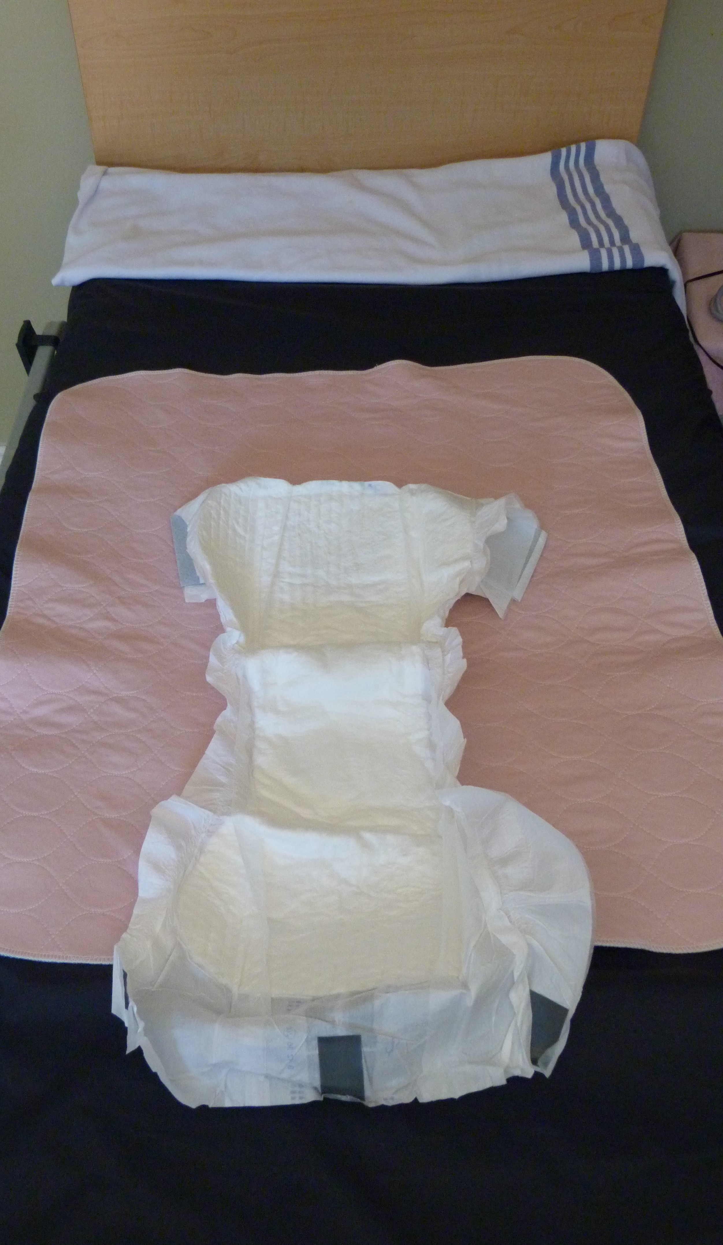
Fecal Incontinence
Fecal incontinence (FI), or in some forms encopresis, is a lack of control over defecation, leading to involuntary loss of bowel contents, both liquid stool elements and mucus, or solid feces. When this loss includes flatus (gas), it is referred to as anal incontinence. FI is a sign or a symptom, not a diagnosis. Incontinence can result from different causes and might occur with either constipation or diarrhea. Continence is maintained by several interrelated factors, including the anal sampling mechanism, and incontinence usually results from deficiency of multiple mechanisms. The most common causes are thought to be immediate or delayed damage from childbirth, complications from prior anorectal surgery (especially involving the anal sphincters or hemorrhoidal vascular cushions), altered bowel habits (e.g., caused by irritable bowel syndrome, Crohn's disease, ulcerative colitis, food intolerance, or constipation with overflow incontinence), and receptive anal sex. An estimated ... [...More Info...] [...Related Items...] OR: [Wikipedia] [Google] [Baidu] |
Neurogenic Bladder Dysfunction
Neurogenic bladder dysfunction, or neurogenic bladder, refers to urinary bladder problems due to disease or injury of the central nervous system or peripheral nerves involved in the control of urination. There are multiple types of neurogenic bladder depending on the underlying cause and the symptoms. Symptoms include overactive bladder, urinary urgency, frequency, incontinence or difficulty passing urine. A range of diseases or conditions can cause neurogenic bladder including spinal cord injury, multiple sclerosis, stroke, brain injury, spina bifida, peripheral nerve damage, Parkinson's disease, or other neurodegenerative diseases. Neurogenic bladder can be diagnosed through a history and physical as well as imaging and more specialized testing. Treatment depends on underlying disease as well as symptoms and can be managed with behavioral changes, medications, surgeries, or other procedures. The symptoms of neurogenic bladder, especially incontinence, can have a significant impact ... [...More Info...] [...Related Items...] OR: [Wikipedia] [Google] [Baidu] |
Vagus Nerve
The vagus nerve, also known as the tenth cranial nerve, cranial nerve X, or simply CN X, is a cranial nerve that interfaces with the parasympathetic control of the heart, lungs, and digestive tract. It comprises two nerves—the left and right vagus nerves—but they are typically referred to collectively as a single subsystem. The vagus is the longest nerve of the autonomic nervous system in the human body and comprises both sensory and motor fibers. The sensory fibers originate from neurons of the nodose ganglion, whereas the motor fibers come from neurons of the dorsal motor nucleus of the vagus and the nucleus ambiguus. The vagus was also historically called the pneumogastric nerve. Structure Upon leaving the medulla oblongata between the olive and the inferior cerebellar peduncle, the vagus nerve extends through the jugular foramen, then passes into the carotid sheath between the internal carotid artery and the internal jugular vein down to the neck, chest, and abdom ... [...More Info...] [...Related Items...] OR: [Wikipedia] [Google] [Baidu] |
Gastrointestinal Tract
The gastrointestinal tract (GI tract, digestive tract, alimentary canal) is the tract or passageway of the digestive system that leads from the mouth to the anus. The GI tract contains all the major organ (biology), organs of the digestive system, in humans and other animals, including the esophagus, stomach, and intestines. Food taken in through the mouth is digestion, digested to extract nutrients and absorb energy, and the waste expelled at the anus as feces. ''Gastrointestinal'' is an adjective meaning of or pertaining to the stomach and intestines. Nephrozoa, Most animals have a "through-gut" or complete digestive tract. Exceptions are more primitive ones: sponges have small pores (ostium (sponges), ostia) throughout their body for digestion and a larger dorsal pore (osculum) for excretion, comb jellies have both a ventral mouth and dorsal anal pores, while cnidarians and acoels have a single pore for both digestion and excretion. The human gastrointestinal tract consists o ... [...More Info...] [...Related Items...] OR: [Wikipedia] [Google] [Baidu] |
Transverse Colon
In human anatomy, the transverse colon is the longest and most movable part of the colon. Anatomical position It crosses the abdomen from the ascending colon at the right colic flexure (hepatic flexure) with a downward convexity to the descending colon where it curves sharply on itself beneath the lower end of the spleen forming the left colic flexure (splenic flexure). In its course, it describes an arch, the concavity of which is directed backward and a little upward. Toward its splenic end there is often an abrupt U-shaped curve which may descend lower than the main curve. It is almost completely invested by the peritoneum, and is connected to the inferior border of the pancreas by a large and wide duplicature of that membrane, the transverse mesocolon. It is in relation, by its upper surface, with the liver and gall-bladder, the greater curvature of the stomach, and the lower end of the spleen; by its under surface, with the small intestine; by its anterior surface, with t ... [...More Info...] [...Related Items...] OR: [Wikipedia] [Google] [Baidu] |
Cervix
The cervix or cervix uteri (Latin, 'neck of the uterus') is the lower part of the uterus (womb) in the human female reproductive system. The cervix is usually 2 to 3 cm long (~1 inch) and roughly cylindrical in shape, which changes during pregnancy. The narrow, central cervical canal runs along its entire length, connecting the uterine cavity and the lumen of the vagina. The opening into the uterus is called the internal os, and the opening into the vagina is called the external os. The lower part of the cervix, known as the vaginal portion of the cervix (or ectocervix), bulges into the top of the vagina. The cervix has been documented anatomically since at least the time of Hippocrates, over 2,000 years ago. The cervical canal is a passage through which sperm must travel to fertilize an egg cell after sexual intercourse. Several methods of contraception, including cervical caps and cervical diaphragms, aim to block or prevent the passage of sperm through the cervical ... [...More Info...] [...Related Items...] OR: [Wikipedia] [Google] [Baidu] |
Erection
An erection (clinically: penile erection or penile tumescence) is a physiological phenomenon in which the penis becomes firm, engorged, and enlarged. Penile erection is the result of a complex interaction of psychological, neural, vascular, and endocrine factors, and is often associated with sexual arousal or sexual attraction, although erections can also be spontaneous. The shape, angle, and direction of an erection varies considerably between humans. Physiologically, an erection is required for a male to effect vaginal penetration or sexual intercourse and is triggered by the parasympathetic division of the autonomic nervous system, causing the levels of nitric oxide (a vasodilator) to rise in the trabecular arteries and smooth muscle of the penis. The arteries dilate causing the corpora cavernosa of the penis (and to a lesser extent the corpus spongiosum) to fill with blood; simultaneously the ischiocavernosus and bulbospongiosus muscles compress the veins of the ... [...More Info...] [...Related Items...] OR: [Wikipedia] [Google] [Baidu] |
Rectum
The rectum is the final straight portion of the large intestine in humans and some other mammals, and the Gastrointestinal tract, gut in others. The adult human rectum is about long, and begins at the rectosigmoid junction (the end of the sigmoid colon) at the level of the third sacral vertebra or the sacral promontory depending upon what definition is used. Its diameter is similar to that of the sigmoid colon at its commencement, but it is dilated near its termination, forming the rectal ampulla. It terminates at the level of the anorectal ring (the level of the puborectalis sling) or the dentate line, again depending upon which definition is used. In humans, the rectum is followed by the anal canal which is about long, before the gastrointestinal tract terminates at the anal verge. The word rectum comes from the Latin ''Wikt:rectum, rectum Wikt:intestinum, intestinum'', meaning ''straight intestine''. Structure The rectum is a part of the lower gastrointestinal tract ... [...More Info...] [...Related Items...] OR: [Wikipedia] [Google] [Baidu] |
Internal Urethral Sphincter
The internal urethral sphincter is a urethral sphincter muscle which constricts the internal urethral orifice. It is located at the junction of the urethra with the urinary bladder and is continuous with the detrusor muscle, but anatomically and functionally fully independent from it., page 29Preview Amazon It is composed of smooth muscle, so it is under the control of the autonomic nervous system, specifically the sympathetic nervous system. Function This is the primary muscle for maintaining continence of urine, a function shared with the external urethral sphincter which is under voluntary control. It prevents urine leakage as the muscle is tonically contracted via sympathetic fibers traveling through the inferior hypogastric plexus and vesical nervous plexus. Specifically, it is controlled by the hypogastric nerve, predominantly via the alpha-1 receptor. During urination, the preganglionic neurons of this sympathetic pathway are inhibited via signals arising in the pontine ... [...More Info...] [...Related Items...] OR: [Wikipedia] [Google] [Baidu] |


