|
Myoglobinuria
Myoglobinuria is the presence of myoglobin in the urine, which usually results from rhabdomyolysis or muscle injury. Myoglobin is present in muscle cells as a reserve of oxygen. Signs and symptoms Signs and symptoms of myoglobinuria are usually nonspecific and needs some clinical prudence. Therefore, among the possible signs and symptoms to look for would be: *Swollen and painful muscles *Fever, nausea * Delirium (elderly individuals) * Myalgia * Dark urine * Calcium ion loss Causes Trauma, vascular problems, malignant hyperthermia, certain drugs and other situations can destroy or damage the muscle, releasing myoglobin to the circulation and thus to the kidneys. Under ideal situations myoglobin will be filtered and excreted with the urine, but if too much myoglobin is released into the circulation or in case of kidney problems, it can occlude the kidneys' filtration system leading to acute tubular necrosis and acute kidney injury. Other causes of myoglobinuria includ ... [...More Info...] [...Related Items...] OR: [Wikipedia] [Google] [Baidu] |
Rhabdomyolysis
Rhabdomyolysis (also called rhabdo) is a condition in which damaged skeletal muscle breaks down rapidly. Symptoms may include muscle pains, weakness, vomiting, and confusion. There may be tea-colored urine or an irregular heartbeat. Some of the muscle breakdown products, such as the protein myoglobin, are harmful to the kidneys and can cause acute kidney injury. The muscle damage is mostly caused by a crush injury, strenuous exercise, medications, or a substance use disorder. Other causes include infections, electrical injury, heat stroke, prolonged immobilization, lack of blood flow to a limb, or snake bites. Statins (prescription drugs to lower cholesterol) are considered a small risk. Some people have inherited muscle conditions that increase the risk of rhabdomyolysis. The diagnosis is supported by a urine test strip which is positive for "blood" but the urine contains no red blood cells when examined with a microscope. Blood tests show a creatine kinase activi ... [...More Info...] [...Related Items...] OR: [Wikipedia] [Google] [Baidu] |
McArdle's Disease
Glycogen storage disease type V (GSD5, GSD-V), also known as McArdle's disease, is a metabolic disorder, one of the metabolic myopathies, more specifically a muscle glycogen storage disease, caused by a deficiency of myophosphorylase. Its incidence is reported as one in 100,000, roughly the same as glycogen storage disease type I. The disease was first reported in 1951 by Dr. Brian McArdle of Guy's Hospital, London. Signs and symptoms The onset of this disease is usually noticed in childhood, but often not diagnosed until the third or fourth decade of life. Symptoms include exercise intolerance with muscle pain, early fatigue, painful cramps, inappropriate rapid heart rate response to exercise, and may include myoglobin in the urine (often provoked by a bout of exercise).Lucia A, Martinuzzi A, Nogales-Gadea G, Quinlivan R, Reason S; International Association for Muscle Glycogen Storage Disease study group. Clinical practice guidelines for glycogen storage disease V & VII (McA ... [...More Info...] [...Related Items...] OR: [Wikipedia] [Google] [Baidu] |
Myoglobin
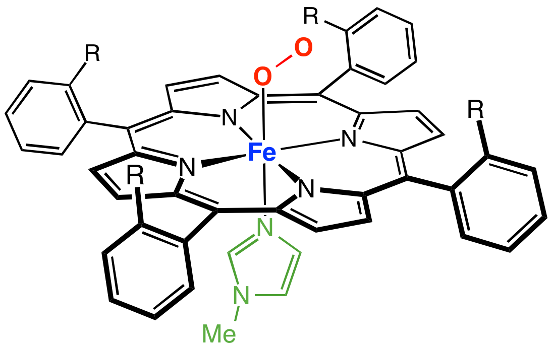
Myoglobin (symbol Mb or MB) is an iron- and oxygen-binding protein found in the cardiac and skeletal muscle tissue of vertebrates in general and in almost all mammals. Myoglobin is distantly related to hemoglobin. Compared to hemoglobin, myoglobin has a higher affinity for oxygen and does not have cooperative binding with oxygen like hemoglobin does. In humans, myoglobin is only found in the bloodstream after muscle injury. (Google books link is the 2008 edition) High concentrations of myoglobin in muscle cells allow organisms to hold their breath for a longer period of time. Diving mammals such as whales and seals have muscles with particularly high abundance of myoglobin. Myoglobin is found in Type I muscle, Type II A, and Type II B; although many texts consider myoglobin not to be found in smooth muscle, this has proved erroneous: there is also myoglobin in smooth muscle cells. Myoglobin was the first protein to have its three-dimensional structure revealed by X-ray cryst ... [...More Info...] [...Related Items...] OR: [Wikipedia] [Google] [Baidu] |
Lactate Dehydrogenase
Lactate dehydrogenase (LDH or LD) is an enzyme found in nearly all living cells. LDH catalyzes the conversion of lactate to pyruvate and back, as it converts NAD+ to NADH and back. A dehydrogenase is an enzyme that transfers a hydride from one molecule to another. LDH exists in four distinct enzyme classes. This article is specifically about the NAD(P)-dependent L-lactate dehydrogenase. Other LDHs act on D-lactate and/or are dependent on cytochrome c: D-lactate dehydrogenase (cytochrome) and L-lactate dehydrogenase (cytochrome). LDH is expressed extensively in body tissues, such as blood cells and heart muscle. Because it is released during tissue damage, it is a marker of common injuries and disease such as heart failure. Reaction Lactate dehydrogenase catalyzes the interconversion of pyruvate and lactate with concomitant interconversion of NADH and NAD+. It converts pyruvate, the final product of glycolysis, to lactate when oxygen is absent or in short supply, a ... [...More Info...] [...Related Items...] OR: [Wikipedia] [Google] [Baidu] |
Myoglobin
Myoglobin (symbol Mb or MB) is an iron- and oxygen-binding protein found in the cardiac and skeletal muscle tissue of vertebrates in general and in almost all mammals. Myoglobin is distantly related to hemoglobin. Compared to hemoglobin, myoglobin has a higher affinity for oxygen and does not have cooperative binding with oxygen like hemoglobin does. In humans, myoglobin is only found in the bloodstream after muscle injury. (Google books link is the 2008 edition) High concentrations of myoglobin in muscle cells allow organisms to hold their breath for a longer period of time. Diving mammals such as whales and seals have muscles with particularly high abundance of myoglobin. Myoglobin is found in Type I muscle, Type II A, and Type II B; although many texts consider myoglobin not to be found in smooth muscle, this has proved erroneous: there is also myoglobin in smooth muscle cells. Myoglobin was the first protein to have its three-dimensional structure revealed by X-ray cryst ... [...More Info...] [...Related Items...] OR: [Wikipedia] [Google] [Baidu] |
Ionized Calcium
Calcium ions (Ca2+) contribute to the physiology and biochemistry of organisms' cells. They play an important role in signal transduction pathways, where they act as a second messenger, in neurotransmitter release from neurons, in contraction of all muscle cell types, and in fertilization. Many enzymes require calcium ions as a cofactor, including several of the coagulation factors. Extracellular calcium is also important for maintaining the potential difference across excitable cell membranes, as well as proper bone formation. Plasma calcium levels in mammals are tightly regulated, electronic-book electronic- with bone acting as the major mineral storage site. Calcium ions, Ca2+, are released from bone into the bloodstream under controlled conditions. Calcium is transported through the bloodstream as dissolved ions or bound to proteins such as serum albumin. Parathyroid hormone secreted by the parathyroid gland regulates the resorption of Ca2+ from bone, reabsorption ... [...More Info...] [...Related Items...] OR: [Wikipedia] [Google] [Baidu] |
Myocyte
A muscle cell is also known as a myocyte when referring to either a cardiac muscle cell (cardiomyocyte), or a smooth muscle cell as these are both small cells. A skeletal muscle cell is long and threadlike with many nuclei and is called a muscle fiber. Muscle cells (including myocytes and muscle fibers) develop from embryonic precursor cells called myoblasts. Myoblasts fuse to form multinucleated skeletal muscle cells known as syncytia in a process known as myogenesis. Skeletal muscle cells and cardiac muscle cells both contain myofibrils and sarcomeres and form a striated muscle tissue. Cardiac muscle cells form the cardiac muscle in the walls of the heart chambers, and have a single central nucleus. Cardiac muscle cells are joined to neighboring cells by intercalated discs, and when joined in a visible unit they are described as a ''cardiac muscle fiber''. Smooth muscle cells control involuntary movements such as the peristalsis contractions in the esophagus and stoma ... [...More Info...] [...Related Items...] OR: [Wikipedia] [Google] [Baidu] |
Burn
A burn is an injury to skin, or other tissues, caused by heat, cold, electricity, chemicals, friction, or ultraviolet radiation (like sunburn). Most burns are due to heat from hot liquids (called scalding), solids, or fire. Burns occur mainly in the home or the workplace. In the home, risks are associated with domestic kitchens, including stoves, flames, and hot liquids. In the workplace, risks are associated with fire and chemical and electric burns. Alcoholism and smoking are other risk factors. Burns can also occur as a result of self-harm or violence between people (assault). Burns that affect only the superficial skin layers are known as superficial or first-degree burns. They appear red without blisters and pain typically lasts around three days. When the injury extends into some of the underlying skin layer, it is a partial-thickness or second-degree burn. Blisters are frequently present and they are often very painful. Healing can require up to eight weeks and sc ... [...More Info...] [...Related Items...] OR: [Wikipedia] [Google] [Baidu] |
Adenosine Monophosphate Deaminase Deficiency Type 1
Adenosine monophosphate deaminase deficiency type 1 or AMPD1, is a human metabolic disorder in which the body consistently lacks the enzyme AMP deaminase, in sufficient quantities. This may result in exercise intolerance, muscle pain and muscle cramping. The disease was formerly known as myoadenylate deaminase deficiency. In virtually all cases, the deficiency has been caused by an SNP mutation, known as ''rs17602729'' or ''C34T''. While it was initially regarded as a recessive (or purely homozygous) disorder, some researchers have reported the existence of similarly deleterious effects from the heterozygous form of the SNP. In the homozygous form of the mutation, a single genetic base (character) has been changed from cytosine ("C") to thymine ("T") on both strands of Chromosome 1 – in other words, "C;C" has been replaced by "T;T". A rarer but analogous condition, in which two guanine bases ("G;G") bases (in the unmutated form) have been changed to adenine ("A;A") has also been ... [...More Info...] [...Related Items...] OR: [Wikipedia] [Google] [Baidu] |
Acute Tubular Necrosis
Acute tubular necrosis (ATN) is a medical condition involving the death of tubular epithelial cells that form the renal tubules of the kidneys. Because necrosis is often not present, the term acute tubular injury (ATI) is preferred by pathologists over the older name acute tubular necrosis (ATN). ATN presents with acute kidney injury (AKI) and is one of the most common causes of AKI. Common causes of ATN include low blood pressure and use of nephrotoxic drugs. The presence of "muddy brown casts" of epithelial cells found in the urine during urinalysis is pathognomonic for ATN. Management relies on aggressive treatment of the factors that precipitated ATN (e.g. hydration and cessation of the offending drug). Because the tubular cells continually replace themselves, the overall prognosis for ATN is quite good if the underlying cause is corrected, and recovery is likely within 7 to 21 days. Classification ATN may be classified as either ''toxic'' or ''ischemic''. Toxic ATN occurs whe ... [...More Info...] [...Related Items...] OR: [Wikipedia] [Google] [Baidu] |
Polymyositis
Polymyositis (PM) is a type of chronic inflammation of the muscles (inflammatory myopathy) related to dermatomyositis and inclusion body myositis. Its name means "inflammation of many muscles" ('' poly-'' + '' myos-'' + '' -itis''). The inflammation of polymyositis is mainly found in the endomysial layer of skeletal muscle, whereas dermatomyositis is characterized primarily by inflammation of the perimysial layer of skeletal muscles. Signs and symptoms The hallmark of polymyositis is weakness and/or loss of muscle mass in the proximal musculature, as well as flexion of the neck and torso. These symptoms can be associated with marked pain in these areas as well. The hip extensors are often severely affected, leading to particular difficulty in climbing stairs and rising from a seated position. The skin involvement of dermatomyositis is absent in polymyositis. Dysphagia (difficulty swallowing) or other problems with esophageal motility occur in as many as 1/3 of patients. Lo ... [...More Info...] [...Related Items...] OR: [Wikipedia] [Google] [Baidu] |
Malignant Hyperthermia
Malignant hyperthermia (MH) is a type of severe reaction that occurs in response to particular medications used during general anesthesia, among those who are susceptible. Symptoms include muscle rigidity, fever, and a fast heart rate. Complications can include muscle breakdown and high blood potassium. Most people who are susceptible to MH are generally unaffected when not exposed to triggering agents. Exposure to triggering agents (certain volatile anesthetic agents or succinylcholine) can lead to the development of MH in those who are susceptible. Susceptibility can occur due to at least six genetic mutations, with the most common one being of the RYR1 gene. These genetic variations are often inherited from a person's parents in an autosomal dominant manner. The condition may also occur as a new mutation or be associated with a number of inherited muscle diseases, such as central core disease. In susceptible individuals, the medications induce the release of stored ca ... [...More Info...] [...Related Items...] OR: [Wikipedia] [Google] [Baidu] |


