|
Muscles Of The Thumb
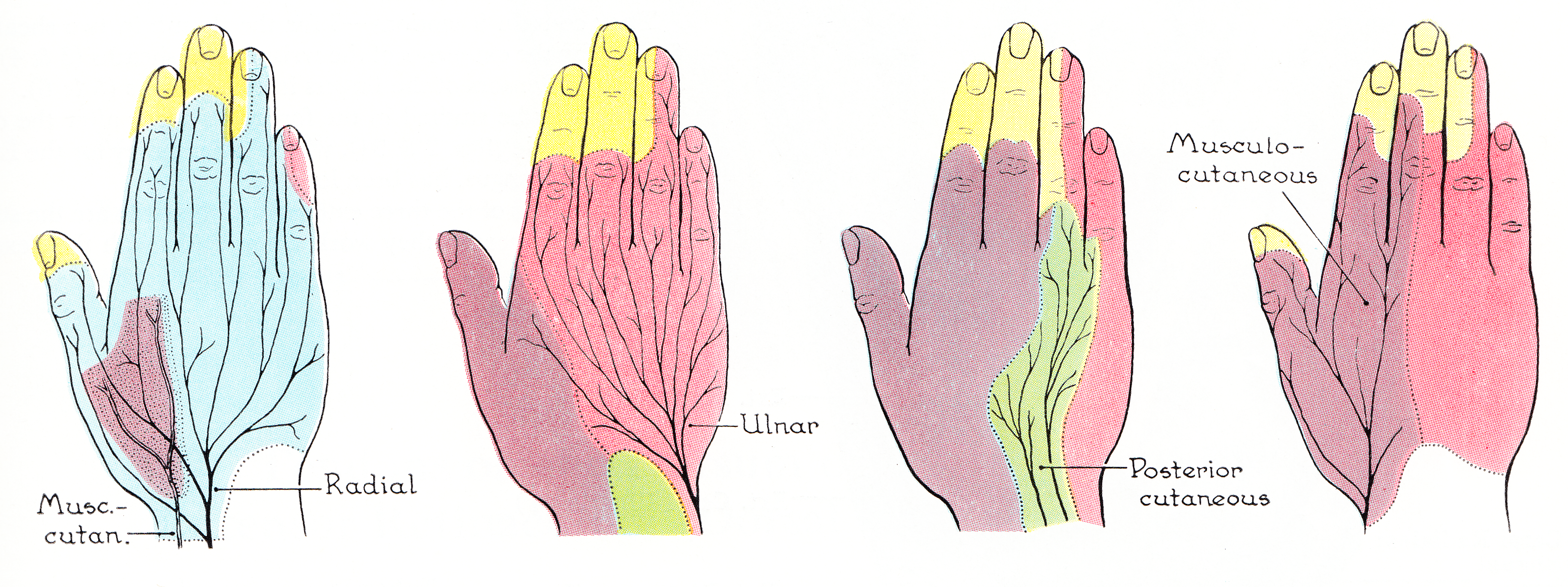
The muscles of the thumb are nine skeletal muscles located in the hand and forearm. The muscles allow for flexion, extension, adduction, abduction and opposition of the thumb. The muscles acting on the thumb can be divided into two groups: The extrinsic hand muscles, with their muscle bellies located in the forearm, and the intrinsic hand muscles, with their muscles bellies located in the hand proper. The muscles can be compared to guy-wires supporting a flagpole; tension from these muscular guy-wires must be provided in all directions to maintain stability in the articulated column formed by the bones of the thumb. Because this stability is actively maintained by muscles rather than by articular constraints, most muscles attached to the thumb tend to be active during most thumb motions. Extrinsic A ventral forearm muscle, the flexor pollicis longus originates on the anterior side of the radius distal to the radial tuberosity and from the interosseous membrane. It passes through ... [...More Info...] [...Related Items...] OR: [Wikipedia] [Google] [Baidu] |
Hand
A hand is a prehensile, multi-fingered appendage located at the end of the forearm or forelimb of primates such as humans, chimpanzees, monkeys, and lemurs. A few other vertebrates such as the koala (which has two opposable thumbs on each "hand" and fingerprints extremely similar to human fingerprints) are often described as having "hands" instead of paws on their front limbs. The raccoon is usually described as having "hands" though opposable thumbs are lacking. Some evolutionary anatomists use the term ''hand'' to refer to the appendage of digits on the forelimb more generally—for example, in the context of whether the three digits of the bird hand involved the same homologous loss of two digits as in the dinosaur hand. The human hand usually has five digits: four fingers plus one thumb; these are often referred to collectively as five fingers, however, whereby the thumb is included as one of the fingers. It has 27 bones, not including the sesamoid bone, the numbe ... [...More Info...] [...Related Items...] OR: [Wikipedia] [Google] [Baidu] |
Skeletal Muscles
Skeletal muscles (commonly referred to as muscles) are organs of the vertebrate muscular system and typically are attached by tendons to bones of a skeleton. The muscle cells of skeletal muscles are much longer than in the other types of muscle tissue, and are often known as muscle fibers. The muscle tissue of a skeletal muscle is striated – having a striped appearance due to the arrangement of the sarcomeres. Skeletal muscles are voluntary muscles under the control of the somatic nervous system. The other types of muscle are cardiac muscle which is also striated and smooth muscle which is non-striated; both of these types of muscle tissue are classified as involuntary, or, under the control of the autonomic nervous system. A skeletal muscle contains multiple muscle fascicle, fascicles – bundles of muscle fibers. Each individual fiber, and each muscle is surrounded by a type of connective tissue layer of fascia. Muscle fibers are formed from the cell fusion, fusion of dev ... [...More Info...] [...Related Items...] OR: [Wikipedia] [Google] [Baidu] |
Ulna
The ulna (''pl''. ulnae or ulnas) is a long bone found in the forearm that stretches from the elbow to the smallest finger, and when in anatomical position, is found on the medial side of the forearm. That is, the ulna is on the same side of the forearm as the little finger. It runs parallel to the radius, the other long bone in the forearm. The ulna is usually slightly longer than the radius, but the radius is thicker. Therefore, the radius is considered to be the larger of the two. Structure The ulna is a long bone found in the forearm that stretches from the elbow to the smallest finger, and when in anatomical position, is found on the medial side of the forearm. It is broader close to the elbow, and narrows as it approaches the wrist. Close to the elbow, the ulna has a bony process, the olecranon process, a hook-like structure that fits into the olecranon fossa of the humerus. This prevents hyperextension and forms a hinge joint with the trochlea of the humerus. Ther ... [...More Info...] [...Related Items...] OR: [Wikipedia] [Google] [Baidu] |
Abductor Pollicis Longus Muscle
In human anatomy, the abductor pollicis longus (APL) is one of the extrinsic muscles of the hand. Its major function is to abduct the thumb at the wrist. Its tendon forms the anterior border of the anatomical snuffbox. Structure The abductor pollicis longus lies immediately below the supinator and is sometimes united with it. It arises from the lateral part of the dorsal surface of the body of the ulna, below the insertion of the anconeus, from the interosseous membrane, and from the middle third of the dorsal surface of the body of the radius.''Gray's Anatomy'' (1918), see infobox Passing obliquely downward and lateralward, it ends in a tendon, which runs through a groove on the lateral side of the lower end of the radius, accompanied by the tendon of the extensor pollicis brevis. The insertion is divided into a distal, superficial part and a proximal, deep part. The superficial part is inserted with one or more tendons into the radial side of the base of the first meta ... [...More Info...] [...Related Items...] OR: [Wikipedia] [Google] [Baidu] |
Cervical Spinal Nerve 8
The cervical spinal nerve 8 (C8) is a spinal nerve of the cervical segment. Nervous System -- Groups of Nerves It originates from the spinal column from below the (C7). Innervation The C8 nerve forms part of the and s via the , and the ...[...More Info...] [...Related Items...] OR: [Wikipedia] [Google] [Baidu] |
Median Nerve
The median nerve is a nerve in humans and other animals in the upper limb. It is one of the five main nerves originating from the brachial plexus. The median nerve originates from the lateral and medial cords of the brachial plexus, and has contributions from ventral roots of C5-C7 (lateral cord) and C8 and T1 (medial cord). The median nerve is the only nerve that passes through the carpal tunnel. Carpal tunnel syndrome is the disability that results from the median nerve being pressed in the carpal tunnel. Structure The median nerve arises from the branches from lateral and medial cords of the brachial plexus, courses through the anterior part of arm, forearm, and hand, and terminates by supplying the muscles of the hand. Arm After receiving inputs from both the lateral and medial cords of the brachial plexus, the median nerve enters the arm from the axilla at the inferior margin of the teres major muscle. It then passes vertically down and courses lateral to the brachial a ... [...More Info...] [...Related Items...] OR: [Wikipedia] [Google] [Baidu] |
Anterior Interosseous Nerve
The anterior interosseous nerve (volar interosseous nerve) is a branch of the median nerve that supplies the deep muscles on the anterior of the forearm, except the ulnar (medial) half of the flexor digitorum profundus. Its nerve roots come from C8 and T1. It accompanies the anterior interosseous artery along the anterior of the interosseous membrane of the forearm, in the interval between the flexor pollicis longus and flexor digitorum profundus, supplying the whole of the former and (most commonly) the radial half of the latter, and ending below in the pronator quadratus and wrist joint. Note that the median nerve supplies all flexor muscles of the forearm except for the ulnar half of flexor digitorum profundus and the flexor carpi ulnaris, which is a superficial muscle of the forearm. Innervation The anterior interosseous nerve classically innervates 2.5 muscles: which are deep muscles of the forearm * flexor pollicis longus * pronator quadratus * the radial (lateral) half of ... [...More Info...] [...Related Items...] OR: [Wikipedia] [Google] [Baidu] |
Tendon Sheath
A tendon sheath is a layer of synovial membrane around a tendon. It permits the tendon to stretch and not adhere to the surrounding fascia. It has two layers: * synovial sheath A synovial sheath is one of the two membranes of a tendon sheath which covers a tendon. The other membrane is the outer fibrous tendon sheath. The tendon invaginates the synovial sheath from one side so that the tendon is suspended from the membra ... * fibrous tendon sheath Fibroma of the tendon sheath has been described. References Musculoskeletal system {{anatomy-stub ... [...More Info...] [...Related Items...] OR: [Wikipedia] [Google] [Baidu] |
Carpal Tunnel
In the human body, the carpal tunnel or carpal canal is the passageway on the palmar side of the wrist that connects the forearm to the hand. The tunnel is bounded by the bones of the wrist and flexor retinaculum from connective tissue. Normally several tendons from the flexor group of forearm muscles and the median nerve pass through it. There are described cases of variable median artery occurrence. When any of the nine long flexor tendons passing through the narrow carpal canal swell or degenerate, the narrowing of the canal may result in the median nerve becoming entrapped or compressed, a common medical condition known as carpal tunnel syndrome (CTS). Structure The carpal bones that make up the wrist form an arch which is convex on the dorsal side of the hand and concave on the palmar side. The groove on the palmar side, the ''sulcus carpi'', is covered by the flexor retinaculum, a sheath of tough connective tissue, thus forming the carpal tunnel. On the side of ... [...More Info...] [...Related Items...] OR: [Wikipedia] [Google] [Baidu] |
Interosseous Membrane Of Forearm
The interosseous membrane of the forearm (rarely middle or intermediate radioulnar joint) is a fibrous sheet that connects the interosseous margins of the radius and the ulna. It is the main part of the radio-ulnar syndesmosis, a fibrous joint between the two bones. Function The interosseous membrane divides the forearm into anterior and posterior compartments, serves as a site of attachment for muscles of the forearm, and transfers loads placed on the forearm. The interosseous membrane is designed to shift compressive loads (as in doing a hand-stand) from the distal radius to the proximal ulna. The fibers within the interosseous membrane are oriented obliquely so that when force is applied the fibers are drawn taut, shifting more of the load to the ulna. This reduces the wear and tear of placing the whole load on a single joint. The role of the membrane in load shifting is illustrated when the interosseous membrane is cut; the forces on each bone equalize from their natural pro ... [...More Info...] [...Related Items...] OR: [Wikipedia] [Google] [Baidu] |
Radial Tuberosity
Beneath the neck of the radius, on the medial side, is an eminence, the radial tuberosity; its surface is divided into: * a ''posterior, rough portion'', for the insertion of the tendon of the biceps brachii. * an ''anterior, smooth portion'', on which a bursa ( grc-gre, Προῦσα, Proûsa, Latin: Prusa, ota, بورسه, Arabic:بورصة) is a city in northwestern Turkey and the administrative center of Bursa Province. The fourth-most populous city in Turkey and second-most populous in the ... is interposed between the tendon and the bone. Ligaments that support the elbow joint also attach to the radial tuberosity. References External links * * () Additional images File:Sobo 1909 124.png, Radial tuberosity shown. File:Radius.jpg, Anterior View. Radial tuberosity. File:Radius2.jpg, Posterior View. Radial tuberosity. File:Radius_post.jpg, Radial tuberosity below neck. File:Radius_ant.jpg, Radial tuberosity below neck. File:Left_radius_-_close-up_-_animation. ... [...More Info...] [...Related Items...] OR: [Wikipedia] [Google] [Baidu] |
Radius (bone)
The radius or radial bone is one of the two large bones of the forearm, the other being the ulna. It extends from the lateral side of the elbow to the thumb side of the wrist and runs parallel to the ulna. The ulna is usually slightly longer than the radius, but the radius is thicker. Therefore the radius is considered to be the larger of the two. It is a long bone, prism-shaped and slightly curved longitudinally. The radius is part of two joints: the elbow and the wrist. At the elbow, it joins with the capitulum of the humerus, and in a separate region, with the ulna at the radial notch. At the wrist, the radius forms a joint with the ulna bone. The corresponding bone in the lower leg is the fibula. Structure The long narrow medullary cavity is enclosed in a strong wall of compact bone. It is thickest along the interosseous border and thinnest at the extremities, same over the cup-shaped articular surface (fovea) of the head. The trabeculae of the spongy ti ... [...More Info...] [...Related Items...] OR: [Wikipedia] [Google] [Baidu] |


