|
Middle Rectal Vein
The middle rectal veins (or middle hemorrhoidal vein) take origin in the hemorrhoidal plexus and receive tributaries from the bladder, prostate, and seminal vesicle. They run lateralward on the pelvic surface of the levator ani to end in the internal iliac vein. Veins superior to the middle rectal vein in the colon and rectum drain via the portal system In the circulatory system of animals, a portal venous system occurs when a capillary bed pools into another capillary bed through veins, without first going through the heart. Both capillary beds and the blood vessels that connect them are co ... to the liver. Veins inferior, and including, the middle rectal vein drain into systemic circulation and are returned to the heart, bypassing the liver. References Veins of the torso {{circulatory-stub ... [...More Info...] [...Related Items...] OR: [Wikipedia] [Google] [Baidu] |
Rectum
The rectum is the final straight portion of the large intestine in humans and some other mammals, and the Gastrointestinal tract, gut in others. The adult human rectum is about long, and begins at the rectosigmoid junction (the end of the sigmoid colon) at the level of the third sacral vertebra or the sacral promontory depending upon what definition is used. Its diameter is similar to that of the sigmoid colon at its commencement, but it is dilated near its termination, forming the rectal ampulla. It terminates at the level of the anorectal ring (the level of the puborectalis sling) or the dentate line, again depending upon which definition is used. In humans, the rectum is followed by the anal canal which is about long, before the gastrointestinal tract terminates at the anal verge. The word rectum comes from the Latin ''Wikt:rectum, rectum Wikt:intestinum, intestinum'', meaning ''straight intestine''. Structure The rectum is a part of the lower gastrointestinal tract ... [...More Info...] [...Related Items...] OR: [Wikipedia] [Google] [Baidu] |
Middle Rectal Artery
The middle rectal artery is an artery in the pelvis that supplies blood to the rectum. Structure The middle rectal artery usually arises from the internal iliac artery. It is distributed to the rectum above the pectinate line. It anastomoses with the inferior vesical artery, superior rectal artery, and inferior rectal artery. In males, the middle rectal artery may give off branches to the prostate and the seminal vesicles. In females, the middle rectal artery gives off branches to the vagina. Function The middle rectal artery supplies the rectum above the pectinate line. Additional images File:Gray538.png, Sigmoid colon and rectum, showing distribution of branches of inferior mesenteric artery and their anastomoses. File:Middle rectal artery.jpg, Middle rectal artery See also * Superior rectal artery * Inferior rectal artery The inferior rectal artery (inferior hemorrhoidal artery) is an artery that supplies blood to the lower third of the anal canal below the pectin ... [...More Info...] [...Related Items...] OR: [Wikipedia] [Google] [Baidu] |
Hemorrhoidal Plexus
The rectal venous plexus (or hemorrhoidal plexus) surrounds the rectum, and communicates in front with the vesical venous plexus in the male, and the vaginal venous plexus in the female. A free communication between the portal and systemic venous systems is established through the rectal venous plexus. This allows administration of some medications normally given by mouth to be given rectally, while still bypassing first pass metabolism. Examples include rectal Diazepam and orally dissolving medications. Parts It consists of two parts, an internal in the submucosa, and an external outside the muscular coat. Internal plexus The internal plexus presents a series of dilated pouches which are arranged in a circle around the tube, immediately above the anal orifice, and are connected by transverse branches. This internal plexus is also known in some medical communities as the Irving plexus. External plexus * The upper part of the external plexus is drained by the superior rectal ve ... [...More Info...] [...Related Items...] OR: [Wikipedia] [Google] [Baidu] |
Urinary Bladder
The urinary bladder, or simply bladder, is a hollow organ in humans and other vertebrates that stores urine from the kidneys before disposal by urination. In humans the bladder is a distensible organ that sits on the pelvic floor. Urine enters the bladder via the ureters and exits via the urethra. The typical adult human bladder will hold between 300 and (10.14 and ) before the urge to empty occurs, but can hold considerably more. The Latin phrase for "urinary bladder" is ''vesica urinaria'', and the term ''vesical'' or prefix ''vesico -'' appear in connection with associated structures such as vesical veins. The modern Latin word for "bladder" – ''cystis'' – appears in associated terms such as cystitis (inflammation of the bladder). Structure In humans, the bladder is a hollow muscular organ situated at the base of the pelvis. In gross anatomy, the bladder can be divided into a broad , a body, an apex, and a neck. The apex (also called the vertex) is directed forward ... [...More Info...] [...Related Items...] OR: [Wikipedia] [Google] [Baidu] |
Prostate
The prostate is both an Male accessory gland, accessory gland of the male reproductive system and a muscle-driven mechanical switch between urination and ejaculation. It is found only in some mammals. It differs between species anatomically, chemically, and physiologically. Anatomically, the prostate is found below the Urinary bladder, bladder, with the urethra passing through it. It is described in gross anatomy as consisting of lobes and in microanatomy by zone. It is surrounded by an elastic, fibromuscular capsule and contains glandular tissue as well as connective tissue. The prostate glands produce and contain fluid that forms part of semen, the substance emitted during ejaculation as part of the male Human sexual response cycle, sexual response. This prostatic fluid is slightly alkaline, milky or white in appearance. The alkalinity of semen helps neutralize the acidity of the vagina, vaginal tract, prolonging the lifespan of sperm. The prostatic fluid is expelled in the ... [...More Info...] [...Related Items...] OR: [Wikipedia] [Google] [Baidu] |
Seminal Vesicle
The seminal vesicles (also called vesicular glands, or seminal glands) are a pair of two convoluted tubular glands that lie behind the urinary bladder of some male mammals. They secrete fluid that partly composes the semen. The vesicles are 5–10 cm in size, 3–5 cm in diameter, and are located between the bladder and the rectum. They have multiple outpouchings which contain secretory glands, which join together with the vas deferens at the ejaculatory duct. They receive blood from the vesiculodeferential artery, and drain into the vesiculodeferential veins. The glands are lined with column-shaped and cuboidal cells. The vesicles are present in many groups of mammals, but not marsupials, monotremes or carnivores. Inflammation of the seminal vesicles is called seminal vesiculitis, most often is due to bacterial infection as a result of a sexually transmitted disease or following a surgical procedure. Seminal vesiculitis can cause pain in the lower abdomen, scrotum, ... [...More Info...] [...Related Items...] OR: [Wikipedia] [Google] [Baidu] |
Levator Ani
The levator ani is a broad, thin muscle group, situated on either side of the pelvis. It is formed from three muscle components: the pubococcygeus, the iliococcygeus, and the puborectalis. It is attached to the inner surface of each side of the lesser pelvis, and these unite to form the greater part of the pelvic floor. The coccygeus muscle completes the pelvic floor, which is also called the ''pelvic diaphragm''. It supports the viscera in the pelvic cavity, and surrounds the various structures that pass through it. The levator ani is the main pelvic floor muscle and painfully contracts during vaginismus. It also contracts rhythmically during orgasm. Structure The levator ani is made up of 3 parts: * Iliococcygeus muscle * Pubococcygeus muscle * Puborectalis muscle The iliococcygeus arises from the inner side of the ischium (the lower and back part of the hip bone) and from the posterior part of the tendinous arch of the obturator fascia, and is attached to the coccyx and a ... [...More Info...] [...Related Items...] OR: [Wikipedia] [Google] [Baidu] |
Portal System
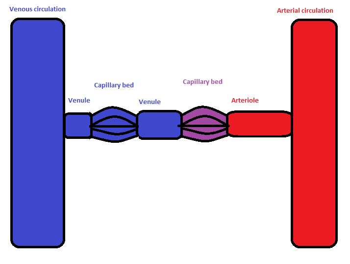
In the circulatory system of animals, a portal venous system occurs when a capillary bed pools into another capillary bed through veins, without first going through the heart. Both capillary beds and the blood vessels that connect them are considered part of the portal venous system. They are relatively uncommon as the majority of capillary beds drain into veins which then drain into the heart, not into another capillary bed. Portal venous systems are considered venous because the blood vessels that join the two capillary beds are either veins or venules. Examples of such systems include the hepatic portal system, the hypophyseal portal system, and (in non-mammals) the renal portal system. Unqualified, ''portal venous system'' often refers to the hepatic portal system. For this reason, ''portal vein'' most commonly refers to the hepatic portal vein. The functional significance of such a system is that it transports products of one region directly to another region in relati ... [...More Info...] [...Related Items...] OR: [Wikipedia] [Google] [Baidu] |


