|
Middle Frontal Gyrus
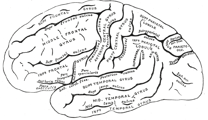
The middle frontal gyrus makes up about one-third of the frontal lobe of the human brain. (A ''gyrus'' is one of the prominent "bumps" or "ridges" on the surface of the human brain.) The middle frontal gyrus, like the inferior frontal gyrus and the superior frontal gyrus, is more of a region in the frontal gyrus than a true gyrus. The borders of the middle frontal gyrus are the ''inferior frontal sulcus'' below; the ''superior frontal sulcus'' above; and the precentral sulcus The precentral sulcus is a part of the human brain that lies parallel to, and in front of, the central sulcus. (A ''sulcus'' is one of the prominent grooves on the surface of the human brain.) The precentral sulcus divides the inferior, midd ... behind. Additional images File:Middle frontal gyrus animation small.gif, Position of middle frontal gyrus (shown in red). File:Gray725 middle frontal gyrus.png, Left cerebral hemisphere seen from above. File:Gray726 middle frontal gyrus.png, Lateral ... [...More Info...] [...Related Items...] OR: [Wikipedia] [Google] [Baidu] |
Frontal Lobe
The frontal lobe is the largest of the four major lobes of the brain in mammals, and is located at the front of each cerebral hemisphere (in front of the parietal lobe and the temporal lobe). It is parted from the parietal lobe by a groove between tissues called the central sulcus and from the temporal lobe by a deeper groove called the lateral sulcus (Sylvian fissure). The most anterior rounded part of the frontal lobe (though not well-defined) is known as the frontal pole, one of the three poles of the cerebrum. The frontal lobe is covered by the frontal cortex. The frontal cortex includes the premotor cortex, and the primary motor cortex – parts of the motor cortex. The front part of the frontal cortex is covered by the prefrontal cortex. There are four principal gyri in the frontal lobe. The precentral gyrus is directly anterior to the central sulcus, running parallel to it and contains the primary motor cortex, which controls voluntary movements of specific body par ... [...More Info...] [...Related Items...] OR: [Wikipedia] [Google] [Baidu] |
Middle Cerebral Artery
The middle cerebral artery (MCA) is one of the three major paired cerebral arteries that supply blood to the cerebrum. The MCA arises from the internal carotid artery and continues into the lateral sulcus where it then branches and projects to many parts of the lateral cerebral cortex. It also supplies blood to the anterior temporal lobes and the insular cortices. The left and right MCAs rise from trifurcations of the internal carotid arteries and thus are connected to the anterior cerebral arteries and the posterior communicating arteries, which connect to the posterior cerebral arteries. The MCAs are not considered a part of the Circle of Willis. Structure The middle cerebral artery divides into four segments, named by the region they supply as opposed to order of branching as the latter can be somewhat variable: *M1: The ''sphenoidal'' segment (stem), receiving its name due to its course along the adjacent sphenoid bone. It is also referred to as the ''horizontal'' seg ... [...More Info...] [...Related Items...] OR: [Wikipedia] [Google] [Baidu] |
Frontal Lobe
The frontal lobe is the largest of the four major lobes of the brain in mammals, and is located at the front of each cerebral hemisphere (in front of the parietal lobe and the temporal lobe). It is parted from the parietal lobe by a groove between tissues called the central sulcus and from the temporal lobe by a deeper groove called the lateral sulcus (Sylvian fissure). The most anterior rounded part of the frontal lobe (though not well-defined) is known as the frontal pole, one of the three poles of the cerebrum. The frontal lobe is covered by the frontal cortex. The frontal cortex includes the premotor cortex, and the primary motor cortex – parts of the motor cortex. The front part of the frontal cortex is covered by the prefrontal cortex. There are four principal gyri in the frontal lobe. The precentral gyrus is directly anterior to the central sulcus, running parallel to it and contains the primary motor cortex, which controls voluntary movements of specific body par ... [...More Info...] [...Related Items...] OR: [Wikipedia] [Google] [Baidu] |
Human Brain
The human brain is the central organ of the human nervous system, and with the spinal cord makes up the central nervous system. The brain consists of the cerebrum, the brainstem and the cerebellum. It controls most of the activities of the body, processing, integrating, and coordinating the information it receives from the sense organs, and making decisions as to the instructions sent to the rest of the body. The brain is contained in, and protected by, the skull bones of the head. The cerebrum, the largest part of the human brain, consists of two cerebral hemispheres. Each hemisphere has an inner core composed of white matter, and an outer surface – the cerebral cortex – composed of grey matter. The cortex has an outer layer, the neocortex, and an inner allocortex. The neocortex is made up of six neuronal layers, while the allocortex has three or four. Each hemisphere is conventionally divided into four lobes – the frontal, temporal, parietal, and oc ... [...More Info...] [...Related Items...] OR: [Wikipedia] [Google] [Baidu] |
Inferior Frontal Gyrus
The inferior frontal gyrus (IFG), (gyrus frontalis inferior), is the lowest positioned gyrus of the frontal gyri, of the frontal lobe, and is part of the prefrontal cortex. Its superior border is the inferior frontal sulcus (which divides it from the middle frontal gyrus), its inferior border is the lateral sulcus (which divides it from the superior temporal gyrus) and its posterior border is the inferior precentral sulcus. Above it is the middle frontal gyrus, behind it is the precentral gyrus. The inferior frontal gyrus contains Broca's area, which is involved in language processing and speech production. Structure The inferior frontal gyrus is highly convoluted and has three cytoarchitecturally diverse regions. The three subdivisions are an opercular part, a triangular part, and an orbital part. These divisions are marked by two rami arising from the lateral sulcus. The ascending ramus separates the opercular and triangular parts. The anterior (horizontal) ramus sep ... [...More Info...] [...Related Items...] OR: [Wikipedia] [Google] [Baidu] |
Superior Frontal Gyrus
In neuroanatomy, the superior frontal gyrus (SFG, also marginal gyrus) is a gyrus – a ridge on the brain's cerebral cortex – which makes up about one third of the frontal lobe. It is bounded laterally by the superior frontal sulcus. The superior frontal gyrus is one of the frontal gyri. Function Self-awareness In fMRI experiments, Goldberg ''et al.'' have found evidence that the superior frontal gyrus is involved in self-awareness, in coordination with the action of the sensory system. Laughter In 1998, neurosurgeon Itzhak Fried described a 16-year-old female patient (referred to as "patient AK") who laughed when her SFG was stimulated with electric current during treatment for epilepsy. Electrical stimulation was applied to the cortical surface of AK's left frontal lobe while an attempt was made to locate the focus of her epileptic seizures (which were never accompanied by laughter). Fried identified a 2 cm by 2 cm area on the left SFG where stimulation produ ... [...More Info...] [...Related Items...] OR: [Wikipedia] [Google] [Baidu] |
Frontal Gyrus
The frontal gyri are four gyri of the frontal lobe in the brain. These are four horizontally oriented, parallel convolutions, of the frontal lobe. The other main gyrus of the frontal lobe is the precentral gyrus which is vertically oriented, and runs parallel with the precentral sulcus. The uppermost of the four gyri is the superior frontal gyrus, below this is the middle frontal gyrus, and below this is the inferior frontal gyrus. Continuing from the superior frontal gyrus on the lateral surface, into the uppermost part of the medial surface of the hemisphere is the medial frontal gyrus. The inferior frontal gyrus includes Broca's area. The lowest part of the inferior frontal gyrus rests on the orbital part of the frontal bone. Gyri Superior The superior frontal gyrus makes up about a third of the frontal lobe. It is bounded laterally by the superior frontal sulcus. Medial Middle It has been recently found that this region participates during linguistic tasks. Mo ... [...More Info...] [...Related Items...] OR: [Wikipedia] [Google] [Baidu] |
Precentral Sulcus
The precentral sulcus is a part of the human brain that lies parallel to, and in front of, the central sulcus. (A ''sulcus'' is one of the prominent grooves on the surface of the human brain.) The precentral sulcus divides the inferior, middle and superior frontal gyri from the precentral gyrus. In most brains, the precentral sulcus is divided into two parts: the inferior precentral sulcus and the superior precentral sulcus. However, the precentral sulcus may sometimes be divided into three parts or form one continuous sulcus. Additional images File:Precentral sulcus animation small.gif, Position of precentral sulcus (shown in red). File:FrontalCaptsLateral.png, Lateral surface of right frontal lobe The frontal lobe is the largest of the four major lobes of the brain in mammals, and is located at the front of each cerebral hemisphere (in front of the parietal lobe and the temporal lobe). It is parted from the parietal lobe by a groove be .... Precentral sulcus is ... [...More Info...] [...Related Items...] OR: [Wikipedia] [Google] [Baidu] |
Gyri
In neuroanatomy, a gyrus (pl. gyri) is a ridge on the cerebral cortex. It is generally surrounded by one or more sulci (depressions or furrows; sg. ''sulcus''). Gyri and sulci create the folded appearance of the brain in humans and other mammals. Structure The gyri are part of a system of folds and ridges that create a larger surface area for the human brain and other mammalian brains. Because the brain is confined to the skull, brain size is limited. Ridges and depressions create folds allowing a larger cortical surface area, and greater cognitive function, to exist in the confines of a smaller cranium. Development The human brain undergoes gyrification during fetal and neonatal development. In embryonic development, all mammalian brains begin as smooth structures derived from the neural tube. A cerebral cortex without surface convolutions is lissencephalic, meaning 'smooth-brained'. As development continues, gyri and sulci begin to take shape on the fetal brain, ... [...More Info...] [...Related Items...] OR: [Wikipedia] [Google] [Baidu] |


