|
Maxillary Nerve
In neuroanatomy, the maxillary nerve (V) is one of the three branches or divisions of the trigeminal nerve, the fifth (CN V) cranial nerve. It comprises the principal functions of sensation from the maxilla, nasal cavity, sinuses, the palate and subsequently that of the mid-face, and is intermediate, both in position and size, between the ophthalmic nerve and the mandibular nerve.Illustrated Anatomy of the Head and Neck, Fehrenbach and Herring, Elsevier, 2012, page 180 Structure It begins at the middle of the trigeminal ganglion as a flattened plexiform band then it passes through the lateral wall of the cavernous sinus. It leaves the skull through the foramen rotundum, where it becomes more cylindrical in form, and firmer in texture. After leaving foramen rotundum it gives two branches to the pterygopalatine ganglion. It then crosses the pterygopalatine fossa, inclines lateralward on the back of the maxilla, and enters the orbit through the inferior orbital fissure. It t ... [...More Info...] [...Related Items...] OR: [Wikipedia] [Google] [Baidu] |
Dental Alveolus
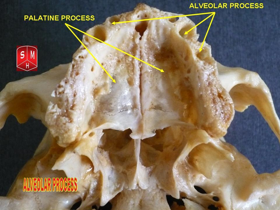
Dental alveoli (singular ''alveolus'') are sockets in the jaws in which the roots of teeth are held in the alveolar process with the periodontal ligament. The lay term for dental alveoli is tooth sockets. A joint that connects the roots of the teeth and the alveolus is called '' gomphosis'' (plural ''gomphoses''). Alveolar bone is the bone that surrounds the roots of the teeth forming bone sockets. In mammals, tooth sockets are found in the maxilla, the premaxilla, and the mandible. Etymology 1706, "a hollow," especially "the socket of a tooth," from Latin alveolus "a tray, trough, basin; bed of a small river; small hollow or cavity," diminutive of alvus "belly, stomach, paunch, bowels; hold of a ship," from PIE root *aulo- "hole, cavity" (source also of Greek aulos "flute, tube, pipe;" Serbo-Croatian, Polish, Russian ulica "street," originally "narrow opening;" Old Church Slavonic uliji, Lithuanian aulys "beehive" (hollow trunk), Armenian yli "pregnant"). The word was extende ... [...More Info...] [...Related Items...] OR: [Wikipedia] [Google] [Baidu] |
Ophthalmic Nerve
The ophthalmic nerve (V1) is a sensory nerve of the face. It is one of three divisions of the trigeminal nerve (CN V). It has three branches that provide sensory innervation to the eye, the skin of the upper face, and the skin of the anterior scalp. Structure The ophthalmic nerve is the first branch of the trigeminal nerve (CN V). It is joined by filaments from the cavernous plexus of the sympathetic, and communicates with the oculomotor, trochlear, and abducent nerves. It gives off a recurrent (meningeal) filament which passes between the layers of the tentorium. Branches The ophthalmic nerve divides into three major branches as it passes through the superior orbital fissure. *The nasociliary nerve gives off several sensory branches to the orbit. It then continues out through the anterior ethmoidal foramen, where it enters the nasal cavity. It provides innervation for much of the anterior nasal mucosa. It also gives off a branch which exits through the nasal bones to for ... [...More Info...] [...Related Items...] OR: [Wikipedia] [Google] [Baidu] |
Sphenopalatine Foramen
The sphenopalatine foramen is a foramen in the skull that connects the nasal cavity with the pterygopalatine fossa. Structure The processes of the superior border of the palatine bone are separated by the ''sphenopalatine notch'', which is converted into the sphenopalatine foramen by the under surface of the body of the sphenoid. In the articulated skull this foramen leads from the pterygopalatine fossa into the posterior part of the superior meatus of the nose, and transmits the sphenopalatine artery and vein and the posterior superior lateral nasal nerve and nasopalatine nerve The nasopalatine nerve (long sphenopalatine nerve) is a nerve of the head. It is a branch of the pterygopalatine ganglion, a continuation from the maxillary nerve (V2). It supplies parts of the palate and nasal septum. Structure The nasopal ...s. Additional images File:Gray167.png, Articulation of left palatine bone with maxilla. File:Gray168.png, Left palatine bone. Nasal aspect. Enlarged. ... [...More Info...] [...Related Items...] OR: [Wikipedia] [Google] [Baidu] |
Nasopalatine
The nasopalatine nerve (long sphenopalatine nerve) is a nerve of the head. It is a branch of the pterygopalatine ganglion, a continuation from the maxillary nerve (V2). It supplies parts of the palate and nasal septum. Structure The nasopalatine nerve communicates with the corresponding nerve of the opposite side and with the greater palatine nerve. The medial superior posterior nasal branches of the maxillary nerve usually branch from the nasopalatine nerve. Origin The nasopalatine nerve is a branch of the pterygopalatine ganglion, a continuation from the maxillary nerve (V2), itself a branch of the trigeminal nerve. It enters the nasal cavity through the sphenopalatine foramen. Course It passes across the roof of the nasal cavity below the orifice of the sphenoidal sinus to reach the nasal septum. It then runs obliquely downward and forward between the periosteum and mucous membrane of the lower part of the nasal septum. It descends to the roof of the mouth through the in ... [...More Info...] [...Related Items...] OR: [Wikipedia] [Google] [Baidu] |
Inferior Orbital Fissure
The inferior orbital fissure is formed by the sphenoid bone and the maxilla. It is located posteriorly along the boundary of the floor and lateral wall of the orbit. It transmits a number of structures, including: * the zygomatic branch of the maxillary nerve * the ascending branches from the pterygopalatine ganglion * the infraorbital vessels, which travel down the infraorbital groove into the infraorbital canal and exit through the infraorbital foramen * the inferior division of the ophthalmic vein Images File:Gray189.png, Left infratemporal fossa. File:Gray191.png, Horizontal section of nasal and orbital cavities. File:Gray787.png, Dissection showing origins of right ocular muscles, and nerves entering by the superior orbital fissure. File:Slide2rome.JPG, Inferior orbital fissure. See also * Foramina of skull *Superior orbital fissure The superior orbital fissure is a foramen or cleft of the skull between the lesser and greater wings of the sphenoid bone. I ... [...More Info...] [...Related Items...] OR: [Wikipedia] [Google] [Baidu] |
Zygomaticofacial Nerve
The zygomaticofacial nerve or zygomaticofacial branch of zygomatic nerve (malar branch) passes along the infero-lateral angle of the orbit, emerges upon the face through the zygomaticofacial foramen in the zygomatic bone, and, perforating the orbicularis oculi to reach the skin of the malar area. It joins with the zygomatic branches of the facial nerve and with the inferior palpebral branches of the maxillary nerve In neuroanatomy, the maxillary nerve (V) is one of the three branches or divisions of the trigeminal nerve, the fifth (CN V) cranial nerve. It comprises the principal functions of sensation from the maxilla, nasal cavity, sinuses, the palate ... (V2). The area of skin supplied by this nerve is over the prominence of the cheek. References External links * * Maxillary nerve {{Neuroanatomy-stub ... [...More Info...] [...Related Items...] OR: [Wikipedia] [Google] [Baidu] |
Zygomaticotemporal Nerve
The zygomaticotemporal nerve (zygomaticotemporal branch, temporal branch) is a small nerve of the face. It is derived from the zygomatic nerve, a branch of the maxillary nerve (CN V2). It is distributed to the skin of the side of the forehead. It communicates with the facial nerve and with the auriculotemporal branch of the mandibular nerve. Structure The zygomaticotemporal nerve is a branch of the zygomatic nerve, a branch of the maxillary nerve. It runs along the lateral wall of the orbit in a groove in the zygomatic bone, receives a branch of communication from the lacrimal nerve, and passes through the zygomaticotemporal foramen in the zygomatic bone to enter the temporal fossa. It ascends between the bone and the substance of the temporalis muscle. It pierces the temporal fascia about 2.5 cm above the zygomatic arch. This is around 17 mm lateral to and 6.5 mm superior to the lateral palpebral fissure. It is distributed to the skin of the side of the forehead. The zygom ... [...More Info...] [...Related Items...] OR: [Wikipedia] [Google] [Baidu] |
Zygomatic Nerve
The zygomatic nerve is a branch of the maxillary nerve, itself a branch of the trigeminal nerve (CN V). It travels through the orbit and divides into the zygomaticotemporal and the zygomaticofacial nerve. It provides sensory supply to skin over the zygomatic bone and the temporal bone. It also carries postganglionic parasympathetic axons to the lacrimal gland. It may be blocked by anaesthetising the maxillary nerve. Structure The zygomatic nerve is a branch of the maxillary nerve (CN V2), itself a branch of the trigeminal nerve (CN V). It branches at the pterygopalatine ganglion. It travels from the pterygopalatine fossa through the inferior orbital fissure to enter the orbit. In the orbit, it travels anteriorly along the lateral wall. Branches Soon after the zygomatic nerve enters the orbit it divides into its branches. These include: * the zygomaticotemporal nerve. This passes through the zygomaticotemporal foramen in the zygomatic bone. * the zygomaticofacial nerve ... [...More Info...] [...Related Items...] OR: [Wikipedia] [Google] [Baidu] |
Meninges
In anatomy, the meninges (, ''singular:'' meninx ( or ), ) are the three membranes that envelop the brain and spinal cord. In mammals, the meninges are the dura mater, the arachnoid mater, and the pia mater. Cerebrospinal fluid is located in the subarachnoid space between the arachnoid mater and the pia mater. The primary function of the meninges is to protect the central nervous system. Structure Dura mater The dura mater ( la, tough mother) (also rarely called ''meninx fibrosa'' or ''pachymeninx'') is a thick, durable membrane, closest to the skull and vertebrae. The dura mater, the outermost part, is a loosely arranged, fibroelastic layer of cells, characterized by multiple interdigitating cell processes, no extracellular collagen, and significant extracellular spaces. The middle region is a mostly fibrous portion. It consists of two layers: the endosteal layer, which lies closest to the skull, and the inner meningeal layer, which lies closer to the brain. It contains lar ... [...More Info...] [...Related Items...] OR: [Wikipedia] [Google] [Baidu] |
Middle Meningeal Nerve
The middle meningeal nerve (meningeal or dural branch) is given off from the maxillary nerve (CN V2) directly after its origin from the trigeminal ganglion, before CN V2 enters the foramen rotundum. It accompanies the middle meningeal artery and vein as the artery and vein enter the cranium through the foramen spinosum and supplies the dura mater In neuroanatomy, dura mater is a thick membrane made of dense irregular connective tissue that surrounds the brain and spinal cord. It is the outermost of the three layers of membrane called the meninges that protect the central nervous syste .... Additional images File:Gray138.png, Left temporal bone. Inner surface. References Maxillary nerve {{neuroanatomy-stub ... [...More Info...] [...Related Items...] OR: [Wikipedia] [Google] [Baidu] |
Infraorbital Foramen
In human anatomy, the infraorbital foramen is one of two small holes in the skull's upper jawbone ( maxillary bone), located below the eye socket and to the left and right of the nose. Both holes are used for blood vessels and nerves. In anatomical terms, it is located below the infraorbital margin of the orbit. It transmits the infraorbital artery and vein, and the infraorbital nerve, a branch of the maxillary nerve. It is typically from the infraorbital margin. Structure Forming the exterior end of the infraorbital canal, the infraorbital foramen communicates with the infraorbital groove, the canal's opening on the interior side. The ramifications of the three principal branches of the trigeminal nerve—at the supraorbital, infraorbital, and mental foramen—are distributed on a vertical line (in anterior view) passing through the middle of the pupil The pupil is a black hole located in the center of the iris of the eye that allows light to strike the retina.Cassin ... [...More Info...] [...Related Items...] OR: [Wikipedia] [Google] [Baidu] |
Inferior Orbital Fissure
The inferior orbital fissure is formed by the sphenoid bone and the maxilla. It is located posteriorly along the boundary of the floor and lateral wall of the orbit. It transmits a number of structures, including: * the zygomatic branch of the maxillary nerve * the ascending branches from the pterygopalatine ganglion * the infraorbital vessels, which travel down the infraorbital groove into the infraorbital canal and exit through the infraorbital foramen * the inferior division of the ophthalmic vein Images File:Gray189.png, Left infratemporal fossa. File:Gray191.png, Horizontal section of nasal and orbital cavities. File:Gray787.png, Dissection showing origins of right ocular muscles, and nerves entering by the superior orbital fissure. File:Slide2rome.JPG, Inferior orbital fissure. See also * Foramina of skull *Superior orbital fissure The superior orbital fissure is a foramen or cleft of the skull between the lesser and greater wings of the sphenoid bone. I ... [...More Info...] [...Related Items...] OR: [Wikipedia] [Google] [Baidu] |

