|
Myelocytes
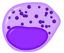
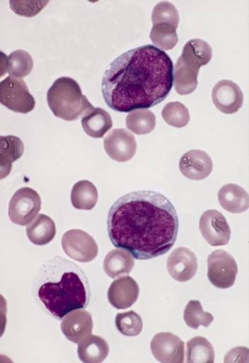
A myelocyte is a young cell of the granulocytic series, occurring normally in bone marrow (can be found in circulating blood when caused by certain diseases). Structure When stained with the usual dyes, the cytoplasm is distinctly basophilic and relatively more abundant than in myeloblasts or promyelocytes, even though myelocytes are smaller cells. Numerous cytoplasmic granules are present in the more mature forms of myelocytes. Neutrophilic and eosinophilic granules are peroxidase-positive, while basophilic granules are not. The nuclear chromatin is coarser than that observed in a promyelocyte, but it is relatively faintly stained and lacks a well-defined membrane. The nucleus is fairly regular in contour (not indented), and seems to be 'buried' beneath the numerous cytoplasmic granules. (If the nucleus were indented, it would likely be a metamyelocyte.) Measurement There is an internationally agreed method of counting blasts, with results from M1 upwards. Development ... [...More Info...] [...Related Items...] OR: [Wikipedia] [Google] [Baidu] |
Basophilic Myelocyte
A myelocyte is a young cell of the granulocytic series, occurring normally in bone marrow (can be found in circulating blood when caused by certain diseases). Structure When stained with the usual dyes, the cytoplasm is distinctly basophilic and relatively more abundant than in myeloblasts or promyelocytes, even though myelocytes are smaller cells. Numerous cytoplasmic granules are present in the more mature forms of myelocytes. Neutrophilic and eosinophilic granules are peroxidase-positive, while basophilic granules are not. The nuclear chromatin is coarser than that observed in a promyelocyte, but it is relatively faintly stained and lacks a well-defined membrane. The nucleus is fairly regular in contour (not indented), and seems to be 'buried' beneath the numerous cytoplasmic granules. (If the nucleus were indented, it would likely be a metamyelocyte.) Measurement There is an internationally agreed method of counting blasts, with results from M1 upwards. Development P ... [...More Info...] [...Related Items...] OR: [Wikipedia] [Google] [Baidu] |
Neutrophilic Myelocyte
A myelocyte is a young cell of the granulocytic series, occurring normally in bone marrow (can be found in circulating blood when caused by certain diseases). Structure When stained with the usual dyes, the cytoplasm is distinctly basophilic and relatively more abundant than in myeloblasts or promyelocytes, even though myelocytes are smaller cells. Numerous cytoplasmic granules are present in the more mature forms of myelocytes. Neutrophilic and eosinophilic granules are peroxidase-positive, while basophilic granules are not. The nuclear chromatin is coarser than that observed in a promyelocyte, but it is relatively faintly stained and lacks a well-defined membrane. The nucleus is fairly regular in contour (not indented), and seems to be 'buried' beneath the numerous cytoplasmic granules. (If the nucleus were indented, it would likely be a metamyelocyte.) Measurement There is an internationally agreed method of counting blasts, with results from M1 upwards. Development P ... [...More Info...] [...Related Items...] OR: [Wikipedia] [Google] [Baidu] |
Promyelocyte
A promyelocyte (or progranulocyte) is a granulocyte precursor, developing from the myeloblast and developing into the myelocyte. Promyelocytes measure 12-20 microns in diameter. The nucleus of a promyelocyte is approximately the same size as a myeloblast but their cytoplasm is much more abundant. They also have less prominent nucleoli than myeloblasts and their chromatin is more coarse and clumped. The cytoplasm is basophilic and contains primary red/purple granules. Additional images File:Cytology of acute promyelocytic leukemia, annotated.png, Bone marrow smear from a patient with acute promyelocytic leukemia, showing characteristic abnormal promyelocytes.Image by Mikael Häggström, MD. Reference for findings: Last author update: 1 February 2013Source image: :File:Faggot cell in AML-M3.jpg froPEIR Digital Library (Pathology image database)(Public Domain) File:Hematopoiesis (human) diagram en.svg, Hematopoiesis File:Basophilic promyelocyte.png, Basophilic promyelocyte File: ... [...More Info...] [...Related Items...] OR: [Wikipedia] [Google] [Baidu] |
Basophilic
Basophilic is a technical term used by pathologists. It describes the appearance of cells, tissues and cellular structures as seen through the microscope after a histological section has been stained with a basic dye. The most common such dye is haematoxylin. The name basophilic refers to the characteristic of these structures to be stained very well by basic dyes. This can be explained by their charges. Basic dyes are cationic, i.e. contain positive charges, and thus they stain anionic structures (i.e. structures containing negative charges), such as the phosphate backbone of DNA in the cell nucleus and ribosomes. "Basophils" are cells that "love" (from greek "-phil") basic dyes, for example haematoxylin, azure and methylene blue. Specifically, this term refers to: * basophil granulocytes * anterior pituitary basophils An abnormal increase in basophil granulocytes is therefore also described as basophilia.https://www.collinsdictionary.com/de/worterbuch/englisch/basophilia ... [...More Info...] [...Related Items...] OR: [Wikipedia] [Google] [Baidu] |
Chromatin
Chromatin is a complex of DNA and protein found in eukaryotic cells. The primary function is to package long DNA molecules into more compact, denser structures. This prevents the strands from becoming tangled and also plays important roles in reinforcing the DNA during cell division, preventing DNA damage, and regulating gene expression and DNA replication. During mitosis and meiosis, chromatin facilitates proper segregation of the chromosomes in anaphase; the characteristic shapes of chromosomes visible during this stage are the result of DNA being coiled into highly condensed chromatin. The primary protein components of chromatin are histones. An octamer of two sets of four histone cores (Histone H2A, Histone H2B, Histone H3, and Histone H4) bind to DNA and function as "anchors" around which the strands are wound.Maeshima, K., Ide, S., & Babokhov, M. (2019). Dynamic chromatin organization without the 30-nm fiber. ''Current opinion in cell biology, 58,'' 95–104. https://doi.o ... [...More Info...] [...Related Items...] OR: [Wikipedia] [Google] [Baidu] |
Peroxidase
Peroxidases or peroxide reductases ( EC numberbr>1.11.1.x are a large group of enzymes which play a role in various biological processes. They are named after the fact that they commonly break up peroxides. Functionality Peroxidases typically catalyze a reaction of the form: :ROOR' + \overset + 2H+ -> ce + R'OH Optimal substrates For many of these enzymes the optimal substrate is hydrogen peroxide, but others are more active with organic hydroperoxides such as lipid peroxides. Peroxidases can contain a heme cofactor in their active sites, or alternately redox-active cysteine or selenocysteine residues. The nature of the electron donor is very dependent on the structure of the enzyme. * For example, horseradish peroxidase can use a variety of organic compounds as electron donors and acceptors. Horseradish peroxidase has an accessible active site, and many compounds can reach the site of the reaction. * On the other hand, for an enzyme such as cytochrome c peroxidase, the co ... [...More Info...] [...Related Items...] OR: [Wikipedia] [Google] [Baidu] |
Specific Granules
Specific granules are secretory vesicles found exclusively in cells of the immune system called granulocytes. It is sometimes described as applying specifically to neutrophils, and sometimes the term is applied to other types of cells. These granules store a mixture of cytotoxic molecules, including many enzymes and antimicrobial peptides, that are released by a process called degranulation following activation of the granulocyte by an immune stimulus. Specific granules are also known as "secondary granules". Contents Examples of cytotoxic molecule stored by specific granules in different granulocytes include: * Neutrophil: alkaline phosphatase, lactoferrin, lysozyme, NADPH oxidase * Eosinophil: cathepsin, major basic protein * Basophil: heparin, histamine (not directly cytotoxic) Clinical significance A specific granule deficiency can be associated with CEBPE CCAAT/enhancer binding protein (C/EBP), epsilon, also known as CEBPE and CRP1, is a type of ccaat-enhancer-binding p ... [...More Info...] [...Related Items...] OR: [Wikipedia] [Google] [Baidu] |
Promyelocyte
A promyelocyte (or progranulocyte) is a granulocyte precursor, developing from the myeloblast and developing into the myelocyte. Promyelocytes measure 12-20 microns in diameter. The nucleus of a promyelocyte is approximately the same size as a myeloblast but their cytoplasm is much more abundant. They also have less prominent nucleoli than myeloblasts and their chromatin is more coarse and clumped. The cytoplasm is basophilic and contains primary red/purple granules. Additional images File:Cytology of acute promyelocytic leukemia, annotated.png, Bone marrow smear from a patient with acute promyelocytic leukemia, showing characteristic abnormal promyelocytes.Image by Mikael Häggström, MD. Reference for findings: Last author update: 1 February 2013Source image: :File:Faggot cell in AML-M3.jpg froPEIR Digital Library (Pathology image database)(Public Domain) File:Hematopoiesis (human) diagram en.svg, Hematopoiesis File:Basophilic promyelocyte.png, Basophilic promyelocyte File: ... [...More Info...] [...Related Items...] OR: [Wikipedia] [Google] [Baidu] |
Myeloblast
The myeloblast is a unipotent stem cell which differentiates into the effectors of the granulocyte series. It is found in the bone marrow. Stimulation of myeloblasts by G-CSF and other cytokines triggers maturation, differentiation, proliferation and cell survival. Structure Myeloblasts reside extravascularly in the bone marrow. Hematopoiesis takes place in the extravascular cavities between the sinuses of the marrow. The wall of the sinuses is composed of two different types of cells, endothelial cells and adventitial reticular cells. The hemopoietic cells are aligned in cords or wedges between these sinuses, with myeloblasts and other granular progenitors concentrated in the subcortical regions of these hemopoietic cords. Myeloblasts are rather small cells with a diameter between 14 and 18μm. The major part is occupied by a large oval nucleus composed of very fine nonaggregated chromatin and possessing 3 or more nucleoli. The cytoplasm has a basophilic character and is devoi ... [...More Info...] [...Related Items...] OR: [Wikipedia] [Google] [Baidu] |
Blood
Blood is a body fluid in the circulatory system of humans and other vertebrates that delivers necessary substances such as nutrients and oxygen to the cells, and transports metabolic waste products away from those same cells. Blood in the circulatory system is also known as ''peripheral blood'', and the blood cells it carries, ''peripheral blood cells''. Blood is composed of blood cells suspended in blood plasma. Plasma, which constitutes 55% of blood fluid, is mostly water (92% by volume), and contains proteins, glucose, mineral ions, hormones, carbon dioxide (plasma being the main medium for excretory product transportation), and blood cells themselves. Albumin is the main protein in plasma, and it functions to regulate the colloidal osmotic pressure of blood. The blood cells are mainly red blood cells (also called RBCs or erythrocytes), white blood cells (also called WBCs or leukocytes) and platelets (also called thrombocytes). The most abundant cells in vertebrate blo ... [...More Info...] [...Related Items...] OR: [Wikipedia] [Google] [Baidu] |
Cytoplasm
In cell biology, the cytoplasm is all of the material within a eukaryotic cell, enclosed by the cell membrane, except for the cell nucleus. The material inside the nucleus and contained within the nuclear membrane is termed the nucleoplasm. The main components of the cytoplasm are cytosol (a gel-like substance), the organelles (the cell's internal sub-structures), and various cytoplasmic inclusions. The cytoplasm is about 80% water and is usually colorless. The submicroscopic ground cell substance or cytoplasmic matrix which remains after exclusion of the cell organelles and particles is groundplasm. It is the hyaloplasm of light microscopy, a highly complex, polyphasic system in which all resolvable cytoplasmic elements are suspended, including the larger organelles such as the ribosomes, mitochondria, the plant plastids, lipid droplets, and vacuoles. Most cellular activities take place within the cytoplasm, such as many metabolic pathways including glycolysis, and proces ... [...More Info...] [...Related Items...] OR: [Wikipedia] [Google] [Baidu] |
Metamyelocyte
A metamyelocyte is a cell undergoing granulopoiesis, derived from a myelocyte, and leading to a band cell. It is characterized by the appearance of a bent cell nucleus, nucleus, cytoplasmic granules, and the absence of visible nucleoli. (If the nucleus is not yet bent, then it is likely a myelocyte.) Additional images File:Hematopoiesis (human) diagram en.svg, Hematopoiesis See also * Pluripotential hemopoietic stem cell External links * - "Bone Marrow and Hemopoiesis: bone marrow smear, neutrophilic metamyelocyte and mature PMN" * * * Interactive diagram at lycos.es Histology Leukocytes {{developmental-biology-stub ... [...More Info...] [...Related Items...] OR: [Wikipedia] [Google] [Baidu] |


