|
Multivesicular Release
Communication between neurons happens primarily through chemical neurotransmission at the synapse. Neurotransmitters are packaged into synaptic vesicles for release from the presynaptic cell into the synapse, from where they diffuse and can bind to postsynaptic receptors. While most presynaptic cells are historically thought to release one vesicle at a time per active site, more recent research has pointed towards the possibility of multiple vesicles being released from the same active site (multivesicular release; MVR) in response to an action potential. Basics of neuronal signaling In the nervous system there are primarily two ways of propagating signals. By far the most common method of intracellular signal propagation is the action potential. The dendrites of neurons contain ionotropic (aka ligand-gated ion channel) and metabotropic neurotransmitter receptors that bind chemical neurotransmitters. At ionotropic receptors, these chemical neurotransmitters cause quick change ... [...More Info...] [...Related Items...] OR: [Wikipedia] [Google] [Baidu] |
Neurotransmission
Neurotransmission (Latin: ''transmissio'' "passage, crossing" from ''transmittere'' "send, let through") is the process by which signaling molecules called neurotransmitters are released by the axon terminal of a neuron (the presynaptic neuron), and bind to and react with the receptors on the dendrites of another neuron (the postsynaptic neuron) a short distance away. A similar process occurs in retrograde neurotransmission, where the dendrites of the postsynaptic neuron release retrograde neurotransmitters (e.g., endocannabinoids; synthesized in response to a rise in intracellular calcium levels) that signal through receptors that are located on the axon terminal of the presynaptic neuron, mainly at GABAergic and glutamatergic synapses. Neurotransmission is regulated by several different factors: the availability and rate-of-synthesis of the neurotransmitter, the release of that neurotransmitter, the baseline activity of the postsynaptic cell, the number of available postsynapti ... [...More Info...] [...Related Items...] OR: [Wikipedia] [Google] [Baidu] |
Memory
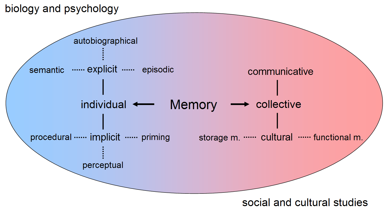
Memory is the faculty of the mind by which data or information is encoded, stored, and retrieved when needed. It is the retention of information over time for the purpose of influencing future action. If past events could not be remembered, it would be impossible for language, relationships, or personal identity to develop. Memory loss is usually described as forgetfulness or amnesia. Memory is often understood as an informational processing system with explicit and implicit functioning that is made up of a sensory processor, short-term (or working) memory, and long-term memory. This can be related to the neuron. The sensory processor allows information from the outside world to be sensed in the form of chemical and physical stimuli and attended to various levels of focus and intent. Working memory serves as an encoding and retrieval processor. Information in the form of stimuli is encoded in accordance with explicit or implicit functions by the working memory processor. ... [...More Info...] [...Related Items...] OR: [Wikipedia] [Google] [Baidu] |
Neuroscience
Neuroscience is the scientific study of the nervous system (the brain, spinal cord, and peripheral nervous system), its functions and disorders. It is a multidisciplinary science that combines physiology, anatomy, molecular biology, developmental biology, cytology, psychology, physics, computer science, chemistry, medicine, statistics, and Mathematical Modeling, mathematical modeling to understand the fundamental and emergent properties of neurons, glia and neural circuits. The understanding of the biological basis of learning, memory, behavior, perception, and consciousness has been described by Eric Kandel as the "epic challenge" of the Biology, biological sciences. The scope of neuroscience has broadened over time to include different approaches used to study the nervous system at different scales. The techniques used by neuroscientists have expanded enormously, from molecular biology, molecular and cell biology, cellular studies of individual neurons to neuroimaging, imaging ... [...More Info...] [...Related Items...] OR: [Wikipedia] [Google] [Baidu] |
Calyx Of Held
The Calyx of Held is a particularly large synapse in the mammalian Auditory system, auditory central nervous system, so named after Hans Held who first described it in his 1893 article ''Die centrale Gehörleitung''Held, H. "Die centrale Gehörleitung" Arch. Anat. Physiol. Anat. Abt, 1893 because of its resemblance to the sepal, calyx of a flower. Globular bushy cells in the anterior cochlear nucleus, anteroventral cochlear nucleus (AVCN) send axons to the contralateral medial nucleus of the trapezoid body (MNTB), where they synapse via these calyces on MNTB principal cells. These principal cells then project to the ipsilateral Superior olivary nucleus, lateral superior olive (LSO), where they inhibit postsynaptic neurons and provide a basis for Interaural level difference, interaural level detection (ILD), required for high frequency sound localization. This synapse has been described as the largest in the brain. The related endbulb of Held is also a large axon terminal smaller sy ... [...More Info...] [...Related Items...] OR: [Wikipedia] [Google] [Baidu] |
Purkinje Cell
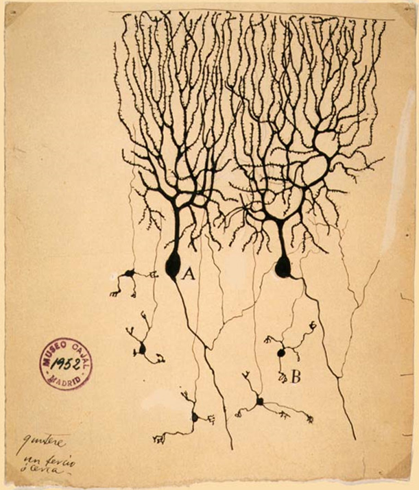
Purkinje cells, or Purkinje neurons, are a class of GABAergic inhibitory neurons located in the cerebellum. They are named after their discoverer, Czech people, Czech anatomist Jan Evangelista Purkyně, who characterized the cells in 1839. Structure These Cell (biology), cells are some of the largest neurons in the human brain (Betz cells being the largest), with an intricately elaborate dendrite, dendritic arbor, characterized by a large number of dendritic spines. Purkinje cells are found within the Cerebellum#Microanatomy, Purkinje layer in the cerebellum. Purkinje cells are aligned like dominos stacked one in front of the other. Their large dendritic arbors form nearly two-dimensional layers through which parallel fibers from the deeper-layers pass. These parallel fibers make relatively weaker excitatory synapse, excitatory (glutamatergic) synapses to spines in the Purkinje cell dendrite, whereas climbing fibers originating from the inferior olivary nucleus in the medull ... [...More Info...] [...Related Items...] OR: [Wikipedia] [Google] [Baidu] |
Climbing Fiber
Climbing fibers are the name given to a series of neuronal projections from the inferior olivary nucleus located in the medulla oblongata. These axons pass through the pons and enter the cerebellum via the inferior cerebellar peduncle where they form synapses with the deep cerebellar nuclei and Purkinje cells. Each climbing fiber will form synapses with 1-10 Purkinje cells. Early in development, Purkinje cells are innervated by multiple climbing fibers, but as the cerebellum matures, these inputs gradually become eliminated resulting in a single climbing fiber input per Purkinje cell. These fibers provide very powerful, excitatory input to the cerebellum which results in the generation of complex spike excitatory postsynaptic potential (EPSP) in Purkinje cells. In this way climbing fibers (CFs) perform a central role in motor behaviors. The climbing fibers carry information from various sources such as the spinal cord, vestibular system, red nucleus, superior collicu ... [...More Info...] [...Related Items...] OR: [Wikipedia] [Google] [Baidu] |
Synaptic Pruning
Synaptic pruning, a phase in the development of the nervous system, is the process of synapse elimination that occurs between early childhood and the onset of puberty in many mammals, including humans. Pruning starts near the time of birth and continues into the late-20s. During pruning, both the axon and dendrite decay and die off. It was traditionally considered to be complete by the time of sexual maturation, but this was discounted by MRI studies. The infant brain will increase in size by a factor of up to 5 by adulthood, reaching a final size of approximately 86 (± 8) billion neurons. Two factors contribute to this growth: the growth of synaptic connections between neurons and the myelination of nerve fibers; the total number of neurons, however, remains the same. After adolescence, the volume of the synaptic connections decreases again due to synaptic pruning. Pruning is influenced by environmental factors and is widely thought to represent learning. Variations Regulato ... [...More Info...] [...Related Items...] OR: [Wikipedia] [Google] [Baidu] |
Ribbon Synapse
The ribbon synapse is a type of neuronal synapse characterized by the presence of an electron-dense structure, the synaptic ribbon, that holds vesicles close to the active zone. It is characterized by a tight vesicle-calcium channel coupling that promotes rapid neurotransmitter release and sustained signal transmission. Ribbon synapses undergo a cycle of exocytosis and endocytosis in response to graded changes of membrane potential. It has been proposed that most ribbon synapses undergo a special type of exocytosis based on coordinated multivesicular release. This interpretation has recently been questioned at the inner hair cell ribbon synapse, where it has been instead proposed that exocytosis is described by uniquantal (i.e., univesicular) release shaped by a flickering vesicle fusion pore. These unique features specialize the ribbon synapse to enable extremely fast, precise and sustained neurotransmission, which is critical for the perception of complex senses such as vision and ... [...More Info...] [...Related Items...] OR: [Wikipedia] [Google] [Baidu] |
NMDA Receptor
The ''N''-methyl-D-aspartate receptor (also known as the NMDA receptor or NMDAR), is a glutamate receptor and ion channel found in neurons. The NMDA receptor is one of three types of ionotropic glutamate receptors, the other two being AMPA receptor, AMPA and kainate receptors. Depending on its subunit composition, its Ligand (biochemistry), ligands are glutamate and glycine (or D-Serine, D-serine). However, the binding of the ligands is typically not sufficient to open the channel as it may be blocked by Magnesium, Mg2+ ions which are only removed when the neuron is sufficiently depolarized. Thus, the channel acts as a “coincidence detector” and only once both of these conditions are met, the channel opens and it allows cation, positively charged ions (cations) to flow through the cell membrane. The NMDA receptor is thought to be very important for controlling synaptic plasticity and mediating learning and memory functions. The NMDA receptor is ionotropic, meaning it is a pr ... [...More Info...] [...Related Items...] OR: [Wikipedia] [Google] [Baidu] |
Neural Facilitation
Neural facilitation, also known as paired-pulse facilitation (PPF), is a phenomenon in neuroscience in which postsynaptic potentials (PSPs) (End-plate potential, EPPs, Excitatory postsynaptic potential, EPSPs or Inhibitory postsynaptic potential, IPSPs) evoked by an impulse are increased when that impulse closely follows a prior impulse. PPF is thus a form of short-term synaptic plasticity. The mechanisms underlying neural facilitation are exclusively pre-synaptic; broadly speaking, PPF arises due to increased presynaptic concentration leading to a greater release of neurotransmitter-containing synaptic vesicles. Neural facilitation may be involved in several neuronal tasks, including simple learning, information processing, and sound-source localization. Mechanisms Overview plays a significant role in transmitting signals at chemical synapses. Voltage-dependent calcium channel, Voltage-gated channels are located within the presynaptic terminal. When an action potential invades ... [...More Info...] [...Related Items...] OR: [Wikipedia] [Google] [Baidu] |
AMPA Receptor
The α-amino-3-hydroxy-5-methyl-4-isoxazolepropionic acid receptor (also known as AMPA receptor, AMPAR, or quisqualate receptor) is an ionotropic receptor, ionotropic transmembrane receptor for glutamate (iGluR) that mediates fast synapse, synaptic transmission in the central nervous system (CNS). It has been traditionally classified as a non-NMDA_receptor, NMDA-type receptor, along with the kainate receptor. Its name is derived from its ability to be activated by the artificial glutamate analog AMPA. The receptor was first named the "quisqualate receptor" by Watkins and colleagues after a naturally occurring agonist quisqualic acid, quisqualate and was only later given the label "AMPA receptor" after the selective agonist developed by Tage Honore and colleagues at the Royal Danish School of Pharmacy in Copenhagen. The ''GRIA2''-encoded AMPA receptor ligand binding core (GluA2 LBD) was the first glutamate receptor ion channel domain to be protein crystal, crystallized. Structure ... [...More Info...] [...Related Items...] OR: [Wikipedia] [Google] [Baidu] |
Glutamate Transporter
Glutamate transporters are a family of neurotransmitter transporter proteins that move glutamate – the principal excitatory neurotransmitter – across a membrane. The family of glutamate transporters is composed of two primary subclasses: the excitatory amino acid transporter (EAAT) family and vesicular glutamate transporter (VGLUT) family. In the brain, EAATs remove glutamate from the synaptic cleft and extrasynaptic sites via glutamate reuptake into glial cells and neurons, while VGLUTs move glutamate from the cell cytoplasm into synaptic vesicles. Glutamate transporters also transport aspartate and are present in virtually all peripheral tissues, including the heart, liver, Testicle, testes, and bone. They exhibit stereoselectivity for L-glutamate but transport both L-aspartate and D-aspartate. The EAATs are membrane-bound secondary transporters that superficially resemble ion channels. These transporters play the important role of regulating concentrations of glutamate in the ... [...More Info...] [...Related Items...] OR: [Wikipedia] [Google] [Baidu] |


.jpg)