|
Monocentric
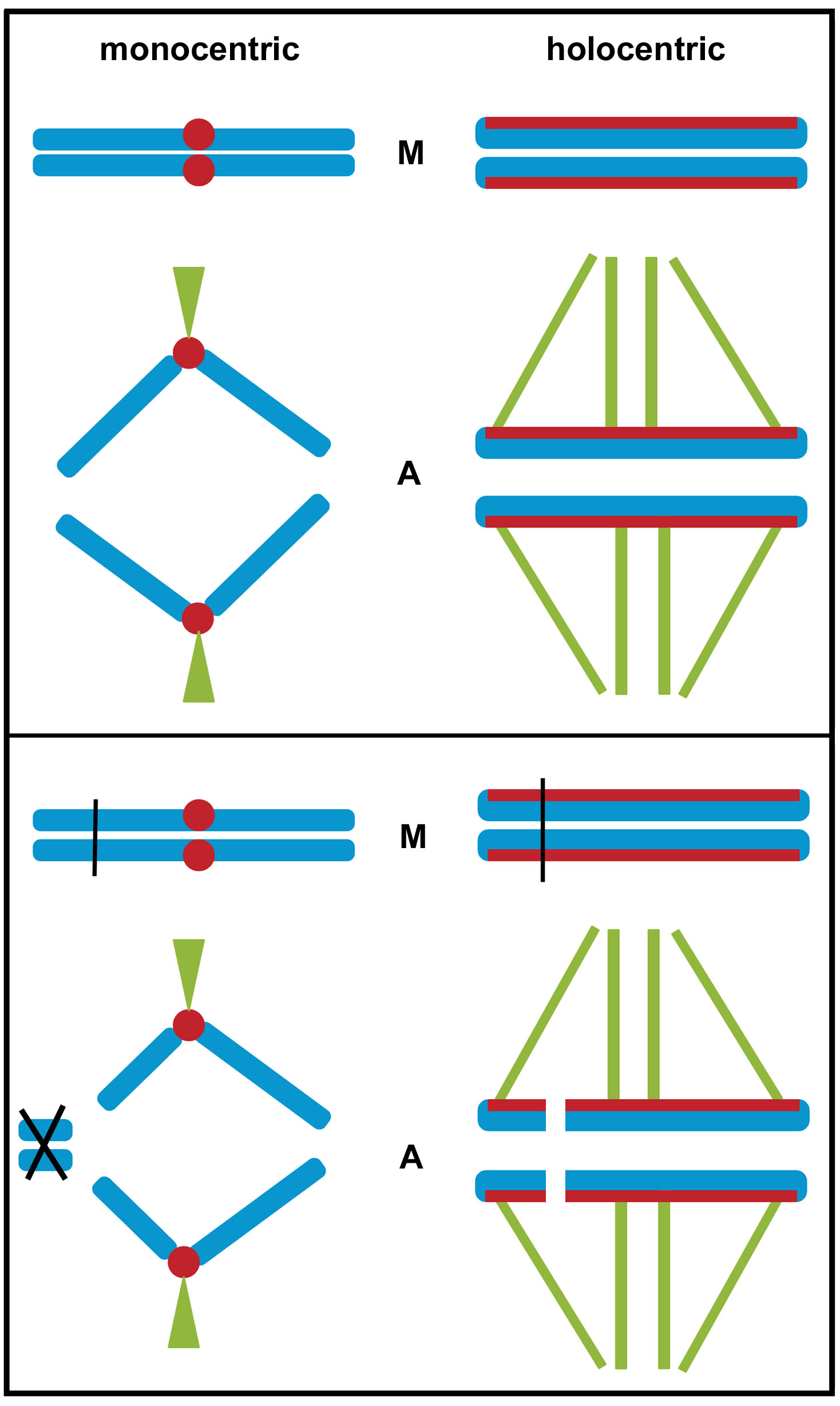
The monocentric chromosome is a chromosome that has only one centromere in a chromosome and forms a narrow constriction. Monocentric centromeres are the most common structure on highly repetitive DNA in plants and animals. Structure Monocentric chromosomes as compared to holocentric chromosomes where the entire length of the chromosome acts as the centromere. In monocentric chromosomes there is one primary constriction and the centromere its CenH3 loci at this location. Holocentric chromosomes are found throughout the plant and animal kingdoms such as the nematode '' Caenorhabditis elegans''. Holocentric chromosomes do have an evolutionary advantage by preventing the loss of chromosome after a DNA double-strand break. The centromere is the point of attachment for the mitotic apparatus Chromosomal aberrations Deletions, duplications and translocations can produce a polycentric chromosome. This is troublesome for cells that divide often since at the time of anaphase the ... [...More Info...] [...Related Items...] OR: [Wikipedia] [Google] [Baidu] |
Holocentric Chromosomes
Holocentric chromosomes are chromosomes that possess multiple kinetochores along their length rather than the single centromere typical of other chromosomes. They were first described in cytogenetic experiments in 1935. Since this first observation, the term holocentric chromosome has referred to chromosomes that: i) lack the primary constriction corresponding to the centromere observed in monocentric chromosomes; and ii) possess multiple kinetochores dispersed along the entire chromosomal axis, such that microtubules bind to the chromosome along its entire length and move broadside to the pole from the metaphase plate. Holocentric chromosomes are also termed ''holokinetic'', because, during cell division, the sister chromatids move apart in parallel and do not form the classical V-shaped figures typical of monocentric chromosomes. Holocentric chromosomes have evolved several times during both animal and plant evolution, and are currently reported in about eight hundred diverse spec ... [...More Info...] [...Related Items...] OR: [Wikipedia] [Google] [Baidu] |
Centromere
The centromere links a pair of sister chromatids together during cell division. This constricted region of chromosome connects the sister chromatids, creating a short arm (p) and a long arm (q) on the chromatids. During mitosis, spindle fibers attach to the centromere via the kinetochore. The physical role of the centromere is to act as the site of assembly of the kinetochores – a highly complex multiprotein structure that is responsible for the actual events of chromosome segregation – i.e. binding microtubules and signaling to the cell cycle machinery when all chromosomes have adopted correct attachments to the spindle, so that it is safe for cell division to proceed to completion and for cells to enter anaphase. There are, broadly speaking, two types of centromeres. "Point centromeres" bind to specific proteins that recognize particular DNA sequences with high efficiency. Any piece of DNA with the point centromere DNA sequence on it will typically form a centromere ... [...More Info...] [...Related Items...] OR: [Wikipedia] [Google] [Baidu] |
Polycentric Chromosome
In genetics, a polycentric chromosome is any chromosome featuring multiple centromeres. Polycentric chromosomes are produced by chromosomal aberrations such as deletion, duplication, or translocation. Polycentric chromosomes usually result in the death of the cell because polycentric chromosomes may fail to move to opposite poles of spindle fiber during anaphase. As a result, the chromosome is fragmented, which causes the death of the cell. In some algae, such as ''Spirogyra ''Spirogyra'' (common names include water silk, mermaid's tresses, and blanket weed) is a genus of filamentous charophyte green algae of the order Zygnematales, named for the helical or spiral arrangement of the chloroplasts that is character ...'', polycentric chromosomes appear normally. References Chromosomes {{genetics-stub ... [...More Info...] [...Related Items...] OR: [Wikipedia] [Google] [Baidu] |
Chromosome
A chromosome is a long DNA molecule with part or all of the genetic material of an organism. In most chromosomes the very long thin DNA fibers are coated with packaging proteins; in eukaryotic cells the most important of these proteins are the histones. These proteins, aided by chaperone proteins, bind to and condense the DNA molecule to maintain its integrity. These chromosomes display a complex three-dimensional structure, which plays a significant role in transcriptional regulation. Chromosomes are normally visible under a light microscope only during the metaphase of cell division (where all chromosomes are aligned in the center of the cell in their condensed form). Before this happens, each chromosome is duplicated ( S phase), and both copies are joined by a centromere, resulting either in an X-shaped structure (pictured above), if the centromere is located equatorially, or a two-arm structure, if the centromere is located distally. The joined copies are now c ... [...More Info...] [...Related Items...] OR: [Wikipedia] [Google] [Baidu] |
Caenorhabditis Elegans
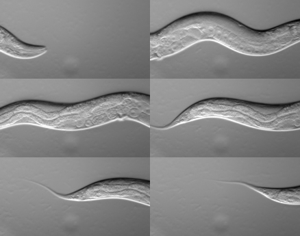
''Caenorhabditis elegans'' () is a free-living transparent nematode about 1 mm in length that lives in temperate soil environments. It is the type species of its genus. The name is a blend of the Greek ''caeno-'' (recent), ''rhabditis'' (rod-like) and Latin ''elegans'' (elegant). In 1900, Maupas initially named it '' Rhabditides elegans.'' Osche placed it in the subgenus ''Caenorhabditis'' in 1952, and in 1955, Dougherty raised ''Caenorhabditis'' to the status of genus. ''C. elegans'' is an unsegmented pseudocoelomate and lacks respiratory or circulatory systems. Most of these nematodes are hermaphrodites and a few are males. Males have specialised tails for mating that include spicules. In 1963, Sydney Brenner proposed research into ''C. elegans,'' primarily in the area of neuronal development. In 1974, he began research into the molecular and developmental biology of ''C. elegans'', which has since been extensively used as a model organism. It was the first mult ... [...More Info...] [...Related Items...] OR: [Wikipedia] [Google] [Baidu] |
Mitotic Apparatus
In cell biology, the spindle apparatus refers to the cytoskeletal structure of eukaryotic cells that forms during cell division to separate sister chromatids between daughter cells. It is referred to as the mitotic spindle during mitosis, a process that produces genetically identical daughter cells, or the meiotic spindle during meiosis, a process that produces gametes with half the number of chromosomes of the parent cell. Besides chromosomes, the spindle apparatus is composed of hundreds of proteins. Microtubules comprise the most abundant components of the machinery. Spindle structure Attachment of microtubules to chromosomes is mediated by kinetochores, which actively monitor spindle formation and prevent premature anaphase onset. Microtubule polymerization and depolymerization dynamic drive chromosome congression. Depolymerization of microtubules generates tension at kinetochores; bipolar attachment of sister kinetochores to microtubules emanating from opposite cell p ... [...More Info...] [...Related Items...] OR: [Wikipedia] [Google] [Baidu] |
Anaphase
Anaphase () is the stage of mitosis after the process of metaphase, when replicated chromosomes are split and the newly-copied chromosomes (daughter chromatids) are moved to opposite poles of the cell. Chromosomes also reach their overall maximum condensation in late anaphase, to help chromosome segregation and the re-formation of the nucleus. Anaphase starts when the anaphase promoting complex marks an inhibitory chaperone called securin for destruction by ubiquinylating it. Securin is a protein which inhibits a protease known as separase. The destruction of securin unleashes separase which then breaks down cohesin, a protein responsible for holding sister chromatids together. At this point, three subclasses of microtubule unique to mitosis are involved in creating the forces necessary to separate the chromatids: kinetochore microtubules, interpolar microtubules, and astral microtubules. The centromeres are split, and the sister chromatids are pulled toward the poles by ... [...More Info...] [...Related Items...] OR: [Wikipedia] [Google] [Baidu] |
Chromosomes
A chromosome is a long DNA molecule with part or all of the genetic material of an organism. In most chromosomes the very long thin DNA fibers are coated with packaging proteins; in eukaryotic cells the most important of these proteins are the histones. These proteins, aided by chaperone proteins, bind to and condense the DNA molecule to maintain its integrity. These chromosomes display a complex three-dimensional structure, which plays a significant role in transcriptional regulation. Chromosomes are normally visible under a light microscope only during the metaphase of cell division (where all chromosomes are aligned in the center of the cell in their condensed form). Before this happens, each chromosome is duplicated ( S phase), and both copies are joined by a centromere, resulting either in an X-shaped structure (pictured above), if the centromere is located equatorially, or a two-arm structure, if the centromere is located distally. The joined copies are now c ... [...More Info...] [...Related Items...] OR: [Wikipedia] [Google] [Baidu] |
Classical Genetics
Classical genetics is the branch of genetics based solely on visible results of reproductive acts. It is the oldest discipline in the field of genetics, going back to the experiments on Mendelian inheritance by Gregor Mendel who made it possible to identify the basic mechanisms of heredity. Subsequently, these mechanisms have been studied and explained at the molecular level. Classical genetics consists of the techniques and methodologies of genetics that were in use before the advent of molecular biology. A key discovery of classical genetics in eukaryotes was genetic linkage. The observation that some genes do not segregate independently at meiosis broke the laws of Mendelian inheritance and provided science with a way to map characteristics to a location on the chromosomes. Linkage maps are still used today, especially in breeding for plant improvement. After the discovery of the genetic code and such tools of cloning as restriction enzymes, the avenues of investigation open t ... [...More Info...] [...Related Items...] OR: [Wikipedia] [Google] [Baidu] |


