|
Microscopy
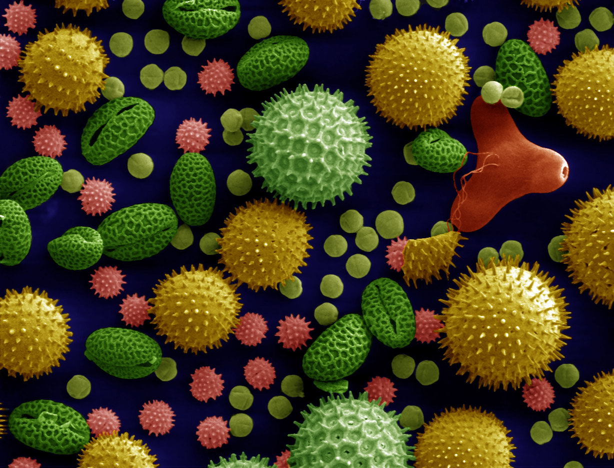
Microscopy is the technical field of using microscopes to view objects and areas of objects that cannot be seen with the naked eye (objects that are not within the resolution range of the normal eye). There are three well-known branches of microscopy: optical, electron, and scanning probe microscopy, along with the emerging field of X-ray microscopy. Optical microscopy and electron microscopy involve the diffraction, reflection, or refraction of electromagnetic radiation/electron beams interacting with the specimen, and the collection of the scattered radiation or another signal in order to create an image. This process may be carried out by wide-field irradiation of the sample (for example standard light microscopy and transmission electron microscopy) or by scanning a fine beam over the sample (for example confocal laser scanning microscopy and scanning electron microscopy). Scanning probe microscopy involves the interaction of a scanning probe with the surface of the objec ... [...More Info...] [...Related Items...] OR: [Wikipedia] [Google] [Baidu] |
Transmission Electron Microscope
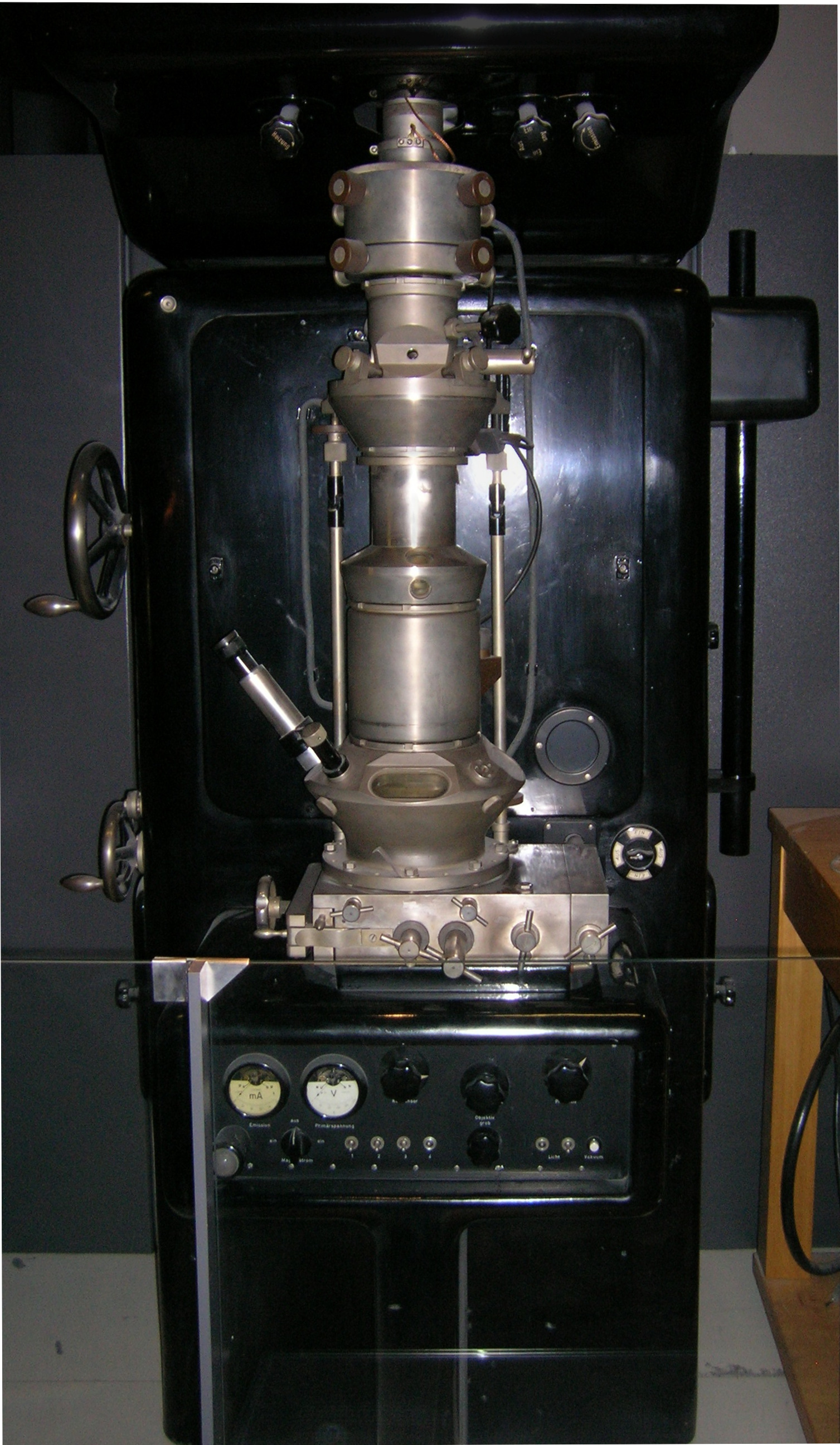
Transmission electron microscopy (TEM) is a microscopy technique in which a beam of electrons is transmitted through a specimen to form an image. The specimen is most often an ultrathin section less than 100 nm thick or a suspension on a grid. An image is formed from the interaction of the electrons with the sample as the beam is transmitted through the specimen. The image is then magnified and focused onto an imaging device, such as a fluorescent screen, a layer of photographic film, or a sensor such as a scintillator attached to a charge-coupled device. Transmission electron microscopes are capable of imaging at a significantly higher resolution than light microscopes, owing to the smaller de Broglie wavelength of electrons. This enables the instrument to capture fine detail—even as small as a single column of atoms, which is thousands of times smaller than a resolvable object seen in a light microscope. Transmission electron microscopy is a major analytical method in ... [...More Info...] [...Related Items...] OR: [Wikipedia] [Google] [Baidu] |
Optical Microscope
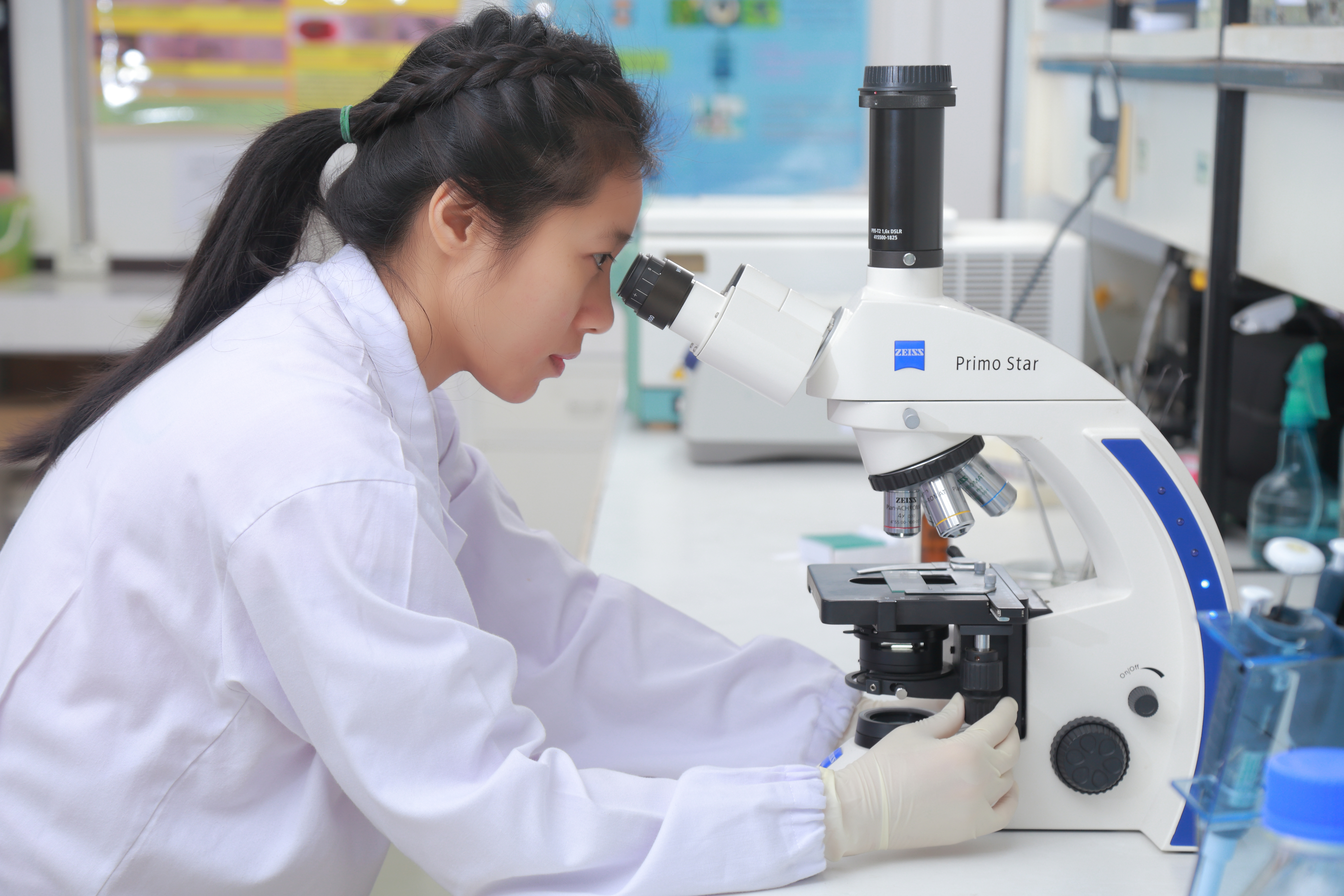
The optical microscope, also referred to as a light microscope, is a type of microscope that commonly uses visible light and a system of lenses to generate magnified images of small objects. Optical microscopes are the oldest design of microscope and were possibly invented in their present compound form in the 17th century. Basic optical microscopes can be very simple, although many complex designs aim to improve resolution and sample contrast. The object is placed on a stage and may be directly viewed through one or two eyepieces on the microscope. In high-power microscopes, both eyepieces typically show the same image, but with a stereo microscope, slightly different images are used to create a 3-D effect. A camera is typically used to capture the image (micrograph). The sample can be lit in a variety of ways. Transparent objects can be lit from below and solid objects can be lit with light coming through ( bright field) or around (dark field) the objective lens. Polarised ... [...More Info...] [...Related Items...] OR: [Wikipedia] [Google] [Baidu] |
Compound Microscope
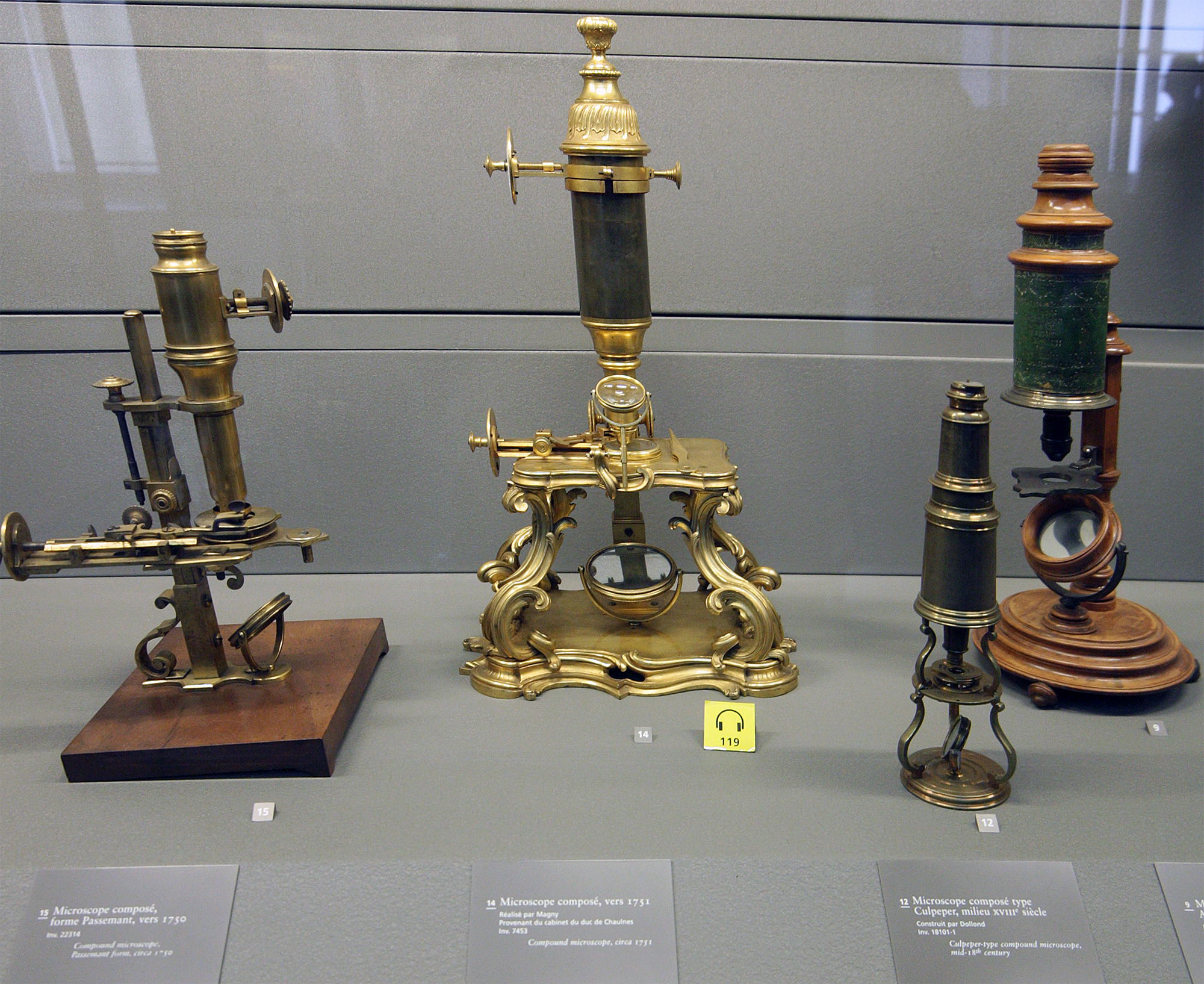
The optical microscope, also referred to as a light microscope, is a type of microscope that commonly uses visible light and a system of lenses to generate magnified images of small objects. Optical microscopes are the oldest design of microscope and were possibly invented in their present compound form in the 17th century. Basic optical microscopes can be very simple, although many complex designs aim to improve resolution and sample contrast. The object is placed on a stage and may be directly viewed through one or two eyepieces on the microscope. In high-power microscopes, both eyepieces typically show the same image, but with a stereo microscope, slightly different images are used to create a 3-D effect. A camera is typically used to capture the image (micrograph). The sample can be lit in a variety of ways. Transparent objects can be lit from below and solid objects can be lit with light coming through ( bright field) or around (dark field) the objective lens. Polarised ... [...More Info...] [...Related Items...] OR: [Wikipedia] [Google] [Baidu] |
Electron Microscope
An electron microscope is a microscope that uses a beam of accelerated electrons as a source of illumination. As the wavelength of an electron can be up to 100,000 times shorter than that of visible light photons, electron microscopes have a higher resolving power than light microscopes and can reveal the structure of smaller objects. A scanning transmission electron microscope has achieved better than 50 pm resolution in annular dark-field imaging mode and magnifications of up to about 10,000,000× whereas most light microscopes are limited by diffraction to about 200 nm resolution and useful magnifications below 2000×. Electron microscopes use shaped magnetic fields to form electron optical lens systems that are analogous to the glass lenses of an optical light microscope. Electron microscopes are used to investigate the ultrastructure of a wide range of biological and inorganic specimens including microorganisms, cells, large molecules, biopsy samples, ... [...More Info...] [...Related Items...] OR: [Wikipedia] [Google] [Baidu] |
Confocal Laser Scanning Microscopy
Confocal microscopy, most frequently confocal laser scanning microscopy (CLSM) or laser confocal scanning microscopy (LCSM), is an optical imaging technique for increasing optical resolution and contrast of a micrograph by means of using a spatial pinhole to block out-of-focus light in image formation. Capturing multiple two-dimensional images at different depths in a sample enables the reconstruction of three-dimensional structures (a process known as optical sectioning) within an object. This technique is used extensively in the scientific and industrial communities and typical applications are in life sciences, semiconductor inspection and materials science. Light travels through the sample under a conventional microscope as far into the specimen as it can penetrate, while a confocal microscope only focuses a smaller beam of light at one narrow depth level at a time. The CLSM achieves a controlled and highly limited depth of field. Basic concept The principle of ... [...More Info...] [...Related Items...] OR: [Wikipedia] [Google] [Baidu] |
X-ray Microscopy
An X-ray microscope uses electromagnetic radiation in the soft X-ray band to produce magnified images of objects. Since X-rays penetrate most objects, there is no need to specially prepare them for X-ray microscopy observations. Unlike visible light, X-rays do not reflect or refract easily and are invisible to the human eye. Therefore, an X-ray microscope exposes film or uses a charge-coupled device (CCD) detector to detect X-rays that pass through the specimen. It is a contrast imaging technology using the difference in absorption of soft X-rays in the water window region (wavelengths: 2.34–4.4 nm, energies: 280–530 eV) by the carbon atom (main element composing the living cell) and the oxygen atom (an element of water). Microfocus X-ray also achieves high magnification by projection. A microfocus X-ray tube produces X-rays from an extremely small focal spot (5 μm down to 0.1 μm). The X-rays are in the more conventional X-ray range (20 to 300 keV) ... [...More Info...] [...Related Items...] OR: [Wikipedia] [Google] [Baidu] |
Microscope
A microscope () is a laboratory instrument used to examine objects that are too small to be seen by the naked eye. Microscopy is the science of investigating small objects and structures using a microscope. Microscopic means being invisible to the eye unless aided by a microscope. There are many types of microscopes, and they may be grouped in different ways. One way is to describe the method an instrument uses to interact with a sample and produce images, either by sending a beam of light or electrons through a sample in its optical path, by detecting photon emissions from a sample, or by scanning across and a short distance from the surface of a sample using a probe. The most common microscope (and the first to be invented) is the optical microscope, which uses lenses to refract visible light that passed through a thinly sectioned sample to produce an observable image. Other major types of microscopes are the fluorescence microscope, electron microscope (both the transmi ... [...More Info...] [...Related Items...] OR: [Wikipedia] [Google] [Baidu] |
Histology
Histology, also known as microscopic anatomy or microanatomy, is the branch of biology which studies the microscopic anatomy of biological tissues. Histology is the microscopic counterpart to gross anatomy, which looks at larger structures visible without a microscope. Although one may divide microscopic anatomy into ''organology'', the study of organs, ''histology'', the study of tissues, and ''cytology'', the study of cells, modern usage places all of these topics under the field of histology. In medicine, histopathology is the branch of histology that includes the microscopic identification and study of diseased tissue. In the field of paleontology, the term paleohistology refers to the histology of fossil organisms. Biological tissues Animal tissue classification There are four basic types of animal tissues: muscle tissue, nervous tissue, connective tissue, and epithelial tissue. All animal tissues are considered to be subtypes of these four principal tissue types ... [...More Info...] [...Related Items...] OR: [Wikipedia] [Google] [Baidu] |
Microscopic Observation, Микроскопирање
The microscopic scale () is the scale of objects and events smaller than those that can easily be seen by the naked eye, requiring a lens or microscope to see them clearly. In physics, the microscopic scale is sometimes regarded as the scale between the macroscopic scale and the quantum scale. Microscopic units and measurements are used to classify and describe very small objects. One common microscopic length scale unit is the micrometre (also called a ''micron'') (symbol: μm), which is one millionth of a metre. History Whilst compound microscopes were first developed in the 1590s, the significance of the microscopic scale was only truly established in the 1600s when Marcello Malphigi and Antonie van Leeuwenhoek microscopically observed frog lungs and microorganisms. As microbiology was established, the significance of making scientific observations at a microscopic level increased. Published in 1665, Robert Hooke’s book Micrographia details his microscopic observations ... [...More Info...] [...Related Items...] OR: [Wikipedia] [Google] [Baidu] |
Antonie Van Leeuwenhoek
Antonie Philips van Leeuwenhoek ( ; ; 24 October 1632 – 26 August 1723) was a Dutch microbiologist and microscopist in the Golden Age of Dutch science and technology. A largely self-taught man in science, he is commonly known as " the Father of Microbiology", and one of the first microscopists and microbiologists. Van Leeuwenhoek is best known for his pioneering work in microscopy and for his contributions toward the establishment of microbiology as a scientific discipline. Raised in Delft, Dutch Republic, van Leeuwenhoek worked as a draper in his youth and founded his own shop in 1654. He became well recognized in municipal politics and developed an interest in lensmaking. In the 1670s, he started to explore microbial life with his microscope. This was one of the notable achievements of the Golden Age of Dutch exploration and discovery (). Using single-lensed microscopes of his own design and make, van Leeuwenhoek was the first to observe and to experiment with micro ... [...More Info...] [...Related Items...] OR: [Wikipedia] [Google] [Baidu] |
Optical Microscopy
Optics is the branch of physics that studies the behaviour and properties of light, including its interactions with matter and the construction of instruments that use or detect it. Optics usually describes the behaviour of visible, ultraviolet, and infrared light. Because light is an electromagnetic wave, other forms of electromagnetic radiation such as X-rays, microwaves, and radio waves exhibit similar properties. Most optical phenomena can be accounted for by using the classical electromagnetic description of light. Complete electromagnetic descriptions of light are, however, often difficult to apply in practice. Practical optics is usually done using simplified models. The most common of these, geometric optics, treats light as a collection of rays that travel in straight lines and bend when they pass through or reflect from surfaces. Physical optics is a more comprehensive model of light, which includes wave effects such as diffraction and interference that cannot be ac ... [...More Info...] [...Related Items...] OR: [Wikipedia] [Google] [Baidu] |
Anthonie Van Leeuwenhoek (1632-1723)
Antonie Philips van Leeuwenhoek ( ; ; 24 October 1632 – 26 August 1723) was a Dutch microbiologist and microscopist in the Golden Age of Dutch science and technology. A largely self-taught man in science, he is commonly known as " the Father of Microbiology", and one of the first microscopists and microbiologists. Van Leeuwenhoek is best known for his pioneering work in microscopy and for his contributions toward the establishment of microbiology as a scientific discipline. Raised in Delft, Dutch Republic, van Leeuwenhoek worked as a draper in his youth and founded his own shop in 1654. He became well recognized in municipal politics and developed an interest in lensmaking. In the 1670s, he started to explore microbial life with his microscope. This was one of the notable achievements of the Golden Age of Dutch exploration and discovery (). Using single-lensed microscopes of his own design and make, van Leeuwenhoek was the first to observe and to experiment with microbe ... [...More Info...] [...Related Items...] OR: [Wikipedia] [Google] [Baidu] |
